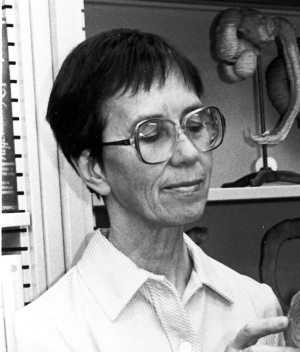
Embryology History - Fabiola Müller
| Embryology - 27 Apr 2024 |
|---|
| Google Translate - select your language from the list shown below (this will open a new external page) |
|
العربية | català | 中文 | 中國傳統的 | français | Deutsche | עִברִית | हिंदी | bahasa Indonesia | italiano | 日本語 | 한국어 | မြန်မာ | Pilipino | Polskie | português | ਪੰਜਾਬੀ ਦੇ | Română | русский | Español | Swahili | Svensk | ไทย | Türkçe | اردو | ייִדיש | Tiếng Việt These external translations are automated and may not be accurate. (More? About Translations) |
Introduction
Cite error: Invalid <ref> tag; refs with no name must have content
- School of Medicine, University of California, Davis, CA, USA.
Fabiola Müller and Ronan O'Rahilly
| Embryologists: William Hunter | Wilhelm Roux | Caspar Wolff | Wilhelm His | Oscar Hertwig | Julius Kollmann | Hans Spemann | Francis Balfour | Charles Minot | Ambrosius Hubrecht | Charles Bardeen | Franz Keibel | Franklin Mall | Florence Sabin | George Streeter | George Corner | James Hill | Jan Florian | Thomas Bryce | Thomas Morgan | Ernest Frazer | Francisco Orts-Llorca | José Doménech Mateu | Frederic Lewis | Arthur Meyer | Robert Meyer | Erich Blechschmidt | Klaus Hinrichsen | Hideo Nishimura | Arthur Hertig | John Rock | Viktor Hamburger | Mary Lyon | Nicole Le Douarin | Robert Winston | Fabiola Müller | Ronan O'Rahilly | Robert Edwards | John Gurdon | Shinya Yamanaka | Embryology History | Category:People | ||
|
Books
O'Rahilly R. and Müller F. Developmental Stages in Human Embryos. Contrib. Embryol., Carnegie Inst. Wash. 637 (1987).
Ronan O'Rahilly and Müller, Fabiola. Human Embryology and Teratology. (1990) Lippincott Williams & Wilkins 400 pages
Vertebral Column Development
The human vertebral column at the end of the embryonic period proper. 1. The column as a whole[1]
- "The present investigation of the vertebral column at 8 post-ovulatory weeks, the first such study based on precise reconstructions, has revealed 33 or 34 cartilaginous vertebrae arranged in flexion and approximately 20--33 mm in total length. At the end of the embryonic period proper, a typical vertebra, such as TV6, consists of a centrum that is continuous with two neural processes. Pedicles, articular and transverse processes, but no spinous processes, are identifiable. The tips of the neural processes, which are formed by the laminae, are connected by fibrous tissue and resemble the condition of total rachischisis. The union of the laminae, the onset of ossification, and the appearance of articular cavities are characteristic of the early fetal period. The variations encountered within a single developmental stage were noted. They were mostly minor, e.g. the number of coccygeal elements and the extent of the dorsal growth of the neural processes."
The human vertebral column at the end of the embryonic period proper. 2. The occipitocervical region[2]
- "The present investigation of the cervical region of the vertebral column at eight post-ovulatory weeks is the first such study based on precise reconstructions of staged embryos. At the end of the embryonic period proper, a typical vertebra is a U-shaped piece of cartilage characterized by spina bifida occulta. The notochord ascends through the centra and leaves the dens to enter the basal plate of the skull. The median column of the axis comprises three parts (designated X, Y, Z) which persist well into the fetal period. They are related to the first, second and third cervical nerves, respectively. Part X may project into the foramen magnum and form an occipito-axial joint. Part Z appears to be the centrum of the axis. The articular columns of the cervical vertebrae are twofold, as in the adult: an anterior (atlanto-occipital and atlanto-axial) and a posterior (from the lower aspect of the axis downwards). Alar and transverse ligaments are present. Cavitation is not found in the embryonic period in either the atlanto-occipital or zygapophysial joints, and is generally not present in the median atlanto-axial joint either. Most of the transverse processes exhibit anterior and posterior tubercles. An 'intertubercular lamella' may or may not be present, i.e. the foramina transversaria are being formed around the vertebral artery. The spinal ganglia are generally partly in the vertebral canal and partly on the neural arches, medial to the articular processes. During the fetal period, the articular processes shift to a coronal position and this alteration appears to be associated with a corresponding change in the location of the spinal ganglia."
The human vertebral column at the end of the embryonic period proper. 3. The thoracicolumbar region[3]
- "The present study of the thoracicolumbar region continues an investigation of the vertebral column at 8 postovulartory weeks (the end of the embryonic period proper) by means of graphic reconstructions. The cartilaginous vertebrae have short neural processes associated with the normal spina bifida occulta present at this time. The separate cartilaginous centres that several authors believe to exist in the cervical and lumbar costal elements, but which have not been observed by the present authors, have been thought to be the forerunners of extrathoracic ribs. A distinction needs to be made, however, between such centres and ribs. Similarly, in the fetal period, ossific loci in the costal elements of CV 7 are very frequent, whereas cervical ribs in the adult are relatively rare. The neurocentral joints, and hence the boundaries between neural arches and centra, are unclear before ossification has begun and has progressed during the fetal period. The sternal bands are almost completely united and the scapula is high in position. Neural relationships aid in the determination of homologous parts within the vertebral column, but clarification of corresponding parts has not previously been possible within the embryonic period. Areas ventral to the dorsal rami are ribs in the thoracic region and costal elements in other regions. Areas underlying the dorsal rami are transverse processes in the thoracic region and minute 'true' transverse elements in the cervical and lumbar regions. Thus, the descriptive lumbar transverse processes correspond to the true transverse processes and the ribs in the thoracic region. The dorsal rami of the thoracic nerves pass between the transverse processes and the tubercles of the ribs and then divide. The ventral rami of lumbar Nerves 1 and 2 resemble the thoracic in their course, whereas those of Nerves 3-5 are similar to the sacral. The thoracic dorsal roots are sloping and, associated with the greater height of the lumbar centra, the lumbar roots even more so. The directions of the various dorsal roots reflect differences in growth gradients between vertebral column and spinal cord. The thoracic and lumbar portions of the column change little in proportion during the embryonic period proper."
The human vertebral column at the end of the embryonic period proper. 4. The sacrococcygeal region[4]
- "The sacral and coccygeal vertebrae at 8 postovulatory weeks (the end of the embryonic period proper) have been studied by means of graphic reconstructions. The cartilaginous sacrum is now a definitive unit composed of five separable vertebrae, each of which consists of a future centrum and bilateral neural processes. The base of each neural process consists of an anterolateral or alar element, not present in the lumbar region, and a posterolateral part, which includes costal and transverse elements. The usual illustrations, in which the costal component is placed in the alar element, are incorrect. The future dorsal foramina (containing dorsal rami) face laterally in the embryo and are in line with the thoracicolumbar intervertebral foramina. Considerable differential growth is required to change the dorsal openings from a lateral to a dorsal positions. The intervertebral foramina transmit ventral rami, but pelvic foramina are not yet present. The lumbosacral plexus is completed by S.N.1-3; S.N.4, 5 and Co.N.1 form the pelvic plexus. The inferior hypogastric plexus and the hypogastric nerves are present. The sacrum takes part in the spina bifida occulta that characterises the entire length of the embryonic vertebral column. The coccygeal vertebrae, which are variable, were 4-6 in number in the present series. The first is the best developed. The ventriculus terminalis ends usually at the level of Co.V.1 and the spinal cord generally at Co.V.5. The coccygeal notochord ends commonly in bifurcation or trifurcation. 'Haemal arches' were not observed."
Occipitocervical segmentation in staged human embryos[5]
- "Serial sections of 108 human embryos from stage 11 to stage 23 were investigated, and 33 reconstructions were prepared. The existence of 4 occipital somites was confirmed. The important developmental distinction between axial (central) and lateral components obtains in the occipital as well as in the vertebral region. The lateral occipital components begin to show dense areas as the cervical region is approached. The lateral occipital and vertebral components arise in registration with the initial sclerotomes. In both the occipital and the vertebral region the related nerves and intersegmental arteries traverse the loose areas of the sclerotomes. The axial occipital region is not segmented, whereas the cervical components develop from perinotochordal loose areas. Three complete centra (known as XYZ) develop in the atlanto-axial region, although they are related to only 2 1/2 sclerotomes and only 2 neural arches. The height of the XYZ complex equals that of 3 centra elsewhere, and not 2 1/2, as previously maintained. The experimental findings in the occipitocervical region of the chick embryo show both similarities to, as well as differences from, the data for the human embryo. A scheme showing the early development of the entire vertebral column is included."
The timing and sequence of events in the development of the human vertebral column during the embryonic period proper.[6]
- "A documented scheme of the early development of the human vertebrae is presented. It is based on (1) reports of workers who personally studied staged human embryos, and (2) personal observations and confirmations. The necessity of studying staged embryos in order to determine the precise sequence of developmental events is stressed."
Carnegie Stage 12
Development of the human brain, the closure of the caudal neuropore, and the beginning of secondary neurulation at stage 12.[7]
References
- ↑ O'Rahilly R, Muller F & Meyer DB. (1980). The human vertebral column at the end of the embryonic period proper. 1. The column as a whole. J. Anat. , 131, 565-75. PMID: 7216919
- ↑ O'Rahilly R, Müller F & Meyer DB. (1983). The human vertebral column at the end of the embryonic period proper. 2. The occipitocervical region. J. Anat. , 136, 181-95. PMID: 6833119
- ↑ O'Rahilly R, Müller F & Meyer DB. (1990). The human vertebral column at the end of the embryonic period proper. 3. The thoracicolumbar region. J. Anat. , 168, 81-93. PMID: 2323997
- ↑ O'Rahilly R, Müller F & Meyer DB. (1990). The human vertebral column at the end of the embryonic period proper. 4. The sacrococcygeal region. J. Anat. , 168, 95-111. PMID: 2182589
- ↑ Müller F & O'Rahilly R. (1994). Occipitocervical segmentation in staged human embryos. J. Anat. , 185 ( Pt 2), 251-8. PMID: 7961131
- ↑ O'Rahilly R & Meyer DB. (1979). The timing and sequence of events in the development of the human vertebral column during the embryonic period proper. Anat. Embryol. , 157, 167-76. PMID: 517765
- ↑ Müller F. and O'Rahilly R. The development of the human brain, the closure of the caudal neuropore, and the beginning of secondary neurulation at stage 12. (1987) Anat Embryol (Berl). 176(4): 413-430. PMID 3688450
Müller F. and O'Rahilly R. The human brain at stages 18-20, including the choroid plexuses and the amygdaloid and septal nuclei. (1990) Anat Embryol (Berl). 182(3): 285-306. PMID 2268071
Search Pubmed
Glossary Links
- Glossary: A | B | C | D | E | F | G | H | I | J | K | L | M | N | O | P | Q | R | S | T | U | V | W | X | Y | Z | Numbers | Symbols | Term Link
Cite this page: Hill, M.A. (2024, April 27) Embryology Embryology History - Fabiola Müller. Retrieved from https://embryology.med.unsw.edu.au/embryology/index.php/Embryology_History_-_Fabiola_M%C3%BCller
- © Dr Mark Hill 2024, UNSW Embryology ISBN: 978 0 7334 2609 4 - UNSW CRICOS Provider Code No. 00098G
