Book - Normal Plates of the Development of Vertebrates 5
| Embryology - 15 Jul 2026 |
|---|
| Google Translate - select your language from the list shown below (this will open a new external page) |
|
العربية | català | 中文 | 中國傳統的 | français | Deutsche | עִברִית | हिंदी | bahasa Indonesia | italiano | 日本語 | 한국어 | မြန်မာ | Pilipino | Polskie | português | ਪੰਜਾਬੀ ਦੇ | Română | русский | Español | Swahili | Svensk | ไทย | Türkçe | اردو | ייִדיש | Tiếng Việt These external translations are automated and may not be accurate. (More? About Translations) |
Minot CS. and Taylor E. Normal Plates of the Development of the Rabbit Embryo (Lepus cuniculus). Vol. 5 in series by Keibel F. Normal plates of the development of vertebrates (Normentafeln zur Entwicklungsgeschichte der Wirbelthiere) Fisher, Jena., Germany.
| Historic Disclaimer - information about historic embryology pages |
|---|
 |
Normal Plates of the Development of the Rabbit Embryo (Lepus cuniculus)

Normentafeln zur Entwicklungsgeschichte der Wirbeltiere - Lepus cuniculus
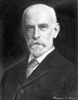
by Charles S. Minot and Edwing Taylor
This is the 1905 Volume 5 of the series that is specific for rabbit development.
| Normal Plates Series: 1897 Pig | 1900 Chicken | 1901 Lungfish | 1904 Sand Lizard | 1905 Rabbit | 1906 Deer | 1907 Tarsiers | 1908 Human | 1909 Northern Lapwing | 1909 South American and African Lungfish | 1910 Salamander | Franz Keibel | Embryology History |
- Links: Franz Keibel | Rabbit Development | Embryology History
Preface
The Normal Plates of the rabbit were originally undertaken by me in 1896, in response to the invitation with which my friend, Professor KEIBEL, the Editor of the Series, honored me. It seemed to me that the rabbit offered particularly favorable opportunities for obtaining stages, which should be really nearly normal, i. e., representative of the median of the variations for each selected age. Accordingly I began collecting litters of embryos of known ages from -nine to twenty-one days, the ages selected being always either even days or half-days. Of each age at least four litters were secured, and of some ages six or seven. The next step was to select for each age by careful comparison of the specimens of that age with one another that litter of embryos which appeared nearest central. Out of this litter three embryos were taken for sectioning as representing the norm for that age. In a few cases the selected embryos were not all from the same litter. Next the selected normal or median embryos of all the ages were compared with one another to make sure that they formed a good progressive series. A typical embryo of each set of three was drawn and thus the series of figures on the plates was prepared. As will be seen the method worked satisfactorily on the whole, though the "normal" embryos of twelve and one half and of thirteen days do not fit perfectly into the series figured. Of the older stages (nine to twenty days), it may be claimed, I think, that we really have considered “normal” embryos.
The three selected embryos of each stage were sectioned, one in the transverse, one in the sagittal and one in the frontal plane. The three series of sections in each case have been added to the Harvard Embryological Collection, where they will be always accessible to competent investigators. The data as to the development of the embryos have been obtained from the study of these series, and similar ones of younger stages. Study soon showed that the three "normal" embryos agree very closely with one another in the details of their development, so that as a rule a correct collective statement as to the condition of each organ could be drawn up with little difficulty so as to be applicable to all three embryos. Exceptions to this rule are not very frequent, and all the important ones observed are noted in the tables.
Under these circumstances Professor KEIBEL consented to my having the collaboration of Dr. Edwing TAYLOR, and it is owing to his steady industry that the work is now completed.
The observations, upon which all the tabulations and descriptions are based, have been made by Dr. TAYLOR, who has been occupied with this labour for two years, during which he has devoted all the time, which could be spared from his duties as Assistant in my Laboratory. My own share has been that of a consultant. To Dr. TAYLOR therefore belongs the chief credit and a large share of the responsibility for this publication. Most of the drudgery of getting together the titles for the Bibliography has also been borne by Dr. TAYLOR. We have endeavored to make the Bibliography complete as regards embryology, extensive as regards anatomy, but have included only the more important systematic and palaeontological papers, which seemed likely to be of interest to embryologists.
Description of Embryos pictured
The embryos were in nearly every case fixed in ZENKER'S fluid, as noted in the separate descriptions. The measurements and drawings were made from the specimens preserved in 80% alcohol, with one exception, the blastodermic vesicle of Fig. I. H. E. C. stands for Harvard Embryological Collection. The numbers in the column marked "Designation" in the Tables are the numbers of the series in this collection.
Figs. 3 (X 20) and 15 (X 5).
The drawing is a reproduction from another drawing made from a specimen which has since been cut. Embryo removed from uterus 7.5, days after coitus. ZENKER fixation. Mesoderm measured 4.oX3.4 mm. The embryonic shield is pear-shaped. Hi<:NsEN’s knot, situated a little anterior to center of shield, is distinct. Notochordal anlage is visible as a more opaque band extending forward from HENsEN’s knot. Primitive groove runs from HENsEN’s knot to posterior end of shield. A more opaque area, corresponding to the extent of the inesoderm, stretches for some distance around the shield. This area reaches the anterior margin of the shield but does not pass in front of it. Over this area, beyond the boundaries of the embryonic shield, the outer ectodermal layer (trophoblast of HUBRECHT) is somewhat thickened. There is a distinct projection at HENSEN’s knot. The embryonic ectoderm over the region of the notochordal anlage is a little thinner than laterally in this part of the shield. The primitive groove is very shallow. H. E. C., No. 622.
Figs. 4 (X 20) and 16 (x 5).
Specimen removed from uterus 8 days, 6 hours after coitus. TELLYESNICKY fixation. Measured 1.8 mm. from anterior end of embryonic shield to posterior end of primitive streak. No segments. Embryonic shield broader at anterior end than at posterior. HENSEN’s knot is distinct: it is situated a little posterior to center of shield. The primitive groove extends from HENSEN’s knot to the posterior end of the shield, ending there in an opaque spot. The notochordal anlage is faintly indicated as a more opaque band extending forward from HENSEN’s knot. The ectoderm of the lateral portion of the shield, anterior to the region of Hr«:NsEN’s knot, is somewhat thicker than the median ectoderm over the notochordal a-nlage. This apparently marks the beginning of the medullary plate with a suggestion of a groove in the center. There is no projection at HENSEN’s knot. There are apparently no blood anlagen. This specimen is only a little more advanced than that of Fig. 3.
Figs. 5 (X 20) and 17 (X 5).
Embryo removed from uterus 8 days, 6 hours after coitus. ZENKER fixation. Measured 2.2 mm. from anterior end of medullary plate to posterior end of primitive streak. (The specimen was, however, a little bent, concave dorsally.) Medullary plate distinct, elongated, flat, slightly expanded at anterior end on each side in a curved manner. Posteriorly it surrounds HENSEN’s knot and merges into region of primitive s eak. Medullary groove is relatively wide and shallow. The notochordal anlage is visible through the
cove. The primitive streak is considerably shorter than the medullary plate. The primitive grooveiisy
HENSEN’s knot conspicuous as a circular opaque spot. Only one pair of segments is,e,l,ear§ .on both sides, but a second pair, posterior to the former, is clearly indicated though no the caudal side. The segments lie under the. narrowest part of the medullary than the anterior end of the plate. There are a few small extra-embryonic blood knot and merges into region of primitive streak 4 Nornieiitufeln zur Eutivicklungsgescliichte def
Notochordal anlage is visible through medullary groove. HENSEN’s knot is distinct as a (‘lI‘L‘Lll.’ll' O]l.'l(]ll4' spot The primitive streak is short There are three distinct pairs of segments but the third l‘-'“i" l‘ “OT
.- completely separated on the posterior aspect. A fourth pair, anterior to these tlirec. IS lndlfltill WI 1 small and not clearly marked ofl‘. There are a few extra-enibryonic blood anlagen and primitive blood cells.
There is a very small coelom.
Figs. 7 (X 20) and 19 (X 5).
The drawing is a reproduction of another drawing made from the specimen before sectioning. The embryo was removed from the uterus 8.5 days after coitus. ZENKER fixation. Measured 3.4 mm. The medullary groove is wide open. There are six distinctly formed pairs of segments; the seventh pair caudad is nearly separated. Primitive streak distinct. The caudal end of the embryo in the region of the primitive streak is somewhat bent. For internal development, see Table No. 2, made from a study of the sections of this embryo.
Figs. 8 (X 20) and 20 (X 5).
Embryo removed from uterus 8.5 days after coitus. ZENKER fixation. Measured 3.2 mm. from tip of head to caudal amnion. There are eight distinctly formed segments. Posterior to the eighth, a ninth is almost completed. The cephalic end of the embryo is raised above the level of the surrounding extra- embryonic disk but dips ventral a little into the proamnion. The back is flat. The medullary groove is open throughout. It is considerably expanded in the region of the optic diverticula; a little expanded in the region of the hind-brain; nearly closed in the region of the future mid-brain. The walls of the medullary groove, between the segments, approach each other, but, posterior to the segments, diverge to form a space in which the remnant of the primitive streak is seen. Laterad of the segments, as seen by transmitted light, is a narrow longitudinal light band, where there is a very small amount of mesoderm. Again laterad, is a broader, darker area, where the mesodenn is thicker and incloses the coelom. Posteriorly, the segments pass into an unsegmented band. The proamniotic area is distinct. The caudal fold of the amnion has
The area of extra-embryonic ectoderm, which was attached to the uterus and torn off on the oval of the specimen, is quite large. It reaches anteriorly as far as the plane of the hind-brain. On ?? view, the msodermal allantoic fold is plain. The pocket of the fore-gut is just discernible. There is not to be made out on external examination. Compare, for internal development, Tables
Figures
Plate 1

Plate 2

Plate 3

| Normal Plates Series: 1897 Pig | 1900 Chicken | 1901 Lungfish | 1904 Sand Lizard | 1905 Rabbit | 1906 Deer | 1907 Tarsiers | 1908 Human | 1909 Northern Lapwing | 1909 South American and African Lungfish | 1910 Salamander | Franz Keibel | Embryology History |
| Historic Disclaimer - information about historic embryology pages |
|---|
|
| Embryologists: William Hunter | Wilhelm Roux | Caspar Wolff | Wilhelm His | Oscar Hertwig | Julius Kollmann | Hans Spemann | Francis Balfour | Charles Minot | Ambrosius Hubrecht | Charles Bardeen | Franz Keibel | Franklin Mall | Florence Sabin | George Streeter | George Corner | James Hill | Jan Florian | Thomas Bryce | Thomas Morgan | Ernest Frazer | Francisco Orts-Llorca | José Doménech Mateu | Frederic Lewis | Arthur Meyer | Robert Meyer | Erich Blechschmidt | Klaus Hinrichsen | Hideo Nishimura | Arthur Hertig | John Rock | Viktor Hamburger | Mary Lyon | Nicole Le Douarin | Robert Winston | Fabiola Müller | Ronan O'Rahilly | Robert Edwards | John Gurdon | Shinya Yamanaka | Embryology History | Category:People | ||
|
Reference
Franz Keibel, Normentafeln zur Entwicklungsgeschichte der Wirbelthiere (Normal plates of the development of vertebrates) Volume Hft.5 (1905) Jena, G. Fischer, Germany.
Glossary Links
- Glossary: A | B | C | D | E | F | G | H | I | J | K | L | M | N | O | P | Q | R | S | T | U | V | W | X | Y | Z | Numbers | Symbols | Term Link
Cite this page: Hill, M.A. (2026, July 15) Embryology Book - Normal Plates of the Development of Vertebrates 5. Retrieved from https://embryology.med.unsw.edu.au/embryology/index.php/Book_-_Normal_Plates_of_the_Development_of_Vertebrates_5
- © Dr Mark Hill 2026, UNSW Embryology ISBN: 978 0 7334 2609 4 - UNSW CRICOS Provider Code No. 00098G