Shark Development: Difference between revisions
mNo edit summary |
mNo edit summary |
||
| (8 intermediate revisions by the same user not shown) | |||
| Line 1: | Line 1: | ||
{{Header}} | {{Header}} | ||
==Introduction== | ==Introduction== | ||
[[File:Model digit origin.png|thumb|alt=Digit origin - Tree shows phylogenetic relationships of shark, paddlefish, zebrafish and mouse|Digit origin - Tree shows phylogenetic relationships of shark, paddlefish, zebrafish and mouse]] | |||
Scyllium | |||
This page gives a brief introduction to {{shark}} development, a species used in many historic comparatively e embryology studies. The largest extant fish is the whale shark (Rhincodon typus). | |||
{{Animals}} | {{Animals}} | ||
==Some Recent Findings== | |||
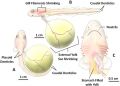
[[File:Catshark egg case stages1-7.jpg|thumb|Catshark egg case stages1-7|Catshark egg case stages 1-7{{#pmid:30399186|PMID30399186}}]] | |||
{| | |||
|-bgcolor="F5FAFF" | |||
| | |||
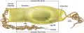
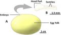
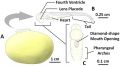
* '''Oviparous elasmobranch development inside the egg case in 7 key stages'''{{#pmid:30399186|PMID30399186}} "Embryological stages of oviparous elasmobranch during development can be difficult to identify, requiring magnification and/or fixation of an anaesthetized embryo. These restrictions are poorly suited for monitoring the development of living elasmobranchs inside their egg cases. There are two major aims of this study. The first was to observe elasmobranch embryonic development non-invasively and produce a non-invasive developmental key for identifying the life stages for an elasmobranch inside the egg case. To this end, 7 key developmental stages were identified for the greater spotted catshark, Scyliorhinus stellaris, and are provided here with diagrams from multiple perspectives to demonstrate the key features of each stage. The physiological and ecological relevance of each stage are discussed in terms of structure and function for embryonic survival in the harsh intertidal zone. Also discussed is the importance of the egg case membrane and the protective embryonic jelly. The second aim of the study was to understand the applicability of the 7 developmental stages from S. stellaris to other oviparous elasmobranchs. Thus, changes in embryonic body size and egg yolk volume at each stage were measured and compared with those of the closely related, lesser spotted catshark, Scyliorhinus canicula. We find nearly identical growth patterns and yolk consumption patterns in both species across the 7 developmental stages. Thus, although the 7 developmental stages have been constructed in reference to the greater spotted catshark, we suggest that it can be applied to other oviparous elasmobranch species with only minor modification." | |||
* '''A staging table for the embryonic development of the brownbanded bamboo shark (Chiloscyllium punctatum)'''{{#pmid:29396887|PMID29396887}} "Studying cartilaginous fishes (chondrichthyans) has helped us understand vertebrate evolution and diversity. However, resources such as genome sequences, embryos, and detailed staging tables are limited for species within this clade. To overcome these limitations, we have focused on a species, the brownbanded bamboo shark (Chiloscyllium punctatum), which is a relatively common aquarium species that lays eggs continuously throughout the year. In addition, because of its relatively small genome size, this species is promising for molecular studies. To enhance biological studies of cartilaginous fishes, we establish a normal staging table for the embryonic development of the brownbanded bamboo shark. Bamboo shark embryos take around 118 days to reach the hatching period at 25°C, which is approximately 1.5 times as fast as the small-spotted catshark (Scyliorhinus canicula) takes. Our staging table divides the embryonic period into 38 stages. Furthermore, we found culture conditions that allow early embryos to grow in partially opened egg cases. In addition to the embryonic staging table, we show that bamboo shark embryos exhibit relatively fast embryonic growth and are amenable to culture, key characteristics that enhance their experimental utility." | |||
|} | |||
= | {| class="wikitable mw-collapsible mw-collapsed" | ||
! More recent papers | |||
|- | |||
| [[File:Mark_Hill.jpg|90px|left]] {{Most_Recent_Refs}} | |||
Search term: [http://www.ncbi.nlm.nih.gov/pubmed/?term=Shark+Development ''Shark Development''] | | |||
[http://www.ncbi.nlm.nih.gov/pubmed/?term=elasmobranch+Development ''Elasmobranch Development''] | |||
|} | |||
{| class="wikitable mw-collapsible mw-collapsed" | |||
! Older papers | |||
|- | |||
| {{Older papers}} | |||
* Development of head and trunk mesoderm in the dogfish, Scyliorhinus torazame: I. Embryology and morphology of the head cavities and related structures{{#pmid:23017073|PMID23017073}} | |||
Vertebrate head segmentation has attracted the attention of comparative and evolutionary morphologists for centuries, given its importance for understanding the developmental body plan of vertebrates and its evolutionary origin. In particular, the segmentation of the mesoderm is central to the problem. The shark embryo has provided a canonical morphological scheme of the head, with its epithelialized coelomic cavities (head cavities), which have often been regarded as head somites. To understand the evolutionary significance of the head cavities, the embryonic development of the mesoderm was investigated at the morphological and histological levels in the shark, Scyliorhinus torazame. Unlike somites and some enterocoelic mesodermal components in other vertebrates, the head cavities in S. torazame appeared as irregular cyst(s) in the originally unsegmented mesenchymal head mesoderm, and not via segmentation of an undivided coelom. The mandibular cavity appeared first in the paraxial part of the mandibular mesoderm, followed by the hyoid cavity, and the premandibular cavity was the last to form. The prechordal plate was recognized as a rhomboid roof of the preoral gut, continuous with the rostral notochord, and was divided anteroposteriorly into two parts by the growth of the hypothalamic primordium. Of those, the posterior part was likely to differentiate into the premandibular cavity, and the anterior part disappeared later. The head cavities and somites in the trunk exhibited significant differences, in terms of histological appearance and timing of differentiation. The mandibular cavity developed a rostral process secondarily; its homology to the anterior cavity reported in some elasmobranch embryos is discussed." | |||
|} | |||
==Catshark Early Stages== | |||
A recent paper has categorised early catshark ''cyliorhinus stellaris'' development into 7 identifiable stages.{{#pmid:30399186|PMID30399186}} | |||
<gallery> | |||
File:Catshark egg case stage 1.jpg|Stage 1 | |||
File:Catshark egg case stage 2.jpg|Stage 2 | |||
File:Catshark egg case stage 3.jpg|Stage 3 | |||
File:Catshark egg case stage 4.jpg|Stage 4 | |||
File:Catshark egg case stage 5.jpg|Stage 5 | |||
File:Catshark egg case stage 6.jpg|Stage 6 | |||
File:Catshark egg case stage 6.jpg|Stage 7 | |||
</gallery> | |||
| Line 33: | Line 65: | ||
==References== | ==References== | ||
<references/> | |||
===Reviews=== | |||
===Articles=== | |||
Awruch, C. A., Pankhurst, N. W., Frusher, S. D., and Stevens, J. D. (2009). Reproductive seasonality and embryo development in the draughtboard shark Cephaloscyllium laticeps. Marine and Freshwater Research 60, 1265–1272.http://dx.doi.org/10.1071/MF090 http://www.publish.csiro.au/paper/MF09030.htm | |||
de Beer, G. R. The Development of the Skull of Scyllium (Scyliorhinus) canicula L. http://jcs.biologists.org/content/s2-74/296/591.full.pdf | de Beer, G. R. The Development of the Skull of Scyllium (Scyliorhinus) canicula L. http://jcs.biologists.org/content/s2-74/296/591.full.pdf | ||
| Line 39: | Line 77: | ||
==External Links== | ==External Links== | ||
{{External Links}} | {{External Links}} | ||
* Shark Foundation - [http://www.shark.ch/Information/Reproduction Reproduction] | * Shark Foundation - [http://www.shark.ch/Information/Reproduction Reproduction] | ||
| Line 46: | Line 83: | ||
{{Footer}} | {{Footer}} | ||
[[Category:Animal Development]][[Category:Shark]] | |||
Latest revision as of 10:28, 23 November 2019
| Embryology - 26 Apr 2024 |
|---|
| Google Translate - select your language from the list shown below (this will open a new external page) |
|
العربية | català | 中文 | 中國傳統的 | français | Deutsche | עִברִית | हिंदी | bahasa Indonesia | italiano | 日本語 | 한국어 | မြန်မာ | Pilipino | Polskie | português | ਪੰਜਾਬੀ ਦੇ | Română | русский | Español | Swahili | Svensk | ไทย | Türkçe | اردو | ייִדיש | Tiếng Việt These external translations are automated and may not be accurate. (More? About Translations) |
Introduction

Scyllium
This page gives a brief introduction to shark development, a species used in many historic comparatively e embryology studies. The largest extant fish is the whale shark (Rhincodon typus).
| Animal Development: axolotl | bat | cat | chicken | cow | dog | dolphin | echidna | fly | frog | goat | grasshopper | guinea pig | hamster | horse | kangaroo | koala | lizard | medaka | mouse | opossum | pig | platypus | rabbit | rat | salamander | sea squirt | sea urchin | sheep | worm | zebrafish | life cycles | development timetable | development models | K12 |
Some Recent Findings

|
| More recent papers |
|---|
 This table allows an automated computer search of the external PubMed database using the listed "Search term" text link.
More? References | Discussion Page | Journal Searches | 2019 References | 2020 References Search term: Shark Development | Elasmobranch Development |
| Older papers |
|---|
| These papers originally appeared in the Some Recent Findings table, but as that list grew in length have now been shuffled down to this collapsible table.
See also the Discussion Page for other references listed by year and References on this current page.
Vertebrate head segmentation has attracted the attention of comparative and evolutionary morphologists for centuries, given its importance for understanding the developmental body plan of vertebrates and its evolutionary origin. In particular, the segmentation of the mesoderm is central to the problem. The shark embryo has provided a canonical morphological scheme of the head, with its epithelialized coelomic cavities (head cavities), which have often been regarded as head somites. To understand the evolutionary significance of the head cavities, the embryonic development of the mesoderm was investigated at the morphological and histological levels in the shark, Scyliorhinus torazame. Unlike somites and some enterocoelic mesodermal components in other vertebrates, the head cavities in S. torazame appeared as irregular cyst(s) in the originally unsegmented mesenchymal head mesoderm, and not via segmentation of an undivided coelom. The mandibular cavity appeared first in the paraxial part of the mandibular mesoderm, followed by the hyoid cavity, and the premandibular cavity was the last to form. The prechordal plate was recognized as a rhomboid roof of the preoral gut, continuous with the rostral notochord, and was divided anteroposteriorly into two parts by the growth of the hypothalamic primordium. Of those, the posterior part was likely to differentiate into the premandibular cavity, and the anterior part disappeared later. The head cavities and somites in the trunk exhibited significant differences, in terms of histological appearance and timing of differentiation. The mandibular cavity developed a rostral process secondarily; its homology to the anterior cavity reported in some elasmobranch embryos is discussed." |
Catshark Early Stages
A recent paper has categorised early catshark cyliorhinus stellaris development into 7 identifiable stages.[1]
Stage 1
Stage 2
Stage 3
Stage 4
Stage 5
Stage 6
Stage 7


Additional Images
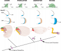
Digit origin - Tree shows phylogenetic relationships of shark, paddlefish, zebrafish and mouse.
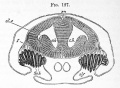
Fig. 127. Section through the brain and olfactory organ of an embryo of scyllium
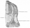
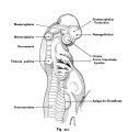
Fig. 395. From a transverse section through a shark (Scyllium) embryo of 15 mm
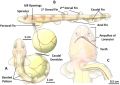
Branchial gill arch and columns, fissures branchiales in an embryo Shark
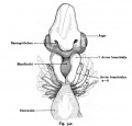
Fig. 342. The anterior end of an embryo of Shark
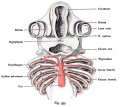
Fig. 535. The aortic arch in the shark embryo (Pristiurus)
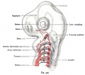
Fig. 536. The arteries of the gill arch region of a shark embryo (Pristiurus)


References
- ↑ 1.0 1.1 1.2 Musa SM, Czachur MV & Shiels HA. (2018). Oviparous elasmobranch development inside the egg case in 7 key stages. PLoS ONE , 13, e0206984. PMID: 30399186 DOI.
- ↑ Onimaru K, Motone F, Kiyatake I, Nishida K & Kuraku S. (2018). A staging table for the embryonic development of the brownbanded bamboo shark (Chiloscyllium punctatum). Dev. Dyn. , 247, 712-723. PMID: 29396887 DOI.
- ↑ Adachi N & Kuratani S. (2012). Development of head and trunk mesoderm in the dogfish, Scyliorhinus torazame: I. Embryology and morphology of the head cavities and related structures. Evol. Dev. , 14, 234-56. PMID: 23017073 DOI.
Reviews
Articles
Awruch, C. A., Pankhurst, N. W., Frusher, S. D., and Stevens, J. D. (2009). Reproductive seasonality and embryo development in the draughtboard shark Cephaloscyllium laticeps. Marine and Freshwater Research 60, 1265–1272.http://dx.doi.org/10.1071/MF090 http://www.publish.csiro.au/paper/MF09030.htm
de Beer, G. R. The Development of the Skull of Scyllium (Scyliorhinus) canicula L. http://jcs.biologists.org/content/s2-74/296/591.full.pdf
External Links
External Links Notice - The dynamic nature of the internet may mean that some of these listed links may no longer function. If the link no longer works search the web with the link text or name. Links to any external commercial sites are provided for information purposes only and should never be considered an endorsement. UNSW Embryology is provided as an educational resource with no clinical information or commercial affiliation.
- Shark Foundation - Reproduction
Glossary Links
- Glossary: A | B | C | D | E | F | G | H | I | J | K | L | M | N | O | P | Q | R | S | T | U | V | W | X | Y | Z | Numbers | Symbols | Term Link
Cite this page: Hill, M.A. (2024, April 26) Embryology Shark Development. Retrieved from https://embryology.med.unsw.edu.au/embryology/index.php/Shark_Development
- © Dr Mark Hill 2024, UNSW Embryology ISBN: 978 0 7334 2609 4 - UNSW CRICOS Provider Code No. 00098G