The printable version is no longer supported and may have rendering errors. Please update your browser bookmarks and please use the default browser print function instead.
Introduction
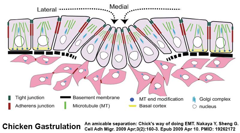
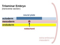
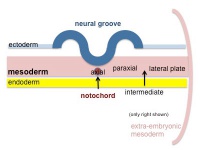
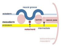
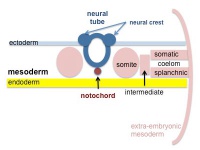
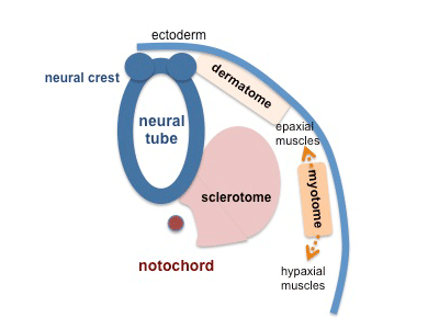
The middle layer of the early trilaminar embryo germ layers (ectoderm, mesoderm and endoderm) formed by gastrulation. The segmentation of the initial mesoderm into somites, and their regular addition, is often used to stage embryonic development (23 somite embryo).
This middle germ layer forms connective tissues and muscle throughout the body, with the exception of in the head region where some of these structures have a neural crest (ectoderm) origin.
- connective tissues - cartilage, bone, blood, blood vessel endothelium, dermis, etc.
- muscle - cardiac, skeletal, smooth.
Students often mix-up the terms mesoderm (middle layer) with mesenchyme (embryonic connective tissue). It is true that mesoderm initially does have a mesenchymal cellular organisation, but can also form a range of epithelial structures (surrounding somites, mesothelium lining of body cavities).
Some Recent Findings
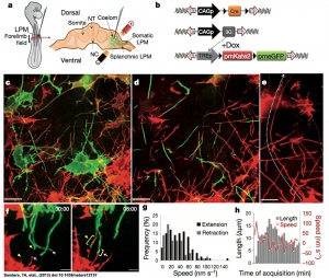
Mesenchymal cells of the developing limb bud possess long and highly dynamic cytoplasmic extensions.
[1]
- STRIP1, a core component of STRIPAK complexes, is essential for normal mesoderm migration in the mouse embryo[2] "Regulated mesoderm migration is necessary for the proper morphogenesis and organ formation during embryonic development. Cell migration and its dependence on the cytoskeleton and signaling machines have been studied extensively in cultured cells; in contrast, remarkably little is known about the mechanisms that regulate mesoderm cell migration in vivo. Here, we report the identification and characterization of a mouse mutation in striatin-interacting protein 1 (Strip1) that disrupts migration of the mesoderm after the gastrulation epithelial-to-mesenchymal transition (EMT). STRIP1 is a core component of the biochemically defined mammalian striatin-interacting phosphatases and kinase (STRIPAK) complexes that appear to act through regulation of protein phosphatase 2A (PP2A), but their functions in mammals in vivo have not been examined. Strip1-null mutants arrest development at midgestation with profound disruptions in the organization of the mesoderm and its derivatives, including a complete failure of the anterior extension of axial mesoderm. Analysis of cultured mesoderm explants and mouse embryonic fibroblasts from null mutants shows that the mesoderm migration defect is correlated with decreased cell spreading, abnormal focal adhesions, changes in the organization of the actin cytoskeleton, and decreased velocity of cell migration. The results show that STRIPAK complexes are essential for cell migration and tissue morphogenesis in vivo." (More? Cell Migration | NCBI Gene - STRIP1)
- A role for Vg1/Nodal signaling in specification of the intermediate mesoderm[3] "The intermediate mesoderm (IM) is the embryonic source of all kidney tissue in vertebrates. The factors that regulate the formation of the IM are not yet well understood. Through investigations in the chick embryo, the current study identifies and characterizes Vg1/Nodal signaling (henceforth referred to as 'Nodal-like signaling') as a novel regulator of IM formation. ... We postulate that Nodal-like signaling regulates IM formation by modulating the IM-inducing effects of BMP signaling." Renal System Development
- Signaling gradients during paraxial mesoderm development[4] "These studies indicate that high levels of Wnt and FGF signaling are required for the segmentation clock activity. Furthermore, we discuss how these signaling gradients act in a dose-dependent manner in the progenitors of the paraxial mesoderm, partly by regulating cell movements during gastrulation. Finally, links between the process of axial specification of vertebral segments and Hox gene expression are discussed."
- Transcriptional profiling of the nucleus pulposus: say yes to notochord[5]"This editorial addresses the debate concerning the origin of adult nucleus pulposus cells in the light of profiling studies by Minogue and colleagues. In their report of several marker genes that distinguish nucleus pulposus cells from other related cell types, the authors provide novel insights into the notochordal nature of the former. Together with recently published work, their work lends support to the view that all cells present within the nucleus pulposus are derived from the notochord. Hence, the choice of an animal model for disc research should be based on considerations other than the cell loss and replacement by non-notochordal cells."
|
| More recent papers
|
|
This table allows an automated computer search of the external PubMed database using the listed "Search term" text link.
- This search now requires a manual link as the original PubMed extension has been disabled.
- The displayed list of references do not reflect any editorial selection of material based on content or relevance.
- References also appear on this list based upon the date of the actual page viewing.
References listed on the rest of the content page and the associated discussion page (listed under the publication year sub-headings) do include some editorial selection based upon both relevance and availability.
More? References | Discussion Page | Journal Searches | 2019 References | 2020 References
Search term: Mesoderm Development | Images
<pubmed limit=5>Mesoderm+Development</pubmed>
|
Mesoderm Movies
Mesoderm Formation during Gastrulation
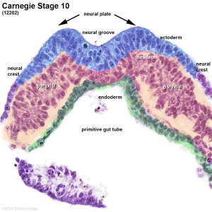
Human embryo (stage 10) mesoderm
- Links: Gastrulation
Patterning
Notochord secreting sonic hedgehog, shown in white

Somite patterning
Molecular Factors
References
- ↑ Timothy A. Sanders, Esther Llagostera, Maria Barna. Specialized filopodia direct long-range transport of SHH during vertebrate tissue patterning. Nature Apr 28, 2013.
- ↑ <pubmed>29203676</pubmed>
- ↑ <pubmed>23533180</pubmed>
- ↑ <pubmed>20182616</pubmed>
- ↑ <pubmed>20497604</pubmed>
Reviews
<pubmed>20568241</pubmed>
<pubmed>17705304</pubmed>
Articles
<pubmed>21159819</pubmed>
<pubmed>20565707</pubmed>
<pubmed>7956820</pubmed>
Historic
<pubmed>17104422</pubmed>
Search PubMed
Search NLM Online Textbooks: "Mesoderm" : Developmental Biology | The Cell- A molecular Approach | Molecular Biology of the Cell | Endocrinology
Search Pubmed: Mesoderm | Notochord
External Links
External Links Notice - The dynamic nature of the internet may mean that some of these listed links may no longer function. If the link no longer works search the web with the link text or name. Links to any external commercial sites are provided for information purposes only and should never be considered an endorsement. UNSW Embryology is provided as an educational resource with no clinical information or commercial affiliation.
Take the Quiz
Glossary Links
- Glossary: A | B | C | D | E | F | G | H | I | J | K | L | M | N | O | P | Q | R | S | T | U | V | W | X | Y | Z | Numbers | Symbols | Term Link
Cite this page: Hill, M.A. (2024, April 26) Embryology Mesoderm. Retrieved from https://embryology.med.unsw.edu.au/embryology/index.php/Mesoderm
- What Links Here?
- © Dr Mark Hill 2024, UNSW Embryology ISBN: 978 0 7334 2609 4 - UNSW CRICOS Provider Code No. 00098G


