Head Development: Difference between revisions
mNo edit summary |
mNo edit summary |
||
| Line 18: | Line 18: | ||
* '''hand2 and Dlx genes specify dorsal, intermediate and ventral domains within zebrafish pharyngeal arches.'''<ref><pubmed>20573696</pubmed></ref> "The ventrally expressed secreted polypeptide endothelin1 (Edn1) patterns the skeleton derived from the first two pharyngeal arches into dorsal, intermediate and ventral domains. ...Collectively, our work indicates that the expression and function of hand2 and Dlx genes specify major patterning domains along the dorsoventral axis of zebrafish pharyngeal arches." | * '''hand2 and Dlx genes specify dorsal, intermediate and ventral domains within zebrafish pharyngeal arches.'''<ref><pubmed>20573696</pubmed></ref> "The ventrally expressed secreted polypeptide endothelin1 (Edn1) patterns the skeleton derived from the first two pharyngeal arches into dorsal, intermediate and ventral domains. ...Collectively, our work indicates that the expression and function of hand2 and Dlx genes specify major patterning domains along the dorsoventral axis of zebrafish pharyngeal arches." | ||
|} | |} | ||
{| class="wikitable collapsible collapsed" | |||
! More recent papers | |||
|- | |||
| [[File:Mark_Hill.jpg|90px|left]] {{Most_Recent_Refs}} | |||
Search term: [http://www.ncbi.nlm.nih.gov/pubmed/?term=Head+Development ''Head Development''] | |||
<pubmed limit=5>Head Development</pubmed> | |||
|} | |||
== Textbooks == | == Textbooks == | ||
[[File:Pharyngeal_arch_cartilages.jpg|thumb]] | [[File:Pharyngeal_arch_cartilages.jpg|thumb]] | ||
| Line 417: | Line 425: | ||
===Search PubMed=== | ===Search PubMed=== | ||
Search | Search term: [http://www.ncbi.nlm.nih.gov/pubmed/?term=Head+Development ''Head Development''] | [http://www.ncbi.nlm.nih.gov/pubmed/?term=Pharyngeal+Arch+Development ''Pharyngeal Arch Development''] | | [http://www.ncbi.nlm.nih.gov/pubmed/?term=Face+Development ''Face Development''] | ||
==Additional Images== | ==Additional Images== | ||
| Line 431: | Line 437: | ||
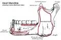
File:Musculoskeletal-_adult_mandible.jpg|adult mandible | File:Musculoskeletal-_adult_mandible.jpg|adult mandible | ||
File:Musculoskeletal-_adult_hyoid.jpg|adult hyoid | File:Musculoskeletal-_adult_hyoid.jpg|adult hyoid | ||
</gallery> | |||
===Historic=== | |||
[[Book_-_Human_Embryology_and_Morphology_1|Development or the Face]] | |||
<gallery> | |||
File:Keith1902 fig001.jpg|Fig. 1. Showing the formation of the face by the Nasal, Maxillary, and Mandibular processes in an embryo of the 4th week. | |||
File:Keith1902 fig002.jpg|Fig. 2. Showing the parts of the face formed from the Nasal, Maxillary and Mandibular processes. | |||
File:Keith1902 fig003.jpg|Fig. 3. Showing the structures formed in the Mesial Nasal Processes. | |||
File:Keith1902 fig004.jpg|Fig. 4. Showing the trough-shaped Vomer of the newly born. | |||
File:Keith1902 fig005.jpg|Fig. 5. Showing the suture on the face between the premaxilla and maxilla in the skull of a young orang. | |||
File:Keith1902 fig006.jpg|Fig. 6. Showing the structures formed in the Lateral Nasal Processes. | |||
File:Keith1902 fig007.jpg|Fig. 7. Coronal section of the skull of a 7th month human foetus to show the cartilages of the Lateral and Mesial Nasal Processes and the bones formed round them. | |||
File:Keith1902 fig008.jpg|Fig. 8. Showing the ingrowth of the palatal plates of the two maxillary processes early in the 2nd month. (After Kollmann.) . | |||
File:Keith1902 fig009.jpg|Fig. 9. Showing the Hard Palate at birth. The premaxillary part is formed from the Mesial Nasal Processes ; the remainder by the Palatal Plates of the Maxillary Processes. | |||
File:Keith1902 fig010a-c.jpg|Fig. 10, a, b, c. Showing what become of the skeletons of the Mandibular Arch (Meckel's Cartilage) and Maxillary Process (Palato-quadrate Cartilage). | |||
File:Keith1902 fig010d.jpg|Fig. 10 D. Illustrating Gadow's view of the origin of the Auditory Ossicles and Tympanic Plate. | |||
File:Keith1902 fig011.jpg|Fig. 11. Showing the manner in which the development of the Maxillary Antrum affects the size of the palate and position of the molar teeth. | |||
File:Keith1902 fig012.jpg|Fig. 12. Showing the Centres of Ossification and age changes in the Lower Jaw. | |||
File:Keith1902 fig013.jpg|Fig. 13. The chief types of the Temporo-Maxillary Articulation. '''A.''' Carnivorous Type. '''B.''' Omnivorous Type. '''C.''' Herbivorous Type. | |||
File:Keith1902 fig014.jpg|Fig. 14. Showing the Chief Changes after birth in the form of the TemporoMaxillary Articulation. | |||
File:Keith1902 fig015a.jpg|Fig. 15 A. Sagittal Section showing the Stomodaeum and position of the Oral Plate in the 3rd week. | |||
File:Keith1902 fig015b.jpg|Fig. 15 B. Showing the parts of the Buccal and Nasal Cavities formed from the Stomodaeum. The relative position of the Oral Plate is indicated. | |||
</gallery> | </gallery> | ||
[[Book_-_Human_Embryology_and_Morphology_3|Development of the Pharynx and Neck]] | |||
<gallery> | |||
File:Keith1902 fig021a.jpg|Fig. 21a. Showing the Visceral Arches and Cleft-depressions in the Pharyngeal Wall of a 4th week human Embryo. Each Visceral Arch contains an Aortic Arch. (After His.) | |||
File:Keith1902 fig021b.jpg|Fig. 21b. Showing the position of the Heart, Visceral and Aortic Arches in a fish. (Diagrammatic — after Gegenbaur.) | |||
File:Keith1902 fig022.jpg|Fig. 22. Showing the Primitive Pharynx of a 3rd week embryo in sagittal section, bounded by the Visceral Arches. (After His.) . | |||
File:Keith1902 fig023.jpg|Fig. 23. Showing the Floor of the Pharynx of a 4th week human embryo. (After His.) | |||
File:Keith1902 fig024.jpg|Fig. 24. Schematic Section of a Visceral Arch. | |||
File:Keith1902 fig025.jpg|Fig. 25. Showing the position of the External Cleft Depressions in the Adult. | |||
File:Keith1902 fig026.jpg|Fig. 26. Showing what become of the Cartilages of the Visceral Arches. | |||
File:Keith1902 fig027.jpg|Fig. 27. Showing what become of the Nerves of the Visceral Arches, hyoid arch) is represented by the chorda tympani and great superficial petrosal. | |||
File:Keith1902 fig028.jpg|Fig. 28. Showing what become of the Aortic Arches in the adult. Only the shaded parts persist. | |||
File:Keith1902 fig029.jpg|Fig. 29. The condition of the Eight and Left Doral Aortae in a 6th week human foetus. | |||
File:Keith1902 fig030.jpg|Fig. 30. Showing the Buccal and Pharyngeal parts of the Tongue. | |||
File:Keith1902 fig031.jpg|Fig. 31. Showing the origin of the tongue in the floor of the primitive pharynx. (After His.) | |||
File:Keith1902 fig032.jpg|Fig. 32. Showing the origin of the Submaxillary and Sublingual Glands from furrows between the gum and tongue during the flth week. (After His.) . | |||
File:Keith1902 fig033.jpg|Fig. 33. Showing the position of the Visceral Clefts in the Adult. | |||
File:Keith1902 fig034.jpg|Fig. 34. Showing the origin of the Tonsil, Thymus, and Thyroid from the Internal Cleft Recesses during the 4th week. (After His.). | |||
</gallery> | |||
==External Links== | ==External Links== | ||
{{External Links}} | {{External Links}} | ||
| Line 441: | Line 486: | ||
---- | ---- | ||
{{Systems}} | {{Systems}} | ||
{{Glossary}} | {{Glossary}} | ||
{{Footer}} | {{Footer}} | ||
[[Category:System Development]] | [[Category:System Development]] | ||
Revision as of 09:45, 21 January 2014
Introduction

The head and neck is not really a "system", but structurally quite different in origin from the body. The head and neck are one of the most complicated structures that the embryo forms, with special intermediate structures (the pharyngeal arches) and contributions from all 3 embryonic layers (ectoderm, mesoderm, endoderm), and significantly, a major contribution from the neural crest. Neural crest contributes jaw skeletal elements,, connective tissues and tendons. The associated muscles derive mainly from cranial mesoderm. These components though will form different structures dependent upon which arch they are within. The cavity within the pharyngeal arches forms the pharynx.
The pharynx contributes to 2 endocrine organs, in the roof the pituitary (hypophysis) and the floor the thyroid. The thyroid gland being one of the first endocrine organs to be formed has an important role in embryonic development. The pharynx floor of all arches also contribute to the formation of the tongue.
Because the head contains many different structures also review notes on: Skull Development, Sensory (placodes, eye, ear, nose, taste), Respiratory (pharynx), Integumentary (teeth, hair) and Endocrine Development (pituitary, thyroid, parathyroid, thymus).
Some Recent Findings
|
| More recent papers |
|---|
 This table allows an automated computer search of the external PubMed database using the listed "Search term" text link.
More? References | Discussion Page | Journal Searches | 2019 References | 2020 References Search term: Head Development <pubmed limit=5>Head Development</pubmed> |
Textbooks

- The Developing Human: Clinically Oriented Embryology (8th Edition) by Keith L. Moore and T.V.N Persaud - Moore & Persaud Chapter Chapter 10 The Pharyngeal Apparatus pp201 - 240.
- Larsen’s Human Embryology by GC. Schoenwolf, SB. Bleyl, PR. Brauer and PH. Francis-West - Chapter 12 Development of the Head, the Neck, the Eyes, and the Ears pp349 - 418.
| UNSW Students | |
|---|---|
| You have access the following online Embryology textbooks through the UNSW Library. | |

|
Moore, K.L. & Persuad, T.V.N. (2008). The Developing Human: clinically oriented embryology (8th ed.). Philadelphia: Saunders. |

|
Schoenwolf, G.C., Bleyl, S.B., Brauer, P.R. and Francis-West, P.H. (2009). Larsen’s Human Embryology (4th ed.). New York; Edinburgh: Churchill Livingstone. |
Objectives
- List the main structures derived from the pharyngeal arches, pouches and clefts.
- Know the stages and structures involved in the development of the face.
- Predict the results of abnormal development of the face and palate.
- Briefly summarise the development of the tongue.
The Pharynx


The cavity within the pharyngeal arches forms the pharynx.
The pharynx contributes to 2 endocrine organs, in the roof the [endocrine7.htm pituitary] (hypophysis) and the floor the thyroid. The thyroid gland being one of the first endocrine organs to be formed has an important role in embryonic development. The pharynx floor of all arches also contribute to the formation of the [head6.htm tongue].
Pharyngeal Arch Components
Major features to identify for each: arch, pouch, groove and membrane. Contribute to the formation of head and neck and in the human appear at the 4th week. The first arch contributes the majority of upper and lower jaw structures.
Early Face and Pharynx
- Pharynx - begins at the buccopharyngeal membrane (oral membrane), apposition of ectoderm with endoderm (no mesoderm between)
Pharyngeal Arch Development


- branchial arch (Gk. branchia= gill)
- arch consists of all 3 trilaminar embryo layers
- ectoderm- outside
- mesoderm- core of mesenchyme
- endoderm- inside
Neural Crest
- Mesenchyme invaded by neural crest generating connective tissue components
- cartilage, bone, ligaments
- arises from midbrain and hindbrain region
Arch Features
Each arch contains: artery, cartilage, nerve, muscular component
Arches and Phanynx Form the face, tongue, lips, jaws, palate, pharynx and neck cranial nerves, sense organ components, glands
- Humans have 5 arches - 1, 2, 3, 4, 6 (Arch 5 does not form or regresses rapidly)
- from in rostro-caudal sequence, Arch 1 to 6 from week 4 onwards
- arch 1 and 2 appear at time of closure of cranial neuropore
- Face - mainly arch 1 and 2
- Neck components - arch 3 and 4 (arch 4 and 6 fuse)
Arch Features
- arch
- groove
- externally separates each arch
- also called a cleft
- only first pair persist as external auditory meatus
- externally separates each arch
- pouch
- internally separates each arch
- pockets from the pharynx
- membrane
- ectoderm and endoderm contact regions
- only first pair persist as tympanic membrane
- Pharyngeal Arch 1 (Mandibular Arch) has 2 prominances
- smaller upper- maxillary forms maxilla, zygomatic bone and squamous part of temporal
- larger lower- mandibular, forms mandible
- Pharyngeal Arch 2 (Hyoid Arch)
- forms most of hyoid bone
- Arch 3 and 4
- neck structures
Arch Arteries
- Arch 1 - mainly lost, form part of maxillary artery
- Arch 2 - stapedial arteries
- Arch 3 - common carotid arteries, internal carotid arteries
- Arch 4 - left forms part of aortic arch, right forms part right subclavian artery
- Arch 6 - left forms part of left pulmonary artery , right forms part of right pulmonary artery
placental vein -> liver -> heart -> truncus arteriosus -> aortic sac -> arch arteries -> dorsal aorta -> placental artery
| Arch 5 Artery? |
|---|
|
Arch Cartilage

- Arch 1 - Meckel's cartilage, horseshoe shaped
- dorsal ends form malleus and incus
- midpart forms ligaments (ant. malleus, sphenomandibular)
- ventral part forms mandible template
- Arch 2 - Reichert's cartilage
- dorsal ends form stapes and Temporal bone styloid process
- ventral part ossifies to form hyoid bone components
- lesser cornu and superior body
- Arch 3- forms greater cornu and inferior part of hyoid
- Arch 4&6- form laryngeal cartilages, except epiglottis (from hypobranchial eminence)
Arch Muscle
- Arch 1 - muscles of mastication, mylohyoid, tensor tympanic, ant. belly digastric
- Arch 2 - muscles of facial expression, stapedius, stylohyoid, post. belly digastric
- Arch 3 - stylopharyngeus
- Arch 4&6 - crycothyroid, pharynx constrictors, larynx muscles, oesophagus (st. muscle)
Arch Nerve
- Arch 1 - CN V trigeminal, caudal 2/3 maxillary and mandibular, cranial 1/3 sensory nerve of heaad and neck, mastication motor
- Arch 2 - CN VII facial
- Arch 3 - CN IX glossopharyngeal
- Arch 4&6 - CN X vagus, arch 4- superior laryngeal, arch 6- recurrent laryngeal
Arch Pouches
- Arch 1 - elongates to form tubotympanic recess, tympanic cavity, mastoid antrum, eustachian tube
- Arch 2 - forms tonsillar sinus, mostly oblierated by palatine tonsil
- Arch 3 - forms inferior parathyroid and thymus
- Arch 4 - forms superior parathyroid, parafollicular cells of Thyroid
Thyroid Gland
- not a pouch structure
- first endocrine organ to develop day 24
- from floor of pharynx
- descends thyroglossal duct (which closes)
- upper end at foramen cecum
Anterior Pituitary

- not a pouch structure
- boundary epitheilal ectoderm in the roof of the pharynx
- forms a pocket (Rathke's pouch) that comes into contact with the ectoderm of developing brain.
- Rathke's pouch is named after German embryologist and anatomist Martin Heinrich Rathke (1793 — 1860).
Face Development

Begins week 4 centered around stomodeum, external depression at oral membrane
5 initial primordia from neural crest mesenchyme
- single frontonasal prominence (FNP) - forms forehead, nose dorsum and apex
- nasal placodes develop later bilateral, pushed medially
- paired maxillary prominences - form upper cheek and upper lip
- paired mandibular prominences - lower cheek, chin and lower lip
Frontonasal Process
The frontonasal process (FNP) forms the majority of the superior part of the early face primordia. It later fuses with the maxillary component of the first pharyngeal arch to form the upper jaw. Failure of this fusion event during the embryonic period leads to cleft lip. Under the surface ectoderm the process mesenchyme consists of two cell populations; neural crest cells, forming the connective tissues; and the mesoderm forming the endothelium of the vascular network.
A chicken developmental model study has identified a specific surface region, the Frontonasal Ectodermal Zone (FEZ), initially induced by bone morphogenetic proteins that appears to regulate the future growth and patterning of the frontonasal process. The specific frontonasal ectodermal zone was located in the frontonasal process ectoderm flanking a boundary between Sonic hedgehog (Shh) and Fibroblast growth factor 8 (Fgf8) expression domains.[4]
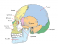
Head/Skull
- chondrocranium forms base of skull
- in lower vertebrates encases brain
- cranial vault
- calveria
- facial skeleton
- pharyngeal arches
Sensory Placodes

- During week 4 a series of thickened surface ectodermal patches form in pairs rostro-caudally in the head region.
- Recent research suggests that all sensory placodes may arise from common panplacodal primordium origin around the neural plate, and then differentiate to eventually have different developmental fates.
- These sensory placodes will later contribute key components of each of our special senses (vision, hearing and smell). Other species have a number of additional placodes which form other sensory structures (fish, lateral line receptor). Note that their initial postion on the developing head is significantly different to their final position in the future sensory system
Otic placode
in the stage 13/14 embryo (shown below) the otic placode has sunk from the surface ectoderm to form a hollow epithelial ball, the otocyst, which now lies beneath the surface surrounded by mesenchyme (mesoderm). The epithelia of this ball varies in thickness and has begun to distort, it will eventually form the inner ear membranous labyrinth.
Lens placode
lies on the surface, adjacent to the outpocketing of the nervous system (which will for the retina) and will form the lens.
Nasal placode
has 2 components (medial and lateral) and will form the nose olefactory epithelium.
- Links: Placodes
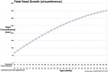
Head Growth

- continues postnatally - fontanelle allow head distortion on birth and early growth
- bone plates remain unfused to allow growth, puberty growth of face

Fetal head growth (circumference)
Second and third trimesters
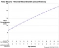
Second trimester

Skull Overview
Chondrocranium - formed from paraxial mesoderm
- cranial end of vertebral column
- modified vertebral elements
- occipital and cervical sclerotome
- bone preformed in cartilage (endochondrial ossification)
Cranial Vault and Facial Skeleton - formed from neural crest
- muscle is paraxial mesoderm
- somitomeres and occipital somites
Calveria - bone has no cartilage (direct ossification of mesenchyme)
- bones do not fuse, fibrous sutures 1. allow distortion to pass through birth canal 2. allow growth of the brain
- 6 fontanelles, posterior closes at 3 months, anterior closes at 18 months
- Links: Skull Development
Palate
The palate has two key stages of development during embryonic and an early fetal involving the fusion of structures (epithelia to mesenchymal).
Embryonic
Primary palate, fusion in the human embryo between stage 17 and 18, from an epithelial seam to the mesenchymal bridge.

Fetal
Secondary palate, fusion in the human embryo in week 9. This requires the early palatal shelves growth, elevation and fusion during the early embryonic period. The fusion event is to both each other and the primary palate. palatal shelf elevation | secondary palate
- Links: Palate Development
Ear Auricles
- form from 6 hillocks (week 5)
- 3 on each of arch 1 and 2
Tongue Development

 ]
]  ]
]
- Ectoderm of the first arch surrounding the stomodeum forms the epithelium lining the buccal cavity.
- Also the salivary glands, enamel of the teeth, epithelium of the body of the tongue.
- As the tongue develops "inside" the floor of the oral cavity, it is not readily visible in the external views of the embryonic (Carnegie) stages of development.
- Contributions from all arches, which changes with time
- begins as swelling rostral to foramen cecum, median tongue bud
- Arch 1 - oral part of tongue (ant 3/2)
- Arch 2 - initial contribution to surface is lost
- Arch 3 - pharyngeal part of tongue (post 1/3)
- Arch 4 - epiglottis and adjacent regions
Tongue muscle

- Tongue muscles originate from the somites.
- Tongue muscles develop before masticatory muscles and is completed by birth.
- Masticatory muscles originate from the somitomeres.
- These muscles develop late and are not complete even at birth.
Salivary Glands
- epithelial buds in oral cavity (week 6 to 7) extend into mesenchyme
- parotid, submandibular, sublingual
Pharyngeal Arch Tables
Structures derived from Arches
| Arch | Nerve | Skeletal Structures | Muscles | Ligaments |
| 1 (maxillary/mandibular) | trigeminal (V) | mandible, maxilla, malleus, incus | ant lig of malleus, sphenomandibular ligament | |
| 2 (hyoid) | facial (VII) | stapes, styloid process, lesser cornu of hyoid, upper part of body of hyoid bone | stylohyoid ligament | |
| 3 | glossopharyngeal (IX) | greater cornu of hyoid, lower part of body of hyoid bone | ||
| 4 & 6 | superior laryngeal and recurrent laryngeal branch of vagus (X) | thyroid, cricoid, arytenoid, corniculate and cuneform cartilages |

|
mandible |

|
hyoid |
| File:Larynx cartilage sm.jpg |
Structures derived from Pouches
Each pouch is lined with endoderm and generates specific structures.
| Overall Structure | Specific Structures | |
| tubotympanic recess | tympanic membrane, tympanic cavity, mastoid antrum, auditory tube | |
| intratonsillar cleft | crypts of palatine tonsil, lymphatic nodules of palatine tonsil | |
| inferior parathyroid gland, thymus gland | ||
| superior parathyroid gland, ultimobranchial body | ||
| becomes part of 4th pouch |
Structures derived from Grooves
Only the first groove differentiates into an adult structure and forms part of the external acoustic meatus.
Structures derived from Membranes
At the bottom of each groove lies the membrane which is formed from the contact region of ectodermal groove and endodermal pouch. Only the first membrane differentiates into an adult structure and forms the tympanic membrane.
Movies
|
|
|
|
|
- Links: Quicktime Movies | Flash Movies
References
Reviews
<pubmed>20607135</pubmed>| PMC2895339 | Integr Comp Biol.
Articles
Search PubMed
Search term: Head Development | Pharyngeal Arch Development | | Face Development
Additional Images

Adult axial skeleton
Adult skull (lateral simplified)

Fetal head lateral (12 weeks)

Fetal head medial (12 weeks)

Fetal head section (12 weeks)
adult mandible

adult hyoid


Historic
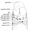
Fig. 1. Showing the formation of the face by the Nasal, Maxillary, and Mandibular processes in an embryo of the 4th week.
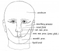
Fig. 2. Showing the parts of the face formed from the Nasal, Maxillary and Mandibular processes.
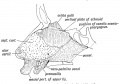
Fig. 3. Showing the structures formed in the Mesial Nasal Processes.
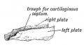
Fig. 4. Showing the trough-shaped Vomer of the newly born.
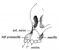
Fig. 5. Showing the suture on the face between the premaxilla and maxilla in the skull of a young orang.
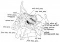
Fig. 6. Showing the structures formed in the Lateral Nasal Processes.
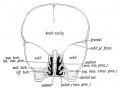
Fig. 7. Coronal section of the skull of a 7th month human foetus to show the cartilages of the Lateral and Mesial Nasal Processes and the bones formed round them.
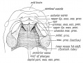
Fig. 8. Showing the ingrowth of the palatal plates of the two maxillary processes early in the 2nd month. (After Kollmann.) .
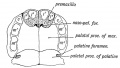
Fig. 9. Showing the Hard Palate at birth. The premaxillary part is formed from the Mesial Nasal Processes ; the remainder by the Palatal Plates of the Maxillary Processes.
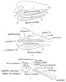
Fig. 10, a, b, c. Showing what become of the skeletons of the Mandibular Arch (Meckel's Cartilage) and Maxillary Process (Palato-quadrate Cartilage).
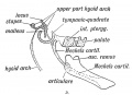
Fig. 10 D. Illustrating Gadow's view of the origin of the Auditory Ossicles and Tympanic Plate.
Fig. 11. Showing the manner in which the development of the Maxillary Antrum affects the size of the palate and position of the molar teeth.
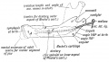
Fig. 12. Showing the Centres of Ossification and age changes in the Lower Jaw.
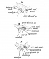
Fig. 13. The chief types of the Temporo-Maxillary Articulation. A. Carnivorous Type. B. Omnivorous Type. C. Herbivorous Type.
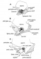
Fig. 14. Showing the Chief Changes after birth in the form of the TemporoMaxillary Articulation.
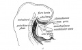
Fig. 15 A. Sagittal Section showing the Stomodaeum and position of the Oral Plate in the 3rd week.
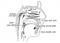
Fig. 15 B. Showing the parts of the Buccal and Nasal Cavities formed from the Stomodaeum. The relative position of the Oral Plate is indicated.


Development of the Pharynx and Neck
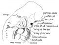
Fig. 21a. Showing the Visceral Arches and Cleft-depressions in the Pharyngeal Wall of a 4th week human Embryo. Each Visceral Arch contains an Aortic Arch. (After His.)
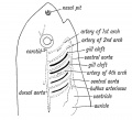
Fig. 21b. Showing the position of the Heart, Visceral and Aortic Arches in a fish. (Diagrammatic — after Gegenbaur.)
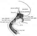
Fig. 22. Showing the Primitive Pharynx of a 3rd week embryo in sagittal section, bounded by the Visceral Arches. (After His.) .
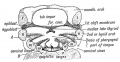
Fig. 23. Showing the Floor of the Pharynx of a 4th week human embryo. (After His.)
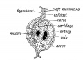
Fig. 24. Schematic Section of a Visceral Arch.
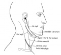
Fig. 25. Showing the position of the External Cleft Depressions in the Adult.
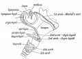
Fig. 26. Showing what become of the Cartilages of the Visceral Arches.
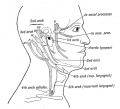
Fig. 27. Showing what become of the Nerves of the Visceral Arches, hyoid arch) is represented by the chorda tympani and great superficial petrosal.
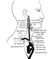
Fig. 28. Showing what become of the Aortic Arches in the adult. Only the shaded parts persist.
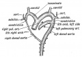
Fig. 29. The condition of the Eight and Left Doral Aortae in a 6th week human foetus.
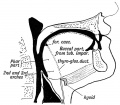
Fig. 30. Showing the Buccal and Pharyngeal parts of the Tongue.
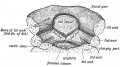
Fig. 31. Showing the origin of the tongue in the floor of the primitive pharynx. (After His.)
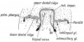
Fig. 32. Showing the origin of the Submaxillary and Sublingual Glands from furrows between the gum and tongue during the flth week. (After His.) .
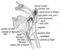
Fig. 33. Showing the position of the Visceral Clefts in the Adult.
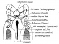
Fig. 34. Showing the origin of the Tonsil, Thymus, and Thyroid from the Internal Cleft Recesses during the 4th week. (After His.).


External Links
External Links Notice - The dynamic nature of the internet may mean that some of these listed links may no longer function. If the link no longer works search the web with the link text or name. Links to any external commercial sites are provided for information purposes only and should never be considered an endorsement. UNSW Embryology is provided as an educational resource with no clinical information or commercial affiliation.
- Essentials of Facial Growth Enlow and Hans
| System Links: Introduction | Cardiovascular | Coelomic Cavity | Endocrine | Gastrointestinal Tract | Genital | Head | Immune | Integumentary | Musculoskeletal | Neural | Neural Crest | Placenta | Renal | Respiratory | Sensory | Birth |
Glossary Links
- Glossary: A | B | C | D | E | F | G | H | I | J | K | L | M | N | O | P | Q | R | S | T | U | V | W | X | Y | Z | Numbers | Symbols | Term Link
Cite this page: Hill, M.A. (2024, May 21) Embryology Head Development. Retrieved from https://embryology.med.unsw.edu.au/embryology/index.php/Head_Development
- © Dr Mark Hill 2024, UNSW Embryology ISBN: 978 0 7334 2609 4 - UNSW CRICOS Provider Code No. 00098G