File:Stage9 bf3a.jpg: Difference between revisions
From Embryology
(uploaded a new version of "File:Stage9 bf3a.jpg": == Human embryo == Stage 9 day 20, somites 3-4 Ventrolateral view (cut away from chorion and amniotic sac) showing the embryo and yolk sac. Note: # the relative size of the embryo and the assoc) |
(uploaded a new version of "File:Stage9 bf3a.jpg": == Human embryo == Stage 9 day 20, somites 3-4 Ventrolateral view (cut away from chorion and amniotic sac) showing the embryo and yolk sac. Note: # the shape of the early folded embryonic disc ) |
(No difference)
| |
Revision as of 16:43, 22 August 2009
Human embryo
Stage 9 day 20, somites 3-4
Ventrolateral view (cut away from chorion and amniotic sac) showing the embryo and yolk sac.
Note:
- the relative size of the embryo and the associated extra-embryonic coeloms.
- the shape of the early folded embryonic disc and rostro-caudal bendings.
- the relative thicknesses of the embryo and extra-embryonic membranes.
- the position of the prechordal plate (cardiogenic region) ventral to brain fold.
Original file name: Stage9day20somites3-4ldorsalbf3-800px.jpg
- Carnegie Stages: 1 | 2 | 3 | 4 | 5 | 6 | 7 | 8 | 9 | 10 | 11 | 12 | 13 | 14 | 15 | 16 | 17 | 18 | 19 | 20 | 21 | 22 | 23 | About Stages | Timeline
Image Source: Scanning electron micrographs of the Carnegie stages of the early human embryos are reproduced with the permission of Prof Kathy Sulik, from embryos collected by Dr. Vekemans and Tania Attié-Bitach. Images are for educational purposes only and cannot be reproduced electronically or in writing without permission.
File history
Click on a date/time to view the file as it appeared at that time.
| Date/Time | Thumbnail | Dimensions | User | Comment | |
|---|---|---|---|---|---|
| current | 16:43, 22 August 2009 |  | 536 × 800 (20 KB) | S8600021 (talk | contribs) | == Human embryo == Stage 9 day 20, somites 3-4 Ventrolateral view (cut away from chorion and amniotic sac) showing the embryo and yolk sac. Note: # the shape of the early folded embryonic disc and rostro-caudal bendings. # the relative thicknesses of |
| 16:15, 22 August 2009 | 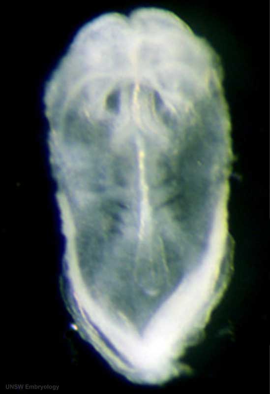 | 549 × 800 (18 KB) | S8600021 (talk | contribs) | == Human embryo == Stage 9 day 20, somites 3-4 Ventrolateral view (cut away from chorion and amniotic sac) showing the embryo and yolk sac. Note: # the relative size of the embryo and the associated extra-embryonic coeloms. # the shape of the early fo | |
| 16:07, 22 August 2009 | 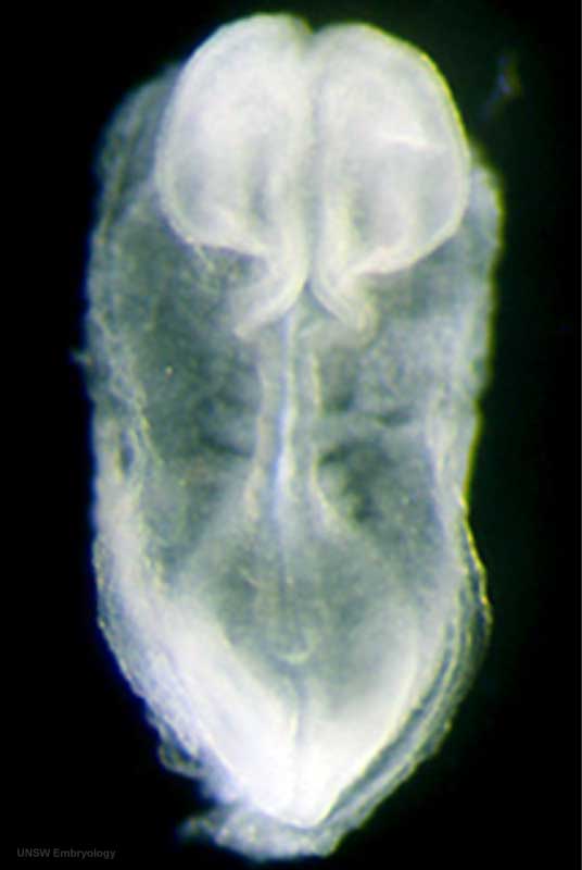 | 536 × 800 (20 KB) | S8600021 (talk | contribs) | == Human embryo == Stage 9 day 20, somites 3-4 Ventrolateral view (cut away from chorion and amniotic sac) showing the embryo and yolk sac. Note: # the relative size of the embryo and the associated extra-embryonic coeloms. # the shape of the early fo |
You cannot overwrite this file.
File usage
The following 2 pages use this file: