File:Stage9 bf1b.jpg
Stage9_bf1b.jpg (532 × 600 pixels, file size: 19 KB, MIME type: image/jpeg)
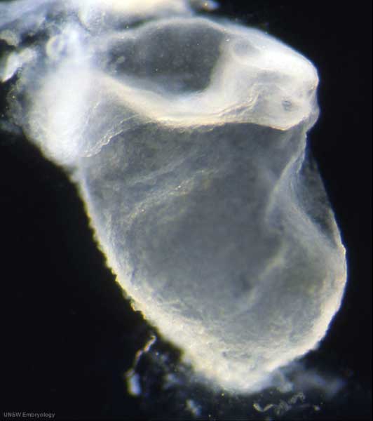
Human Embryo - Carnegie Stage 9
Stage 9 day 20, somites 3-4
Ventrolateral view (cut chorionic) showing the embryo, yolk sac and amniotic sac.
Note:
- the relative size of the embryo and the associated extra-embryonic coeloms.
- the shape of the early folded embryonic disc and rostro-caudal bendings.
- the caudal attachment of the embryo to the chorionic wall.
- the relative thicknesses of the embryo and extra-embryonic membranes.
Original file name: Stage9day20somites3-4lateralbf1-600px.jpg
Carnegie Stage 9 Image Links: (below are listed image size versions of each image)
- Light Image dorsal - Large 1000px | 800px | Medium 600px | Small 400px
- Light Image ventrolateral - Large 1000px | 800px | Medium 600px | Small 400px
- Light Image lateral - Large 1000px | 800px | Medium 600px | Small 400px
- SEM Dorsal - Large 1000px | 800px | Medium 600px | Small 400px
- SEM Cranial Neural fold - Large 1000px | 800px | Medium 600px | Small 400px
- SEM Caudal Region - Large 1000px | 800px | Medium 600px | Small 400px
- SEM Caudal Region cross section- Large 1000px | 800px | Medium 600px | Small 400px
- Carnegie Stages: 1 | 2 | 3 | 4 | 5 | 6 | 7 | 8 | 9 | 10 | 11 | 12 | 13 | 14 | 15 | 16 | 17 | 18 | 19 | 20 | 21 | 22 | 23 | About Stages | Timeline
Image Source: Scanning electron micrographs of the Carnegie stages of the early human embryos are reproduced with the permission of Prof Kathy Sulik, from embryos collected by Dr. Vekemans and Tania Attié-Bitach. Images are for educational purposes only and cannot be reproduced electronically or in writing without permission.
Cite this page: Hill, M.A. (2024, April 27) Embryology Stage9 bf1b.jpg. Retrieved from https://embryology.med.unsw.edu.au/embryology/index.php/File:Stage9_bf1b.jpg
- © Dr Mark Hill 2024, UNSW Embryology ISBN: 978 0 7334 2609 4 - UNSW CRICOS Provider Code No. 00098G
File history
Click on a date/time to view the file as it appeared at that time.
| Date/Time | Thumbnail | Dimensions | User | Comment | |
|---|---|---|---|---|---|
| current | 11:43, 22 August 2009 | | 532 × 600 (19 KB) | S8600021 (talk | contribs) | Human embryo Stage 9 day 20, somites 3-4 Ventrolateral view (cut chorionic) showing the embryo, yolk sac and amniotic sac. Note: # the relative size of the embryo and the associated extra-embryonic coeloms. # the shape of the early folded embryonic di |
You cannot overwrite this file.
File usage
The following page uses this file: