File:Stage7-sem2.jpg

Original file (590 × 800 pixels, file size: 98 KB, MIME type: image/jpeg)
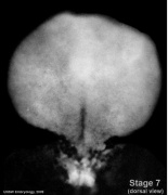
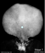
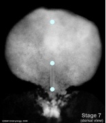
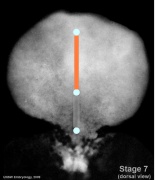
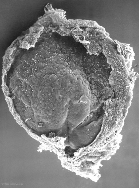
Human Embryo Carnegie stage 7
17 days, pre-somite, scanning electron micrograph image
Embryonic disc (epiblast/ectoderm layer) dorsal view, with amniotic membrane partially removed.
Primitive node (Henson's node) in centre of disc and primitive streak, shown as indentation in disc is extending to the bottom of image. Connecting stalk is shown at the bottom of image.
See also Stage7-sem1.jpg image original orientation.
- Stage 7 Links: Carnegie stage 7 | Gastrulation | Notochord | Week 3 | Image 1 - Stage 7 SEM | Image 2 - Stage 7 SEM
- Embryo Stage 7 (dorsal)
Dorsal view
Primitive streak and node
Oral and cloacal membranes
Axial mesoderm
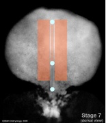
Paraxial mesoderm
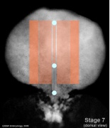
Intermediate mesoderm
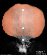
Lateral plate


- Carnegie Stages: 1 | 2 | 3 | 4 | 5 | 6 | 7 | 8 | 9 | 10 | 11 | 12 | 13 | 14 | 15 | 16 | 17 | 18 | 19 | 20 | 21 | 22 | 23 | About Stages | Timeline
Image Source: Scanning electron micrographs of the Carnegie stages of the early human embryos are reproduced with the permission of Prof Kathy Sulik, from embryos collected by Dr. Vekemans and Tania Attié-Bitach. Images are for educational purposes only and cannot be reproduced electronically or in writing without permission.
File history
Click on a date/time to view the file as it appeared at that time.
| Date/Time | Thumbnail | Dimensions | User | Comment | |
|---|---|---|---|---|---|
| current | 13:15, 21 August 2009 | | 590 × 800 (98 KB) | MarkHill (talk | contribs) | Human Embryo Carnegie stage 7, 17 days, pre-somite, scanning electron micrograph image Embryonic disc (epiblast/ectoderm layer) dorsal view, with amniotic membrane partially removed. Primitive node (Henson's node) in centre of disc and primitive streak |
You cannot overwrite this file.
File usage
The following 29 pages use this file:
- 2010 BGD Practical 3 - Gastrulation
- 2010 BGD Practical 3 - Week 3 Summary
- 2010 BGD Practical 6 - Week 3
- 2010 Lab 2
- 2010 Lab 3
- 2011 Lab 2 - Week 3
- BGDA Lecture - Development of the Embryo/Fetus 1
- BGDA Lecture - Development of the Embryo/Fetus 2
- BGDA Practical 3 - Gastrulation
- BGDA Practical 3 - Week 3 Summary
- BGDA Practical 7 - Week 3
- Carnegie stage 7
- Fetal ECHO Meeting 2012
- Human Embryo - Scanning electron microscopy
- Human Embryo SEM
- Lecture - Mesoderm Development
- Lecture - Week 3 Development
- Primordial Germ Cell Development
- REI - Reproductive Medicine Seminar 2018
- RPAH Cardiac Embryology 2014
- Royal Hospital for Women - Reproductive Medicine Seminar 2018
- Scanning Electron Microscopy
- Timeline human development
- Week 3
- Talk:2011 Lab 3
- Talk:Timeline human development
- Template:First Trimester Timeline
- Template:First Trimester Timeline collapsable table
- Template talk:First Trimester Timeline