File:Stage15 bf2.jpg

Original file (1,874 × 2,000 pixels, file size: 1.36 MB, MIME type: image/jpeg)
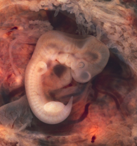
Human embryo - 5 week
External appearance appears to be about a Carnegie stage 15 embryo.
Carnegie Stage 15 Information
- Week 5, 35 - 38 days, 7 - 9 mm
- Lateral view. Amniotic membrane removed.
Events
- Ectoderm: sensory placodes, lens pit, otocyst, nasal pit, primary/secondary vesicles, fourth ventricle of brain,
- Mesoderm: heart prominence
- Head: 1st, 2nd and 3rd pharyngeal arch, forebrain, site of lens placode, site of otic placode, stomodeum
- Body: heart, liver, umbilical cord, mesonephric ridge
- Limb: upper and lower limb buds, hand plate
Identify: midbrain region, nasal pit, lens pit, 1st, 2nd and 3rd pharyngeal arches, 1st pharyngeal groove, maxillary and mandibular components of 1st pharyngeal arch, fourth ventricle of brain, heart prominence, cervical sinus, upper limb bud, mesonephric ridge, lower limb bud, umbilical cord
Image version links: ExtraLarge 1874 x 2000px | Large 959 x 1024px | Medium 468 x 500px | Small 225 x 240px
Related Links: Carnegie stage 15
- Carnegie Stages: 1 | 2 | 3 | 4 | 5 | 6 | 7 | 8 | 9 | 10 | 11 | 12 | 13 | 14 | 15 | 16 | 17 | 18 | 19 | 20 | 21 | 22 | 23 | About Stages | Timeline
Reference
Original Author Legend
- "This photo of an opened oviduct with an ectopic pregnancy features a spectacularly well preserved 10-millimeter embryo. It is uncommon to see any embryo at all in an ectopic, and for one to be this well preserved (and undisturbed by the prosector's knife) is quite unusual.
Even an embryo this tiny shows very distinct anatomic features, including tail, limb buds, heart (which actually protrudes from the chest), eye cups, cornea/lens, brain, and prominent segmentation into somites. The gestational sac is surrounded by a myriad of chorionic villi resembling elongate party balloons. This embryo is about five weeks old (or seven weeks in the biologically misleading but eminently practical dating system used in obstetrics).
The photo was taken on Kodak Elite 200 slide film, with a Minolta X-370 camera and 100mm f/4 Rokkor bellows lens at near-full extension. The formalin-fixed specimen was immersed in tapwater and pinned to a tray lined with black velvet. The exposure was 1/4 second at f/8."
Original file name: 304334264_8cba67ad75_o.jpg
http://www.flickr.com/photos/euthman/304334264
Uploaded to Flickr on November 23, 2006 by Ed Uthman Image (pathologist in Houston, Texas)
http://creativecommons.org/licenses/by/2.0/
Cite this page: Hill, M.A. (2024, May 30) Embryology Stage15 bf2.jpg. Retrieved from https://embryology.med.unsw.edu.au/embryology/index.php/File:Stage15_bf2.jpg
- © Dr Mark Hill 2024, UNSW Embryology ISBN: 978 0 7334 2609 4 - UNSW CRICOS Provider Code No. 00098G
File history
Click on a date/time to view the file as it appeared at that time.
| Date/Time | Thumbnail | Dimensions | User | Comment | |
|---|---|---|---|---|---|
| current | 14:22, 21 July 2010 |  | 1,874 × 2,000 (1.36 MB) | S8600021 (talk | contribs) | ==Human embryo - 5 week== External appearance appears to be about a Carnegie stage 15 embryo. Original Author Legend :"This photo of an opened oviduct with an ectopic pregnancy features a spectacularly well preserved 10-millimeter embryo. It is uncom |
You cannot overwrite this file.
File usage
The following 4 pages use this file: