File:Stage11 sem20.jpg

Original file (668 × 1,000 pixels, file size: 132 KB, MIME type: image/jpeg)
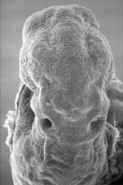
Human Embryo Carnegie stage 11
25 days, 19 somite pairs, week 4 (GA week 6)
Facts: Carnegie stage 11 Week 4, 23 - 26 days, 2.5 - 4.5 mm, Somite Number 13 - 20
View: This is a scanning EM of the embryo superior dorsal view showing the paired otic placodes sinking into the surface at the level of the hindbrain.
Features: surface ectoderm, paired otic placodes, pharyngeal arches heart
In week 4 (GA week 6) of human development, on the embryo head region surface ectoderm specialised placodes ("patches") form associated with later sensory and endocrine development.
The first to appear are the paired otic placodes, forming behind the second pharyngeal arch. These placodes will invaginate and are then eventually lost from the surface. These placodes will form the structures of the inner ear. Other sensory placodes, nasal and optic, can be seen slightly later.
Related Images: Scanning EM image 2 | Scanning EM image 3 | Scanning EM image 4 | Scanning EM image 10
- Carnegie Stages: 1 | 2 | 3 | 4 | 5 | 6 | 7 | 8 | 9 | 10 | 11 | 12 | 13 | 14 | 15 | 16 | 17 | 18 | 19 | 20 | 21 | 22 | 23 | About Stages | Timeline
Image version links: Large 1000px | 800px | Medium 600px
Image Source: Scanning electron micrographs of the Carnegie stages of the early human embryos are reproduced with the permission of Prof Kathy Sulik, from embryos collected by Dr. Vekemans and Tania Attié-Bitach. Images are for educational purposes only and cannot be reproduced electronically or in writing without permission.
File history
Click on a date/time to view the file as it appeared at that time.
| Date/Time | Thumbnail | Dimensions | User | Comment | |
|---|---|---|---|---|---|
| current | 23:21, 3 May 2010 | | 668 × 1,000 (132 KB) | S8600021 (talk | contribs) | '''Human Embryo''' Carnegie stage 11 25 days, 19 somite pairs Facts: Week 4, 23 - 26 days, 2.5 - 4.5 mm, Somite Number 13 - 20 View: This is a scanning EM of the embryo superior dorsal view showing the paired otic placodes sinking into the surface at t |
You cannot overwrite this file.
File usage
The following file is a duplicate of this file (more details):
The following 5 pages use this file: