File:Fetal head section.jpg: Difference between revisions
From Embryology
No edit summary |
mNo edit summary |
||
| (8 intermediate revisions by the same user not shown) | |||
| Line 1: | Line 1: | ||
==Human Fetal Head Week 12== | ==Human Fetal Head Week 12== | ||
This medial (mid-line) section through the fetal head (12 weeks) shows features of the developing skull and the brain, face and mouth. Note extensive nasal cartilage, nasal conchae, pituitary, secondary palate, oral cavity, tongue, mandible, hyoid, choana, oropharynx. Also note the developing tongue musculature and its mandibular attachment site. | |||
{{Fetal head 12 week}} | |||
{| | |||
| valign=top| | |||
===Neural=== | |||
* [[Neural_System_Development|brain and brainstem]]. | |||
* [[Neural_System_Development#Human_Early_Neural_Development|lamina terminalis]] (site of anterior neuropore closure). | |||
* [[Neural_-_Ventricular_System_Development|fourth ventricle]]. | |||
* {{pituitary}} sitting in the sella turcica. | |||
| valign=top| | |||
* | |||
===Musculoskeletal=== | ===Musculoskeletal=== | ||
* cartilage - septum of the nose. | * {{cartilage}} - {{skull}} septum of the nose. | ||
* bone - ossifying nasal concha. | |||
* bone - palate roof of mouth. | * {{bone}} - [[Respiratory_System_-_Upper_Respiratory_Tract|upper respiratory]] ossifying nasal concha. | ||
* cartilage - soft palate back of mouth. | |||
* cartilage - base of skull and vertebra. | * {{bone}} - [[Palate_Development|hard palate]] roof of mouth. | ||
* muscle - | |||
* bone - mandible. | * {{cartilage}} - [[Palate_Development|soft palate]] back of mouth. | ||
* {{cartilage}} - base of skull and vertebra. | |||
* {{muscle}} - {{Tongue}}, attached to mandible, note foramen cecum. | |||
* {{bone}} - mandible. | |||
|} | |||
:'''Links:''' [[:File:Fetal head section.jpg|Image - Head histology]] | [[:File:Fetal head medial.jpg|Image - Head medial cartilage/bone]] | [[:File:Fetal head lateral.jpg|Image - Head lateral cartilage/bone]] | {{Head}} | {{Gastrointestinal tract}} | {{Skull}} | {{Bone}} | |||
{{Fetal Links}} | |||
===Reference=== | |||
{{Virginia Diewert images}} | |||
{{ | {{Footer}} | ||
[[Category:Human Fetus]] [[Category:Week 12]] [[Category:Musculoskeletal]] [[Category:Tongue]] [[Category:Bone]] [[Category:Cartilage]] [[Category:Neural]] [[Category:Head]] [[Category:Skull]] [[Category:Pituitary]] | [[Category:Human Fetus]] [[Category:Week 12]] [[Category:Musculoskeletal]] [[Category:Tongue]] [[Category:Bone]] [[Category:Cartilage]] [[Category:Neural]] [[Category:Head]] [[Category:Skull]] [[Category:Pituitary]] | ||
Latest revision as of 08:09, 13 November 2018
Human Fetal Head Week 12
This medial (mid-line) section through the fetal head (12 weeks) shows features of the developing skull and the brain, face and mouth. Note extensive nasal cartilage, nasal conchae, pituitary, secondary palate, oral cavity, tongue, mandible, hyoid, choana, oropharynx. Also note the developing tongue musculature and its mandibular attachment site.
- 12 Week Images: Sagittal unlabeled | Sagittal labeled | Sagittal medial view | Sagittal lateral view | Pituitary unlabeled | Pituitary labeled | Tongue | Skull Development | Head Development
Neural
|
Musculoskeletal
|
- Links: Image - Head histology | Image - Head medial cartilage/bone | Image - Head lateral cartilage/bone | head | gastrointestinal tract | skull | bone
| Fetal Links: fetal | Week 10 | Week 12 | second trimester | third trimester | fetal neural | Fetal Blood Sampling | fetal growth restriction | birth | birth weight | preterm birth | Developmental Origins of Health and Disease | macrosomia | BGD Practical | Medicine Lecture | Science Lecture | Lecture Movie | Category:Human Fetus | Category:Fetal | |||
|
Reference
Image Source: Prof Virginia Diewert
Cite this page: Hill, M.A. (2024, April 26) Embryology Fetal head section.jpg. Retrieved from https://embryology.med.unsw.edu.au/embryology/index.php/File:Fetal_head_section.jpg
- © Dr Mark Hill 2024, UNSW Embryology ISBN: 978 0 7334 2609 4 - UNSW CRICOS Provider Code No. 00098G
File history
Click on a date/time to view the file as it appeared at that time.
| Date/Time | Thumbnail | Dimensions | User | Comment | |
|---|---|---|---|---|---|
| current | 12:24, 18 March 2012 |  | 1,200 × 821 (167 KB) | Z8600021 (talk | contribs) | |
| 12:18, 18 March 2012 | 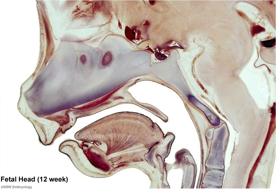 | 965 × 660 (107 KB) | Z8600021 (talk | contribs) | ||
| 15:41, 13 September 2009 |  | 965 × 660 (63 KB) | S8600021 (talk | contribs) | Selected medial head view showing key features of head musculoskeletal and neurological development. Note extensive nasal cartilage, nasal conchae, pituitary, secondary palate, oral cavity, tongue, mandible, hyoid, choana, oropharynx. Also note the devel |
You cannot overwrite this file.
File usage
The following 26 pages use this file:
- 2009 Lecture 13
- 2010 Foundations Lecture - Introduction to Human Development
- 2010 Lab 6
- 2010 Lecture 13
- 2011 Lab 6 - Fetal
- AACP Meeting 2013 - Face Embryology
- ANAT2241 Bone, Bone Formation and Joints
- ANAT2341 Lab 6 - Fetal
- Abnormal Development - Fetal Growth Restriction
- BGDB Face and Ear - Fetal
- Bone Development
- Bone Histology
- Cartilage Histology
- Fetal Development
- Fetal Development - 12 Weeks
- Foundations Lecture - Introduction to Human Development
- Head Development
- Joint Development - Temporomandibular Joint
- Lecture - Musculoskeletal Development
- Musculoskeletal System - Abnormalities
- Musculoskeletal System - Axial Skeleton Development
- Musculoskeletal System - Bone Development
- Musculoskeletal System - Cartilage Development
- Musculoskeletal System - Joint Development
- Musculoskeletal System Development
- Pre-Medicine Program - Embryology