File:Dandy1910-plate01.jpg

Original file (1,738 × 2,359 pixels, file size: 541 KB, MIME type: image/jpeg)
Plate I
(Figs. 1—7).
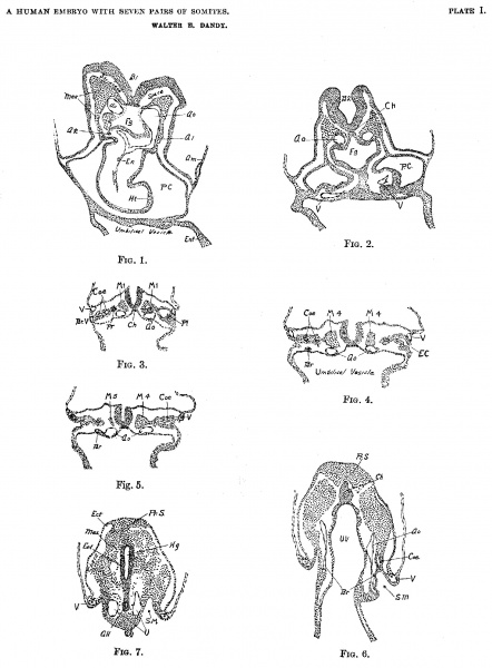
Fig. 1. Section 22, X 50. B1, First primary brain vesicle; Oo, Aorta; A1, Complete View of longitudinal section of tirst aortic arch; A2, Cross section of stub of second aortic arch; En, Endothelial heart; Ht, Mesodermal wall of heart; P.C., Pericardial cavity; Fg, foregut; Am, Amnion; Mes, Mesoderm; Ent, Entoderm.
Fig. 2. Section 53, X 50. Ch, Chorda; Ao, Dorsal aorta; V, Umbilical vein; Fg, Foregut; P.C., Pericardial cavity; B2, Second primary vesicle of the brain.
Fig. 3. Section 76, X 50. Pr, Pronephros; Coe, Coelom; Pl, Pleural Cuelom; V, Umbilical vein; Br V, Branch of umbilical vein, only suggestion of a vitelline vein, it unites with umbilical vein two sections below; Ch, Chorda; M1, First Somite in each side.
Fig. 4. Section 93, X 50. Br, Cross section of lateral branch of dorsal aorta; V, Umbilical vein; M4, Somite IV, with cavity (anterior tip of somite IV on opposite side) ; Coe, Coelom; E.C., Communication of coelom with exterior; Ch, Chorda.
Fig. 5. Section 101, X 50. Br. Lateral branch of Aorta; Ao, Aorta; V, Umbilical vein; M4, Somite IV with cavity; M5, Tip of Somite V; Coe, Coelom.
Fig. 6. Section 137, X 50. A0, Dorsal aorta; Br, Lateral branch of aorta (branch on left side terminates in a mass of mesodermal cells which may be a small blood island); Ch, Chorda at point of greatest development; Coe, Coelom; S.M, Slit in mesoderm. (See also Fig. 11.) Pr. S, Beginning of primitive streak. U. V, Umbilical vesicle.
Fig. 7. Section 152, X 50. Shows primitive streak region (Pr. S.) from fusion of ectoderm, mesoderm and entfoderm. Hg, Hindgut; A11, Allantois, near point of origin from hindgut; U, Umbilical artery; V, Umbilical vein; S. M, Slit in mesoderm. Ect, Ectoderm; Ent, Entoderm; Mes, Meoderm.
Online Editor - embryo corresponds to Carnegie stage 10 in Week 4.
| Stage 10 Links: Week 4 | Gastrulation | Lecture | Practical | Image Gallery | Carnegie Embryos | Embryos | Category:Carnegie Stage 10 | Next Stage 11 |
| Historic Papers: 1910 | 1917 | 1926 | 1939 | 1943 | 1957 | 1985 |
| Week: | 1 | 2 | 3 | 4 | 5 | 6 | 7 | 8 |
| Carnegie stage: | 1 2 3 4 | 5 6 | 7 8 9 | 10 11 12 13 | 14 15 | 16 17 | 18 19 | 20 21 22 23 |
| Historic Disclaimer - information about historic embryology pages |
|---|
| Pages where the terms "Historic" (textbooks, papers, people, recommendations) appear on this site, and sections within pages where this disclaimer appears, indicate that the content and scientific understanding are specific to the time of publication. This means that while some scientific descriptions are still accurate, the terminology and interpretation of the developmental mechanisms reflect the understanding at the time of original publication and those of the preceding periods, these terms, interpretations and recommendations may not reflect our current scientific understanding. (More? Embryology History | Historic Embryology Papers) |
- Links: Plate 1 | Plate 2 | Plate 3 | Plate 4 | Plate 5 | Plate 6 | Dandy 1910 | Carnegie stage 10 | Category:Carnegie Stage 10 | Week 4 | Historic Embryology Papers
Reference
Dandy WE. A human embryo with seven pairs of somites measuring about 2 mm in length. (1910) Amer. J Anat. 10: 85-109.
Cite this page: Hill, M.A. (2024, April 27) Embryology Dandy1910-plate01.jpg. Retrieved from https://embryology.med.unsw.edu.au/embryology/index.php/File:Dandy1910-plate01.jpg
- © Dr Mark Hill 2024, UNSW Embryology ISBN: 978 0 7334 2609 4 - UNSW CRICOS Provider Code No. 00098G
File history
Click on a date/time to view the file as it appeared at that time.
| Date/Time | Thumbnail | Dimensions | User | Comment | |
|---|---|---|---|---|---|
| current | 08:44, 18 September 2015 |  | 1,738 × 2,359 (541 KB) | Z8600021 (talk | contribs) | {{Dandy1910 figures}} |
You cannot overwrite this file.
File usage
The following 2 pages use this file:
