File:Chicken embryo E-cad and P-cad gastrulation.png: Difference between revisions
| Line 1: | Line 1: | ||
==Chicken Embryo Gastrulation and Cadherin Isoforms== | ==Chicken Embryo Gastrulation and Cadherin Isoforms== | ||
[[User:Z8600021|Mark Hill]] ([[User talk:Z8600021|talk]]) 12:43, 3 August 2017 (AEST) The text below has been written by the person who has uploaded the image to the Wiki. It is intended to give a simplified explanation to other students of what the image shows, independent of the original paper context. | [[User:Z8600021|Mark Hill]] ([[User talk:Z8600021|talk]]) 12:43, 3 August 2017 (AEST) The text below has been written by the person who has uploaded the image to the Wiki. It is intended to give a simplified explanation to other students of what the image shows, independent of the original paper context. | ||
Revision as of 13:22, 3 August 2017
Chicken Embryo Gastrulation and Cadherin Isoforms
Mark Hill (talk) 12:43, 3 August 2017 (AEST) The text below has been written by the person who has uploaded the image to the Wiki. It is intended to give a simplified explanation to other students of what the image shows, independent of the original paper context.
Contributor summary
This figure comes from a research paper that was able to show that chicken gastrulation epithelial-mesenchymal transition (EMT) was independent of (did not require) a changing level in P-Cadherin isoform. Also identified for reference is Slug, a zinc finger transcription factor belonging to the Snail family (SLUG) required for epithelial-mesenchymal transitions in development.
Gastrulation is an important developmental process that establishes the 3 main embryonic layers (ectoderm, mesoderm and endoderm).
This image shows the localisation and changing levels of cadherin during chicken embryo gastrulation ( Hamburger Hamilton stage 4 to 7 embryos).
Cadherins are family of transmembrane proteins (isoforms) that found at adherens junctions for cell adhesion that also require calcium to act ("calcium-dependent adhesion").
The naming of the cadherin isoforms is based upon the site of original identification: P-cadherin (placenta cadherin, cadherin 3), E-cadherin (epithelial cadherin cadherin 1), N-cadherin (neural cadherin, cadherin 2).
The upper panel bright field images (A-F) show in situ hybridisation localisation of different mRNAs during chicken gastrulation. The lefthand panels view the whole embryo and the righthand panels are transverse sections through the embryo.
The lower fluorescent images (G, H, I) show protein localisation during chicken embryo gastrulation.
- Green - Slug protein, (a zinc finger transcription factor belonging to the Snail family, SLUG)
- Red - Cadherin protein, both E-cadherin and P-cadherin isoforms)
(G-I) Immunofluorescence localization of Slug (green) and cadherin proteins using an antibody that recognizes E-cad and P-cad (red) in transverse sections through the middle streak region of HH stage 4, 5 and 7 embryos.
Original figure legend
Note that there are several unexplained terms and acronyms in the legend title and description that should be fully explained when uploaded and used in your student project.
Localization of E-cadherin and P-cadherin mRNA, and E-cadherin and Slug proteins, in HH stage 4–7 embryos
(A-C, A’-C’) E-cad (CDH1) mRNA expression in HH stage 4, 5 and 7 embryos (A-C) and in transverse sections (A’-C’). (D-F, D’-F’) P-Cad (CDH3) mRNA expression in embryos at stages 4, 5 and 6 (D-F) and in transverse sections (D’-F’). E-cad mRNAs are downregulated in the pre ingression epiblast and in the primitive streak, while P-cad mRNAs persist in the primitive streak and medial mesoderm (arrows). (G-I) Immunofluorescence localization of Slug (green) and cadherin proteins using an antibody that recognizes E-cad and P-cad (red) in transverse sections through the middle streak region of HH stage 4, 5 and 7 embryos.
Reference
<pubmed>27097030</pubmed>
Moly PK, Cooley JR, Zeltzer SL, Yatskievych TA, Antin PB (2016) Gastrulation EMT Is Independent of P-Cadherin Downregulation. PLoS ONE 11(4): e0153591. doi:10.1371/journal.pone.0153591
Copyright
© 2016 Moly et al. This is an open access article distributed under the terms of the Creative Commons Attribution License, which permits unrestricted use, distribution, and reproduction in any medium, provided the original author and source are credited.
http://dx.doi.org/10.1371/journal.pone.0153591.g001
- Note - This image was originally uploaded as part of an undergraduate science student project and may contain inaccuracies in either description or acknowledgements. Students have been advised in writing concerning the reuse of content and may accidentally have misunderstood the original terms of use. If image reuse on this non-commercial educational site infringes your existing copyright, please contact the site editor for immediate removal.
File history
Click on a date/time to view the file as it appeared at that time.
| Date/Time | Thumbnail | Dimensions | User | Comment | |
|---|---|---|---|---|---|
| current | 15:02, 12 August 2016 | 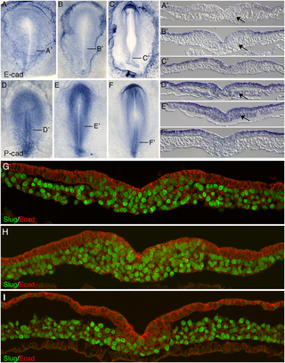 | 320 × 408 (253 KB) | Z8600021 (talk | contribs) | PMID 27097030 |
You cannot overwrite this file.
File usage
The following 51 pages use this file:
- 3460148
- ANAT2341 Lab 1 - Online Assessment
- Dummy
- Student Page
- Student page
- Talk:ANAT2341 Lab 1 - Online Assessment
- Talk:Student Page
- User:Z3414482
- User:Z3416557
- User:Z3417363
- User:Z3439257
- User:Z3460148
- User:Z3462474
- User:Z3491219
- User:Z3516832
- User:Z5014803
- User:Z5015014
- User:Z5015337
- User:Z5015544
- User:Z5015686
- User:Z5017002
- User:Z5017644
- User:Z5018156
- User:Z5018221
- User:Z5018267
- User:Z5019282
- User:Z5019306
- User:Z5019526
- User:Z5019880
- User:Z5020117
- User:Z5020373
- User:Z5020466
- User:Z5039628
- User:Z5059503
- User:Z5059696
- User:Z5059949
- User:Z5059996
- User:Z5062492
- User:Z5075778
- User:Z5076351
- User:Z5076466
- User:Z5093005
- User:Z5113034
- User:Z5114433
- User:Z5117343
- User:Z5177691
- User:Z5177699
- User:Z5178275
- User:Z5178407
- User:Z5178570
- Template:ANAT2341Student2016