Category:Human
From Embryology
This Embryology category shows pages and media related to human development.
Subcategories
This category has the following 72 subcategories, out of 72 total.
C
- Carnegie Embryo
- Carnegie Embryo 1
- Carnegie Embryo 112
- Carnegie Embryo 1134B
- Carnegie Embryo 116
- Carnegie Embryo 1266
- Carnegie Embryo 1455
- Carnegie Embryo 148
- Carnegie Embryo 172
- Carnegie Embryo 19
- Carnegie Embryo 239
- Carnegie Embryo 2393
- Carnegie Embryo 240
- Carnegie Embryo 248
- Carnegie Embryo 256
- Carnegie Embryo 296
- Carnegie Embryo 3527
- Carnegie Embryo 3956
- Carnegie Embryo 4046
- Carnegie Embryo 4059
- Carnegie Embryo 407
- Carnegie Embryo 4148
- Carnegie Embryo 43
- Carnegie Embryo 4405
- Carnegie Embryo 460
- Carnegie Embryo 463
- Carnegie Embryo 523
- Carnegie Embryo 5541
- Carnegie Embryo 5609
- Carnegie Embryo 5652
- Carnegie Embryo 5682
- Carnegie Embryo 5874
- Carnegie Embryo 6032
- Carnegie Embryo 625
- Carnegie Embryo 6426
- Carnegie Embryo 6581
- Carnegie Embryo 7618
- Carnegie Embryo 7669
- Carnegie Embryo 786
- Carnegie Embryo 808
- Carnegie Embryo 8147
- Carnegie Embryo 8239
- Carnegie Embryo 8370
- Carnegie Embryo 858
- Carnegie Embryo 8630
- Carnegie Embryo 8967
- Carnegie Embryo 9296
- Carnegie Embryo 96
- Carnegie Embryo 963
- Carnegie Embryo 966
- Carnegie Embryo 9697
D
F
H
Pages in category 'Human'
The following 200 pages are in this category, out of 332 total.
(previous page) (next page)A
B
- Template:B050966
- Template:B100658
- Template:B220849
- Template:Bardeen1906 figures
- Template:Barniville1914 figures
- Template:Bartelmez1922 figures
- Template:BaxterBoyd1939 figures
- BGDA Practical 7 - Week 6
- Birth MRI Movie
- Book - Contributions to Embryology Carnegie Institution No.10
- Book - Contributions to Embryology Carnegie Institution No.112
- Book - Contributions to Embryology Carnegie Institution No.131
- Book - Contributions to Embryology Carnegie Institution No.159
- Book - Contributions to Embryology Carnegie Institution No.21
- Book - Contributions to Embryology Carnegie Institution No.22
- Book - Contributions to Embryology Carnegie Institution No.27
- Book - Contributions to Embryology Carnegie Institution No.29
- Book - Contributions to Embryology Carnegie Institution No.30
- Book - Contributions to Embryology Carnegie Institution No.32
- Book - Contributions to Embryology Carnegie Institution No.33
- Book - Contributions to Embryology Carnegie Institution No.34
- Book - Contributions to Embryology Carnegie Institution No.35
- Book - Contributions to Embryology Carnegie Institution No.38
- Book - Contributions to Embryology Carnegie Institution No.40
- Book - Contributions to Embryology Carnegie Institution No.42
- Book - Contributions to Embryology Carnegie Institution No.43
- Book - Contributions to Embryology Carnegie Institution No.44
- Book - Contributions to Embryology Carnegie Institution No.46
- Book - Contributions to Embryology Carnegie Institution No.47
- Book - Contributions to Embryology Carnegie Institution No.48
- Book - Contributions to Embryology Carnegie Institution No.52
- Book - Contributions to Embryology Carnegie Institution No.55
- Book - Contributions to Embryology Carnegie Institution No.59
- Book - Contributions to Embryology Carnegie Institution No.61
- Book - Contributions to Embryology Carnegie Institution No.65
- Book - Contributions to Embryology Carnegie Institution No.69
- Book - Contributions to Embryology Carnegie Institution No.72
- Book - Contributions to the Development of the Human Brain (1919)
- History:Book - Contributions to the Development of the Human Brain (1919)
- Book - Human embryos of different ages examined in median sections - a contribution to the mechanics of development
- Template:Braune 1877 Plate 2
C
E
F
H
- Template:Hamilton1944 figures
- Harvard Collection
- Template:Hearing EAM timeline
- Hela Apoptosis Movie
- Template:HillH12inks
- Template:HillH13 links
- Template:HillH145 links
- Template:HillH159 links
- Template:HillH202 links
- Template:HillH257 links
- Template:HillH4 links
- Template:HillH5 links
- Template:HillH52
- Template:HillH58 links
- Template:HillH6 links
- Template:HillH8 links
- Template:Hochstadter plates
- Template:Huber1905 table1
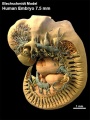
- Template:Human 7.5mm Embryo links
- Human Adult Brain Movie
- Human Development Timeline Movie
- Human Embryo - Scanning electron microscopy
- Template:Human embryo neck links
- Human Embryo SEM
- Human Fertilization Detail Movie
- Human Fertilization Movie
- Template:Human Fertilization Movie 1 frame table
- Template:Human Fertilization Movie 1 frames
- Template:Human Fertilization Movie 2 frame table
- Template:Human follicles lm and em links
- Template:Human ovary - corpus luteum links
- Template:Human Spermatozoa Statistics collapse table
- Template:Human Spermatozoa Statistics table
- Human Sylvian Fissure Movie
- Template:Human timeline
- Hutchinson-Gilford Progeria Syndrome
J
K
L
M
- Template:Macklin1921 figures
- Template:Mall1912 figures
- Template:Mall1916 figures
- Template:Mall1917 figures
- Menstrual Cycle - Histology
- Model Embryo 1.6mm Movie 1
- Model Embryo 10mm Movie 1
- Model Embryo 3.1mm Movie 1
- Model Embryo 7.5mm Movie 1
- Monosomic Embryo Movie 1
- Template:Morton1949 figures
- Template:Mouse Human lung table
- Movie - Neural Sylvian Fissure
N
P
- Palate Development
- Paper - A human embryo before the appearance of the myotomes (1918)
- Paper - A Human Embryo of Twenty-five Somites
- Paper - A Human Embryo of Twenty-seven Pairs of Somites, Embedded in Decidua
- Paper - A human embryo with head-process and commencing arch enteric canal
- Paper - A Human Embryo with Seven Pairs of Somites Measuring about 2 mm in Length
- Paper - A human embryo with seventeen pairs of somites (1930)
- Paper - A morphological study of testicular descent
- Paper - A Note on the Development of the Septum Transversum and the Liver
- Paper - A presomite human embryo (Shaw) - the implantation
- Paper - A presomite human embryo (Shaw) - the implantation (1942)
- Paper - A presomite human embryo showing a yolk-sac duct
- Paper - A presomite human embryo showing an early stage of the primitive streak
- Paper - A presomite human embryo with a neurenteric canal (embryo R.S.)
- Paper - A study of a 7 mm human embryo with special reference to its peculiar spirally twisted form, and its large aortic cell-clusters
- Paper - A study of the development of certain features of the cerebellum (1920)
- Paper - A very Young Human Embryo found embedded in a "Decidual Cast" of the Uterus
- Paper - A well-preserved human embryo of 10 somites (1929)
- Paper - A Young Human Embryo (Embryo Dobbin) with Head-Process and Prochordal Plate
- Paper - An early human embryo (no. 1285, Manchester Collection) with capsular attachment of the connecting stalk (1935)
- Paper - An Early Human Embryo (No. 1285, Manchester Collection), with Capsular Attachment of the Connecting Stalk
- Paper - An Early Human Embryo, with 0.55 mm long Embryonic Shield
- Paper - An Early Human Ovum (Thomson) in situ
- Paper - An iconometrographic representation of the growth of the central nervous system in man
- Paper - Breech fused twin monster (1934)
- Paper - Changes in fetuses due to formalin preservation
- Special:Badtitle/NS501:Paper - Description of a Human Embryo of 13-14 Mesodermic Somites
- Paper - Description of a Human Embryo of Twenty-three Paired Somites
- History:Paper - Description of a Human Embryo of Twenty-two paired Somites
- Paper - Description of a reconstruction of the head of a thirty-millimetre embryo (1910)
- Paper - Development and variation of the nerves and the musculature of the inferior extremity and of the neighboring regions of the trunk in man
- Paper - Development of the human heart from its earliest appearance to the stage found in embryos of twenty paired somites (1927)
- Paper - Developmental Changes in the Pericardium, the Mesocardia, and the Pleural Sacs in the Human Embryo
- Paper - Early development of the cervical vertebrae and the base of the occipital bone in man
- Paper - Early Stages of Human Development
- Paper - Factors Involved In The Formation Of The Filum Terminale
- Paper - Fine structure of the human ovum in the pronuclear stage
- Paper - Further note on the pro-chordal plate in man
- Paper - History of the development of the human ovum (1834)
Media in category 'Human'
The following 200 files are in this category, out of 2,421 total.
(previous page) (next page)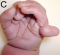 220px-Patauhand.PNG 220 × 200; 69 KB
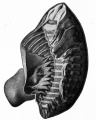 226.jpg 565 × 705; 71 KB
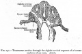 231.jpg 747 × 493; 69 KB
 3D Human pancreatic islet.jpg 1,088 × 1,280; 295 KB
 7.5mm Embryo movie 1 icon.jpg 299 × 400; 38 KB
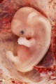 9 Week Human Embryo.jpg 400 × 600; 41 KB
 Abbott 191.jpg 1,034 × 1,000; 234 KB
 Abbott 201.jpg 1,150 × 1,000; 203 KB
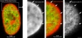
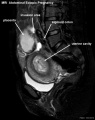 Abdominal ectopic pregnancy MRI.jpg 553 × 700; 45 KB
 Abnormal AusData81-92.png 523 × 358; 10 KB
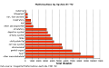 Abnormal AusData81-92Graph.png 509 × 320; 7 KB
 Abnormal81-92-heart.png 481 × 344; 6 KB
 Abnormal81-92-neuron.png 481 × 344; 9 KB
 Accessory renal artery.jpg 800 × 798; 103 KB
 Adrenal and gonad steroidogenic factor 1 expression.jpg 1,000 × 636; 88 KB
 Adult brain 01.mov ; 1.59 MB
- Adult brain 02.mov ; 434 KB
 Adult female fibroblast and lymphocyte nuclear DNA 01.jpg 1,095 × 1,200; 197 KB
 Adult female fibroblast nuclear DNA 01.jpg 2,186 × 1,000; 219 KB
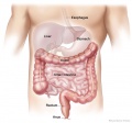 Adult gastrointestinal tract cartoon01.jpg 745 × 698; 55 KB
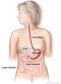 Adult gastrointestinal tract cartoon02.jpg 541 × 738; 42 KB
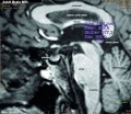 Adult human brain MRI01.jpg 700 × 607; 81 KB
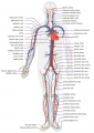 Adult human cardiovascular system.jpg 707 × 1,000; 151 KB
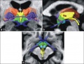 Adult human hypothalamus 01.jpg 917 × 700; 124 KB
 Adult human hypothalamus 02.jpg 1,151 × 343; 100 KB
 Adult human hypothalamus 03.jpg 767 × 854; 176 KB
 Adult human hypothalamus 04.jpg 660 × 500; 69 KB
 Adult renal venous cartoon.jpg 600 × 600; 62 KB
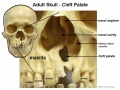
 Adult skull cleft palate 01.jpg 750 × 1,000; 89 KB
 Adult skull cleft palate 02.jpg 1,280 × 720; 139 KB
 Adult skull cleft palate 03.jpg 904 × 678; 104 KB
 Advanced Heart Development Timeline GA.jpg 1,000 × 434; 97 KB
 Advanced Heart Development Timeline.jpg 1,772 × 769; 158 KB
 Agenesis of left lung.jpg 600 × 424; 28 KB
 Anderson2016-fig07a.jpg 800 × 800; 193 KB
 Anderson2016-fig07b.jpg 800 × 800; 304 KB
 Anderson2016-fig09a.jpg 800 × 800; 106 KB
 Anderson2016-fig09b.jpg 800 × 800; 90 KB
 Anderson2016-fig10.jpg 800 × 800; 109 KB
 Anderson2016-fig11a.jpg 800 × 800; 108 KB
 Anderson2016-fig11b.jpg 800 × 800; 98 KB
 Anderson2016-fig12b.jpg 800 × 800; 138 KB
 Anderson2016-fig13b.jpg 800 × 800; 191 KB
 Anderson2016-fig14.jpg 800 × 800; 168 KB
 Anderson2016-fig15a.jpg 800 × 800; 142 KB
 Anderson2016-fig15b.jpg 800 × 800; 148 KB
 Anderson2016-fig16a.jpg 800 × 800; 193 KB
 Anderson2016-fig16b.jpg 800 × 800; 167 KB
 Anderson2016-fig18.jpg 800 × 800; 105 KB
 Anderson2016-fig19.jpg 800 × 800; 99 KB
 Anderson2016-fig20.jpg 800 × 800; 101 KB
 Anderson2016-fig21.jpg 762 × 800; 74 KB
 Anderson2016-fig22.jpg 800 × 779; 90 KB
 Anderson2016-fig23.jpg 783 × 800; 97 KB
 Anderson2016-fig34b.jpg 800 × 800; 92 KB
 Anderson2016-fig35b.jpg 800 × 800; 84 KB
 Anderson2016-fig36.jpg 800 × 800; 112 KB
 Anderson2016-fig37.jpg 800 × 800; 69 KB
 Anderson2016-fig41a.jpg 800 × 755; 92 KB
 Anderson2016-fig43a.jpg 800 × 800; 60 KB
 Anderson2016-fig43b.jpg 800 × 800; 48 KB
 Anderson2016-fig44a.jpg 800 × 800; 83 KB
 Anderson2016-fig44b.jpg 800 × 800; 76 KB
 Anderson2016-fig45a.jpg 800 × 800; 62 KB
 Anderson2016-fig45b.jpg 800 × 800; 57 KB
 Anderson2016-fig47.jpg 800 × 800; 83 KB
 Anderson2016-fig48.jpg 800 × 800; 203 KB
 Anderson2016-fig49.jpg 800 × 800; 90 KB
 Anencephaly ultrasound.jpg 900 × 658; 108 KB
 Aneuploidy model based on fragmentation 1.jpg 946 × 907; 154 KB
 Aneuploidy model based on fragmentation.jpg 668 × 790; 105 KB
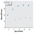 Anogenital distance from birth to 2 years.jpg 565 × 545; 34 KB
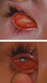 Anophthalmia and microphthalmia.jpg 273 × 477; 30 KB
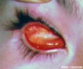 Anophthalmia.jpg 600 × 504; 46 KB
 Anotia 01.jpg 796 × 660; 61 KB
 Anson1934 fig01-8.jpg 1,337 × 888; 126 KB
 Anson1934 fig09.jpg 546 × 272; 16 KB
 Anson1934 fig10.jpg 520 × 612; 31 KB
 Anson1934 fig11.jpg 758 × 870; 52 KB
 Anson1934 fig12.jpg 545 × 968; 44 KB
 Anson1934 fig13.jpg 761 × 1,323; 74 KB
 Anson1934 fig14.jpg 761 × 1,323; 81 KB
 Anson1934 fig15.jpg 693 × 1,003; 62 KB
 Anson1934 fig16.jpg 691 × 985; 47 KB
 Anson1934 fig17.jpg 605 × 1,143; 57 KB
 Anson1934 fig18.jpg 418 × 1,161; 34 KB
 Anson1934 fig19.jpg 524 × 1,218; 48 KB
 Anson1934 plate01.jpg 1,557 × 2,279; 288 KB
 Anson1934 plate02.jpg 1,464 × 2,311; 259 KB
 Arthrogryposis.jpg 800 × 503; 39 KB
 Australian abnormalities 81-92 git.jpg 481 × 344; 43 KB
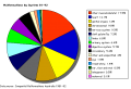
 Australian abnormalities pie skmus.png 481 × 344; 9 KB
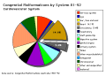
 Australian abnormalities pie urogen.png 481 × 344; 6 KB
 B050966-01.jpg 2,000 × 992; 681 KB
 B100658-01.jpg 1,388 × 1,040; 677 KB
 B100658-02.jpg 1,068 × 800; 458 KB
 B220849-01.jpg 1,311 × 849; 325 KB
 B220849-02.jpg 1,311 × 849; 358 KB
 B220849-03.jpg 1,388 × 1,040; 346 KB
 Bailey001.jpg 850 × 794; 155 KB
 Bailey004.jpg 364 × 1,013; 40 KB
 Bailey008.jpg 838 × 815; 89 KB
 Bailey009.jpg 774 × 766; 68 KB
 Bailey014.jpg 704 × 587; 116 KB
 Bailey073.jpg 705 × 501; 122 KB
 Bailey074.jpg 885 × 700; 176 KB
 Bailey075.jpg 920 × 590; 110 KB
 Bailey076.jpg 603 × 644; 63 KB
 Bailey077.jpg 775 × 686; 86 KB
 Bailey081.jpg 782 × 755; 118 KB
 Bailey082.jpg 879 × 756; 137 KB
 Bailey083.jpg 924 × 774; 129 KB
 Bailey084.jpg 569 × 590; 56 KB
 Bailey085.jpg 505 × 425; 59 KB
 Bailey086.jpg 720 × 662; 85 KB
 Bailey087.jpg 928 × 803; 132 KB
 Bailey088.jpg 888 × 701; 102 KB
 Bailey089.jpg 482 × 690; 55 KB
 Bailey090.jpg 716 × 478; 55 KB
 Bailey091.jpg 884 × 549; 81 KB
 Bailey094.jpg 376 × 689; 27 KB
 Bailey095.jpg 409 × 635; 52 KB
 Bailey096.jpg 693 × 501; 59 KB
 Bailey097.jpg 776 × 674; 71 KB
 Bailey098.jpg 680 × 432; 47 KB
 Bailey099.jpg 704 × 464; 52 KB
 Bailey103.jpg 739 × 738; 89 KB
 Bailey104.jpg 878 × 638; 107 KB
 Bailey105.jpg 545 × 567; 61 KB
 Bailey106.jpg 601 × 610; 86 KB
 Bailey109.jpg 710 × 669; 124 KB
 Bailey113.jpg 813 × 553; 128 KB
 Bailey114.jpg 690 × 433; 63 KB
 Bailey121.jpg 800 × 279; 51 KB
 Bailey122.jpg 514 × 440; 48 KB
 Bailey123.jpg 885 × 618; 78 KB
 Bailey125.jpg 496 × 429; 40 KB
 Bailey127.jpg 597 × 270; 43 KB
 Bailey128.jpg 805 × 356; 46 KB
 Bailey129.jpg 560 × 464; 44 KB
 Bailey130.jpg 714 × 439; 59 KB
 Bailey131.jpg 235 × 560; 39 KB
 Bailey132+133.jpg 940 × 570; 101 KB
 Bailey132.jpg 466 × 413; 43 KB
 Bailey133.jpg 806 × 655; 85 KB
 Bailey135.jpg 940 × 965; 216 KB
 Bailey137.jpg 672 × 539; 73 KB
 Bailey138.jpg 831 × 400; 62 KB
 Bailey139.jpg 961 × 671; 96 KB
 Bailey140.jpg 793 × 505; 58 KB
 Bailey141.jpg 761 × 323; 66 KB
 Bailey142.jpg 778 × 479; 72 KB
 Bailey143.jpg 911 × 673; 114 KB
 Bailey144.jpg 491 × 398; 39 KB
 Bailey145.jpg 777 × 654; 80 KB
 Bailey146.jpg 609 × 476; 42 KB
 Bailey147.jpg 660 × 632; 54 KB
 Bailey148.jpg 574 × 459; 38 KB
 Bailey149.jpg 576 × 520; 38 KB
 Bailey150.jpg 406 × 596; 42 KB
 Bailey151.jpg 585 × 631; 73 KB
 Bailey152.jpg 900 × 803; 300 KB
 Bailey153.jpg 799 × 585; 175 KB
 Bailey154.jpg 898 × 563; 191 KB
 Bailey155.jpg 894 × 833; 209 KB
 Bailey162.jpg 799 × 642; 102 KB
 Bailey163.jpg 757 × 691; 107 KB
 Bailey164.jpg 928 × 862; 149 KB
 Bailey166.jpg 787 × 656; 90 KB
 Bailey167.jpg 610 × 458; 58 KB
 Bailey169.jpg 504 × 264; 27 KB
 Bailey170.jpg 709 × 457; 67 KB
 Bailey171.jpg 954 × 507; 81 KB
 Bailey174.jpg 955 × 542; 88 KB
 Bailey175.jpg 885 × 306; 51 KB
 Bailey176.jpg 918 × 352; 62 KB
 Bailey178.jpg 913 × 653; 125 KB
 Bailey179.jpg 892 × 794; 114 KB
 Bailey180.jpg 938 × 431; 70 KB
 Bailey182.jpg 913 × 718; 103 KB
 Bailey183.jpg 847 × 448; 57 KB
 Bailey184.jpg 625 × 462; 48 KB
 Bailey185.jpg 944 × 499; 98 KB
 Bailey187.jpg 817 × 732; 70 KB
 Bailey188.jpg 906 × 538; 64 KB
 Bailey189.jpg 810 × 632; 63 KB
 Bailey190.jpg 801 × 584; 69 KB
 Bailey191.jpg 863 × 509; 109 KB
 Bailey192.jpg 960 × 806; 133 KB
 Bailey193.jpg 747 × 848; 94 KB
 Bailey194.jpg 841 × 638; 80 KB
 Bailey195.jpg 924 × 781; 79 KB
 Bailey196.jpg 760 × 510; 47 KB
 Bailey197.jpg 878 × 705; 122 KB
 Bailey198.jpg 931 × 623; 83 KB
 Bailey199.jpg 924 × 451; 62 KB
 Bailey200.jpg 898 × 671; 149 KB
 Bailey201.jpg 1,059 × 1,033; 259 KB
 Bailey202.jpg 906 × 848; 138 KB
 Bailey206.jpg 973 × 854; 162 KB


