Category:Human
From Embryology
This Embryology category shows pages and media related to human development.
Subcategories
This category has the following 72 subcategories, out of 72 total.
C
- Carnegie Embryo
- Carnegie Embryo 1
- Carnegie Embryo 112
- Carnegie Embryo 1134B
- Carnegie Embryo 116
- Carnegie Embryo 1266
- Carnegie Embryo 1455
- Carnegie Embryo 148
- Carnegie Embryo 172
- Carnegie Embryo 19
- Carnegie Embryo 239
- Carnegie Embryo 2393
- Carnegie Embryo 240
- Carnegie Embryo 248
- Carnegie Embryo 256
- Carnegie Embryo 296
- Carnegie Embryo 3527
- Carnegie Embryo 3956
- Carnegie Embryo 4046
- Carnegie Embryo 4059
- Carnegie Embryo 407
- Carnegie Embryo 4148
- Carnegie Embryo 43
- Carnegie Embryo 4405
- Carnegie Embryo 460
- Carnegie Embryo 463
- Carnegie Embryo 523
- Carnegie Embryo 5541
- Carnegie Embryo 5609
- Carnegie Embryo 5652
- Carnegie Embryo 5682
- Carnegie Embryo 5874
- Carnegie Embryo 6032
- Carnegie Embryo 625
- Carnegie Embryo 6426
- Carnegie Embryo 6581
- Carnegie Embryo 7618
- Carnegie Embryo 7669
- Carnegie Embryo 786
- Carnegie Embryo 808
- Carnegie Embryo 8147
- Carnegie Embryo 8239
- Carnegie Embryo 8370
- Carnegie Embryo 858
- Carnegie Embryo 8630
- Carnegie Embryo 8967
- Carnegie Embryo 9296
- Carnegie Embryo 96
- Carnegie Embryo 963
- Carnegie Embryo 966
- Carnegie Embryo 9697
D
F
H
Pages in category 'Human'
The following 200 pages are in this category, out of 332 total.
(previous page) (next page)A
- Abnormal Development - Cleft Lip and Palate
- Abnormal Development - Cleft Palate
- Abnormal Development - Toxoplasmosis
- Template:Abnormal Newborn Neural Exam Table
- Template:Adipose Timeline table
- Template:Adrenal GA32 Links
- Template:Anderson2016 collapsetable1
- Template:Anderson2016 table1
- Template:AnsonBlack1934 figures
- Aschheim-Zondek Test 1928 Movie
- Template:Australian GIT abnormalities 2002-2003
- Template:Australian Palate abnormalities 2002-2003
B
- Template:B050966
- Template:B100658
- Template:B220849
- Template:Bardeen1906 figures
- Template:Barniville1914 figures
- Template:Bartelmez1922 figures
- Template:BaxterBoyd1939 figures
- BGDA Practical 7 - Week 6
- Birth MRI Movie
- Book - Contributions to Embryology Carnegie Institution No.10
- Book - Contributions to Embryology Carnegie Institution No.112
- Book - Contributions to Embryology Carnegie Institution No.131
- Book - Contributions to Embryology Carnegie Institution No.159
- Book - Contributions to Embryology Carnegie Institution No.21
- Book - Contributions to Embryology Carnegie Institution No.22
- Book - Contributions to Embryology Carnegie Institution No.27
- Book - Contributions to Embryology Carnegie Institution No.29
- Book - Contributions to Embryology Carnegie Institution No.30
- Book - Contributions to Embryology Carnegie Institution No.32
- Book - Contributions to Embryology Carnegie Institution No.33
- Book - Contributions to Embryology Carnegie Institution No.34
- Book - Contributions to Embryology Carnegie Institution No.35
- Book - Contributions to Embryology Carnegie Institution No.38
- Book - Contributions to Embryology Carnegie Institution No.40
- Book - Contributions to Embryology Carnegie Institution No.42
- Book - Contributions to Embryology Carnegie Institution No.43
- Book - Contributions to Embryology Carnegie Institution No.44
- Book - Contributions to Embryology Carnegie Institution No.46
- Book - Contributions to Embryology Carnegie Institution No.47
- Book - Contributions to Embryology Carnegie Institution No.48
- Book - Contributions to Embryology Carnegie Institution No.52
- Book - Contributions to Embryology Carnegie Institution No.55
- Book - Contributions to Embryology Carnegie Institution No.59
- Book - Contributions to Embryology Carnegie Institution No.61
- Book - Contributions to Embryology Carnegie Institution No.65
- Book - Contributions to Embryology Carnegie Institution No.69
- Book - Contributions to Embryology Carnegie Institution No.72
- Book - Contributions to the Development of the Human Brain (1919)
- History:Book - Contributions to the Development of the Human Brain (1919)
- Book - Human embryos of different ages examined in median sections - a contribution to the mechanics of development
- Template:Braune 1877 Plate 2
C
E
F
H
- Template:Hamilton1944 figures
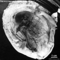
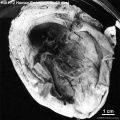
- Harvard Collection
- Template:Hearing EAM timeline
- Hela Apoptosis Movie
- Template:HillH12inks
- Template:HillH13 links
- Template:HillH145 links
- Template:HillH159 links
- Template:HillH202 links
- Template:HillH257 links
- Template:HillH4 links
- Template:HillH5 links
- Template:HillH52
- Template:HillH58 links
- Template:HillH6 links
- Template:HillH8 links
- Template:Hochstadter plates
- Template:Huber1905 table1
- Template:Human 7.5mm Embryo links
- Human Adult Brain Movie
- Human Development Timeline Movie
- Human Embryo - Scanning electron microscopy
- Template:Human embryo neck links
- Human Embryo SEM
- Human Fertilization Detail Movie
- Human Fertilization Movie
- Template:Human Fertilization Movie 1 frame table
- Template:Human Fertilization Movie 1 frames
- Template:Human Fertilization Movie 2 frame table
- Template:Human follicles lm and em links
- Template:Human ovary - corpus luteum links
- Template:Human Spermatozoa Statistics collapse table
- Template:Human Spermatozoa Statistics table
- Human Sylvian Fissure Movie
- Template:Human timeline
- Hutchinson-Gilford Progeria Syndrome
J
K
L
M
- Template:Macklin1921 figures
- Template:Mall1912 figures
- Template:Mall1916 figures
- Template:Mall1917 figures
- Menstrual Cycle - Histology
- Model Embryo 1.6mm Movie 1
- Model Embryo 10mm Movie 1
- Model Embryo 3.1mm Movie 1
- Model Embryo 7.5mm Movie 1
- Monosomic Embryo Movie 1
- Template:Morton1949 figures
- Template:Mouse Human lung table
- Movie - Neural Sylvian Fissure
N
P
- Palate Development
- Paper - A human embryo before the appearance of the myotomes (1918)
- Paper - A Human Embryo of Twenty-five Somites
- Paper - A Human Embryo of Twenty-seven Pairs of Somites, Embedded in Decidua
- Paper - A human embryo with head-process and commencing arch enteric canal
- Paper - A Human Embryo with Seven Pairs of Somites Measuring about 2 mm in Length
- Paper - A human embryo with seventeen pairs of somites (1930)
- Paper - A morphological study of testicular descent
- Paper - A Note on the Development of the Septum Transversum and the Liver
- Paper - A presomite human embryo (Shaw) - the implantation
- Paper - A presomite human embryo (Shaw) - the implantation (1942)
- Paper - A presomite human embryo showing a yolk-sac duct
- Paper - A presomite human embryo showing an early stage of the primitive streak
- Paper - A presomite human embryo with a neurenteric canal (embryo R.S.)
- Paper - A study of a 7 mm human embryo with special reference to its peculiar spirally twisted form, and its large aortic cell-clusters
- Paper - A study of the development of certain features of the cerebellum (1920)
- Paper - A very Young Human Embryo found embedded in a "Decidual Cast" of the Uterus
- Paper - A well-preserved human embryo of 10 somites (1929)
- Paper - A Young Human Embryo (Embryo Dobbin) with Head-Process and Prochordal Plate
- Paper - An early human embryo (no. 1285, Manchester Collection) with capsular attachment of the connecting stalk (1935)
- Paper - An Early Human Embryo (No. 1285, Manchester Collection), with Capsular Attachment of the Connecting Stalk
- Paper - An Early Human Embryo, with 0.55 mm long Embryonic Shield
- Paper - An Early Human Ovum (Thomson) in situ
- Paper - An iconometrographic representation of the growth of the central nervous system in man
- Paper - Breech fused twin monster (1934)
- Paper - Changes in fetuses due to formalin preservation
- Special:Badtitle/NS501:Paper - Description of a Human Embryo of 13-14 Mesodermic Somites
- Paper - Description of a Human Embryo of Twenty-three Paired Somites
- History:Paper - Description of a Human Embryo of Twenty-two paired Somites
- Paper - Description of a reconstruction of the head of a thirty-millimetre embryo (1910)
- Paper - Development and variation of the nerves and the musculature of the inferior extremity and of the neighboring regions of the trunk in man
- Paper - Development of the human heart from its earliest appearance to the stage found in embryos of twenty paired somites (1927)
- Paper - Developmental Changes in the Pericardium, the Mesocardia, and the Pleural Sacs in the Human Embryo
Media in category 'Human'
The following 200 files are in this category, out of 2,421 total.
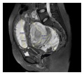
(previous page) (next page)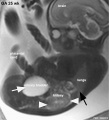 Fetal kidney MRI 01.jpg 797 × 880; 68 KB
 Fetal kidney MRI 02.jpg 797 × 880; 64 KB
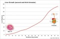
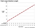 Fetal large Intestine length growth graph.jpg 800 × 653; 50 KB
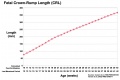 Fetal length change.jpg 972 × 648; 72 KB
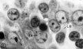 Fetal liver erythroblasts 01.jpg 905 × 534; 69 KB
 Fetal liver weight growth graph.jpg 800 × 521; 34 KB
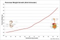 Fetal pancreas weight growth graph.jpg 1,000 × 669; 49 KB
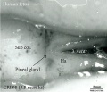 Fetal pineal gland 01.jpg 700 × 603; 67 KB
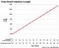 Fetal small Intestine length growth graph.jpg 800 × 653; 51 KB
 Fetal temporomandibular joint 01.jpg 600 × 391; 107 KB
 Fetal temporomandibular joint 02.jpg 600 × 390; 109 KB
 Fetal temporomandibular joint 03.jpg 600 × 393; 48 KB
 Fetal temporomandibular joint 04.jpg 600 × 390; 67 KB
 Fetal temporomandibular joint 05.jpg 600 × 392; 71 KB
 Fetal temporomandibular joint 06.jpg 600 × 389; 50 KB
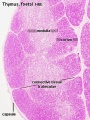 Fetal thymus.jpg 450 × 600; 122 KB
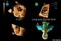 Fetal ultrasound ductal arch 01.jpg 800 × 533; 27 KB
 Finley1923 fig01.jpg 494 × 968; 53 KB
 Finley1923 fig02.jpg 700 × 800; 77 KB
 Finley1923 fig03.jpg 600 × 512; 56 KB
 Finley1923 fig04.jpg 700 × 627; 61 KB
 Finley1923 fig05.jpg 674 × 800; 146 KB
 Finley1923 fig06.jpg 729 × 800; 95 KB
 Finley1923 fig07.jpg 314 × 800; 40 KB
 Finley1923 fig08.jpg 296 × 800; 32 KB
 Finley1923 fig09.jpg 456 × 800; 49 KB
 Finley1923 fig10.jpg 221 × 802; 17 KB
 Finley1923 fig11.jpg 432 × 800; 38 KB
 Finley1923 fig12.jpg 593 × 800; 48 KB
 Finley1923 fig13.jpg 594 × 800; 51 KB
 Finley1923 Plate 1.jpg 776 × 1,000; 151 KB
 Finley1923 Plate 2.jpg 864 × 1,200; 153 KB
 Fleming1927-fig01.jpg 953 × 1,000; 167 KB
 Fleming1927-fig02.jpg 1,000 × 685; 53 KB
 Fleming1927-fig03.jpg 1,200 × 1,908; 602 KB
 Fleming1927-fig03a.jpg 1,179 × 493; 150 KB
 Fleming1927-fig03b.jpg 1,179 × 577; 171 KB
 Fleming1927-fig03c.jpg 1,179 × 800; 266 KB
 Fleming1927-fig04.jpg 800 × 899; 103 KB
 Florian1933 fig01.jpg 532 × 605; 38 KB
 Florian1933 fig02.jpg 580 × 569; 37 KB
 Florian1933 fig03.jpg 602 × 593; 40 KB
 Florian1933 fig04.jpg 754 × 610; 50 KB
 Florian1933 fig05.jpg 682 × 605; 51 KB
 Florian1933 fig06.jpg 731 × 584; 55 KB
 Florian1933 fig07.jpg 1,059 × 590; 60 KB
 Florian1933 fig08.jpg 1,109 × 520; 55 KB
 Foster109.jpg 873 × 372; 70 KB
 Foster110.jpg 924 × 821; 113 KB
 Foster112.jpg 1,002 × 494; 76 KB
 Foster113.jpg 927 × 512; 57 KB
 Foster114.jpg 725 × 1,071; 135 KB
 Foster117.jpg 797 × 707; 97 KB
 Foster118b.jpg 809 × 1,077; 232 KB
 Foster122.jpg 608 × 419; 45 KB
 Foster134.jpg 342 × 503; 37 KB
 Foster138.jpg 755 × 540; 51 KB
 Frazer1926 fig01.jpg 1,200 × 804; 137 KB
 Frazer1926 fig02.jpg 991 × 833; 145 KB
 Frazer1926 fig03.jpg 1,200 × 804; 69 KB
 Frazer1926 fig04.jpg 1,200 × 804; 95 KB
 Frazer1926 fig05.jpg 1,000 × 643; 85 KB
 Frazer1926 fig06.jpg 563 × 811; 35 KB
 Frazer1926 fig07.gif 554 × 600; 134 KB
 Frazer1926 fig07.jpg 1,229 × 996; 95 KB
 Frazer1926 fig08.jpg 616 × 789; 38 KB
 Frazer1926 plate01.jpg 1,914 × 2,681; 469 KB
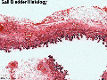
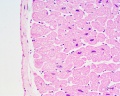
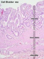 Gall bladder histology 001.jpg 375 × 500; 78 KB
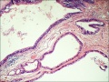
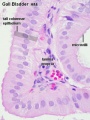 Gall bladder histology 002.jpg 375 × 500; 45 KB
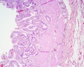 Gall bladder histology 003.jpg 1,280 × 1,024; 577 KB
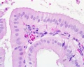 Gall bladder histology 004.jpg 1,280 × 1,024; 254 KB
 Gall bladder histology 005.gif 600 × 450; 683 KB
 Germ cell tumor 02.jpg 800 × 599; 168 KB
 Gillilan1959-fig01.jpg 1,000 × 1,313; 155 KB
 Gillilan1959-fig02.jpg 905 × 1,000; 159 KB
 Gillilan1959-fig03.jpg 1,042 × 1,366; 317 KB
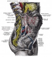
 Gray0054.jpg 800 × 513; 71 KB
 Gray0070.jpg 800 × 796; 182 KB
 Gray0071.jpg 700 × 440; 104 KB
 Gray0178.jpg 617 × 368; 44 KB
 Gray0179.jpg 617 × 368; 47 KB
 Gray0180.jpg 617 × 368; 48 KB
 Gray0181.jpg 617 × 368; 52 KB
 Gray0649.jpg 698 × 700; 72 KB
 Gray0651.jpg 698 × 700; 94 KB
 Gray0652.jpg 698 × 700; 103 KB
 Gray0653.jpg 698 × 700; 108 KB
 Gray0654.jpg 402 × 500; 39 KB
 Gray0655.jpg 500 × 419; 39 KB
 Gray0658.jpg 361 × 450; 29 KB
 Gray0847.jpg 559 × 900; 155 KB
 Gray0848.jpg 800 × 935; 289 KB
 Gray0849.jpg 800 × 885; 258 KB
 Gray0865.jpg 759 × 400; 70 KB
 Gray0867.jpg 495 × 600; 131 KB
 Gray0869.jpg 748 × 600; 126 KB
 Gray0870.jpg 637 × 600; 109 KB
 Gray0871.jpg 450 × 682; 114 KB
 Gray0872.jpg 499 × 600; 93 KB
 Gray0873.jpg 919 × 545; 124 KB
 Gray0874.jpg 600 × 545; 120 KB
 Gray0875.jpg 600 × 537; 83 KB
 Gray0876.jpg 256 × 700; 75 KB
 Gray0877.jpg 500 × 794; 101 KB
 Gray0878.jpg 580 × 550; 120 KB
 Gray0879.jpg 600 × 522; 66 KB
 Gray0880.jpg 800 × 496; 147 KB
 Gray0881.jpg 800 × 536; 87 KB
 Gray0882.jpg 800 × 649; 123 KB
 Gray0883.jpg 616 × 700; 107 KB
 Gray0884.jpg 419 × 400; 58 KB
 Gray0885.jpg 667 × 400; 20 KB
 Gray0886.jpg 500 × 368; 17 KB
 Gray0887.jpg 221 × 800; 67 KB
 Gray0888.jpg 711 × 566; 110 KB
 Gray0889.jpg 800 × 519; 103 KB
 Gray0890.jpg 700 × 700; 104 KB
 Gray0891.jpg 600 × 544; 104 KB
 Gray0892.jpg 500 × 409; 47 KB
 Gray0893.jpg 355 × 700; 93 KB
 Gray0894.jpg 710 × 400; 74 KB
 Gray0895.jpg 700 × 452; 94 KB
 Gray0896.jpg 600 × 638; 68 KB
 Gray0897.jpg 500 × 366; 47 KB
 Gray0904.jpg 226 × 350; 26 KB
 Gray0905.jpg 430 × 275; 19 KB
 Gray0906.jpg 438 × 600; 81 KB
 Gray0907.jpg 679 × 600; 110 KB
 Gray0909.jpg 600 × 437; 56 KB
 Gray0910.jpg 400 × 548; 78 KB
 Gray0911.jpg 651 × 400; 73 KB
 Gray0912.jpg 600 × 540; 87 KB
 Gray0913.jpg 671 × 600; 98 KB
 Gray0914.jpg 708 × 500; 102 KB
 Gray0915.jpg 720 × 600; 94 KB
 Gray0940.jpg 618 × 700; 160 KB
 Gray0941.jpg 700 × 524; 106 KB
 Gray0942.jpg 800 × 515; 127 KB
 Gray0971.jpg 800 × 583; 166 KB
 Gray0978.jpg 483 × 600; 67 KB
 Gray0979.jpg 500 × 446; 56 KB
 Gray0980.jpg 542 × 450; 57 KB
 Gray0981.jpg 538 × 340; 50 KB
 Gray0986.jpg 565 × 606; 56 KB
 Gray1108.jpg 590 × 400; 73 KB
 Gray1109.jpg 464 × 487; 56 KB
 Gray1111.jpg 523 × 600; 66 KB
 Gray1113.jpg 600 × 385; 68 KB
 Gray1114.jpg 450 × 471; 47 KB
 Gray1115.jpg 600 × 474; 73 KB
 Gray1116.jpg 600 × 433; 88 KB
 Gray1117.jpg 581 × 510; 75 KB
 Gray1118.jpg 600 × 403; 45 KB
 Gray1119.jpg 700 × 807; 115 KB
 Gray1174.jpg 782 × 800; 165 KB
 Gray1175.jpg 500 × 500; 31 KB
 Gray1183.jpg 800 × 325; 45 KB
 Gray1184.jpg 800 × 325; 39 KB
 Gray1235.jpg 800 × 389; 41 KB
 Gray1236.jpg 800 × 281; 29 KB
 Gray1237.jpg 371 × 600; 57 KB
 Greater-omentum.jpg 537 × 419; 48 KB
 Haemomonochorial human placenta EM01.jpg 792 × 775; 79 KB
 Hamilton1944-fig01.jpg 732 × 800; 168 KB
 Hamilton1944-fig02.jpg 540 × 556; 74 KB
 Hamilton1944-fig03.jpg 732 × 800; 169 KB
 Hamilton1944-fig04.jpg 714 × 794; 189 KB
 Hamilton1944-fig05.jpg 795 × 782; 101 KB
 Hamilton1944-fig06.jpg 385 × 749; 48 KB
 Hamilton1944-fig07.jpg 873 × 800; 140 KB
 Hamilton1944-plate01.jpg 1,200 × 1,472; 387 KB
 Hamilton1944-plate02.jpg 1,200 × 1,663; 287 KB
 Hamilton1944-table01.jpg 1,200 × 645; 113 KB
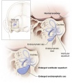 Hearing-vestibular sac abnormality.jpg 432 × 493; 45 KB
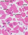
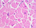
 Heart histology 001.jpg 400 × 500; 83 KB
 Heart histology 002.jpg 400 × 500; 81 KB
 Heart histology 003.jpg 400 × 500; 136 KB
 Heart histology 004.jpg 400 × 500; 97 KB
 Heart histology 101.jpg 1,280 × 1,024; 258 KB
 Heart histology 102.jpg 1,280 × 1,024; 242 KB
 Heart histology 103.jpg 1,280 × 1,024; 281 KB
 Heart histology 104.jpg 1,280 × 1,024; 280 KB
 Heart histology 105.jpg 1,280 × 1,024; 379 KB
 Heart histology 106.jpg 1,280 × 1,024; 347 KB
 Heart histology 107.jpg 1,280 × 1,024; 395 KB
 Heart human embryo CRL10mm 01.jpg 1,000 × 1,389; 537 KB
 Heart innervation 01.jpg 1,280 × 599; 92 KB
 Heart valve histology 01.jpg 1,008 × 1,280; 318 KB
 Heart valve histology 02.jpg 800 × 456; 103 KB
 Heart valve histology 03.jpg 800 × 489; 111 KB
 Heart-cartoon-001.jpg 600 × 697; 40 KB
 Heart-histology-102.jpg 1,280 × 1,024; 242 KB
 Herring1908b fig06.jpg 1,280 × 1,234; 283 KB
 Herring1908b fig07.jpg 1,280 × 974; 290 KB
 Heterotopic pregnancy MRI 01.jpg 600 × 536; 106 KB
 HillFlorian1931a fig01.jpg 594 × 1,372; 74 KB
 HillH12 Stage 23 bf01.jpg 1,500 × 1,500; 227 KB
 HillH12 Stage 23 bf02.jpg 1,500 × 1,500; 219 KB
 HillH12 Stage 23 bf03.jpg 1,500 × 1,500; 275 KB
 HillH12 Stage 23 bf04.jpg 1,500 × 1,500; 316 KB


