BGDA Practical - Male Reproductive Tract Histology: Difference between revisions
mNo edit summary |
mNo edit summary |
||
| Line 14: | Line 14: | ||
# Undertake a microscopical examination of the testis, epididymis, vas deferens, seminal vesicle, prostate gland, penis, and sperm. The functional significance of the various histological structures identified will be discussed. | # Undertake a microscopical examination of the testis, epididymis, vas deferens, seminal vesicle, prostate gland, penis, and sperm. The functional significance of the various histological structures identified will be discussed. | ||
:'''Links:''' | :'''Links:''' [[Testis Development]] | [[Histology]] | ||
==Human Testis (young)== | |||
<gallery> | |||
File:Testis_histology_001.jpg|overview Loupe | |||
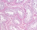
File:Testis_histology_004.jpg|convoluted seminiferous tubules x10 | |||
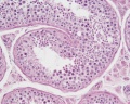
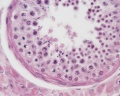
File:Testis_histology_006.jpg|convoluted seminiferous tubules x40 | |||
File:Testis_histology_007.jpg|convoluted seminiferous tubules x40 | |||
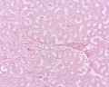
File:Testis_histology_005.jpg|tunica albuginea x20 | |||
</gallery> | |||
==Human Testis (adult)== | |||
<gallery> | |||
File:Testis_histology_002.jpg|overview x2 | |||
File:Testis_histology_010.jpg|convoluted seminiferous tubules x10 | |||
File:Testis_histology_009.jpg|convoluted seminiferous tubules x20 | |||
File:Testis_histology_011.jpg|convoluted seminiferous tubules x40 | |||
File:Testis_histology_012.jpg|convoluted seminiferous tubules x40 | |||
File:Testis_histology_008.jpg|epididymis overview x4 | |||
File:Testis_histology_013.jpg|epididymis x10 | |||
File:Testis_histology_014.jpg|epididymis x20 | |||
File:Testis_histology_015.jpg|epididymis x40 | |||
File:Testis_histology_003.jpg|ductus deferens overview x2 | |||
File:Testis_histology_016.jpg|ductus deferens x10 | |||
File:Testis_histology_017.jpg|ductus deferens x40 | |||
</gallery> | |||
:[[Rabbit Development|'''Rabbit''']]: convoluted seminiferous tubules [[:File:Testis_histology_018.jpg|x20]] | [[:File:Testis_histology_019.jpg|x100]] | |||
:[[Mouse Development|'''Mouse''']]: [[:File:Mouse epididymis development 01.jpg|postnatal epididymis]] | [[:File:Mouse epididymis development 02.jpg|14 days postnatal]] | [[:File:Mouse epididymis development 03.jpg|33 days postnatal]] | [[:File:Mouse epididymis development 04.jpg|45 days postnatal]] | [[:File:Mouse epididymis development 05.jpg|2 months postnatal]] | |||
:'''Spermatozoa human:''' [[:File:Spermatozoa_histology_001.jpg|x20]] | [[:File:Spermatozoa_histology_002.jpg|x40]] | [[:File:Spermatozoa_histology_003.jpg|x100]] | |||
==Terms== | ==Terms== | ||
Revision as of 13:49, 27 May 2013
Introduction


This current page provides background support information for Medicine phase 1 BGD Histology Practical Virtual Slides. Page does not form part of the BGDA practical class virtual slides.
- Virtual Slides: Male Histology (requires zpass login)
- Practical Manual Notes 2013 online PDF (2 pages)
- Draft page (notice removed when completed).
Objectives
- Gain an overview of the microanatomy of the male reproductive system.
- Undertake a microscopical examination of the testis, epididymis, vas deferens, seminal vesicle, prostate gland, penis, and sperm. The functional significance of the various histological structures identified will be discussed.
- Links: Testis Development | Histology
Human Testis (young)
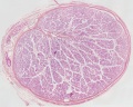
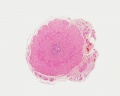
overview Loupe
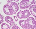
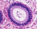
convoluted seminiferous tubules x10

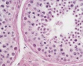
convoluted seminiferous tubules x40
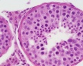
convoluted seminiferous tubules x40
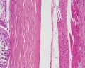
tunica albuginea x20


Human Testis (adult)
overview x2
convoluted seminiferous tubules x10
convoluted seminiferous tubules x20
convoluted seminiferous tubules x40
convoluted seminiferous tubules x40
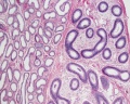
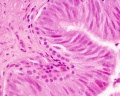
epididymis overview x4
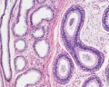
epididymis x10
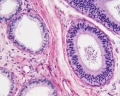
epididymis x20
epididymis x40
ductus deferens overview x2

ductus deferens x10
ductus deferens x40


- Mouse: postnatal epididymis | 14 days postnatal | 33 days postnatal | 45 days postnatal | 2 months postnatal
Terms
- cortex - (Latin = rind, or bark) outer layer of an organ.
- hilum - or hilus (Latin,= a trifle; depression in a seed) a depression at vascular entrance/exit of a gland or organ.
- medulla - (Latin, medulla = pith, marrow) the inner portion of an organ, in contrast to cortex.
- menstrual - (Latin, menstruus = monthly) relating to the monthly female sexual cycle.
- mucosa - (Latin, = mucous membrane) thin layer which lines body cavities and passages formed by epithelium and lamina propria.
- parenchyma - (Greek," + enkeim = to pour in) the essential functional cells of an organ as opposed to its stroma.
- serosa - (Latin, serum = whey; a pale fluid) a serous membrane lining body cavities.
- stroma - (Greek, = a cover, table-cloth, bedding) term for the internal supporting frame-work of a tissue, or organ, as opposed to its parenchyma.
- tunica albuginea - a dense, white, fibrous sheath enclosing a part or organ.
- Histology Glossary: A | B | C | D | E | F | G | H | I | J | K | L | M | N | O | P | Q | R | S | T | U | V | W | X | Y | Z | ANAT2241 Support | Histology | Histology Stains | Embryology Glossary
BGDA: Lecture 1 | Lecture 2 | Practical 3 | Practical 6 | Practical 12 | Lecture Neural | Practical 14 | Histology Support - Female | Male | Tutorial
Glossary Links
- Glossary: A | B | C | D | E | F | G | H | I | J | K | L | M | N | O | P | Q | R | S | T | U | V | W | X | Y | Z | Numbers | Symbols | Term Link
Cite this page: Hill, M.A. (2024, May 28) Embryology BGDA Practical - Male Reproductive Tract Histology. Retrieved from https://embryology.med.unsw.edu.au/embryology/index.php/BGDA_Practical_-_Male_Reproductive_Tract_Histology
- © Dr Mark Hill 2024, UNSW Embryology ISBN: 978 0 7334 2609 4 - UNSW CRICOS Provider Code No. 00098G