BGDA Practical - Male Reproductive Tract Histology: Difference between revisions
mNo edit summary |
mNo edit summary |
||
| (13 intermediate revisions by the same user not shown) | |||
| Line 3: | Line 3: | ||
| [[File:BGDsmall.jpg|left]] This current page provides background support information for Medicine phase 1 BGD Histology Practical Virtual Slides. Page does not form part of the BGDA practical class virtual slides. | | [[File:BGDsmall.jpg|left]] This current page provides background support information for Medicine phase 1 BGD Histology Practical Virtual Slides. Page does not form part of the BGDA practical class virtual slides. | ||
: | <br> | ||
<br> | |||
{| | |||
| [[File:Moodle icon2.jpg|link=http://moodle.telt.unsw.edu.au/mod/book/view.php?id=853422&chapterid=106729]] | |||
| [http://moodle.telt.unsw.edu.au/mod/book/view.php?id=853422&chapterid=106729 Moodle Lab Slides - Male Reproductive Tract] | |||
Small Moodle icon links appearing below on this page go directly to the Lab Slide. (you must be logged in to Moodle) | |||
Slides: [http://moodle.telt.unsw.edu.au/mod/book/view.php?id=853422 Female] | [http://moodle.telt.unsw.edu.au/mod/book/view.php?id=853422&chapterid=106729 Male] | [http://moodle.telt.unsw.edu.au/mod/book/view.php?id=853422&chapterid=106721 Placenta] | |||
|} | |||
:'''Practical Manual Notes 2013''' [[Talk:BGDA_Practical_-_Male_Reproductive_Tract_Histology|online]] [[Media:BGDAPracManual 2013-Prac10.pdf|PDF (2 pages)]] | [[BGDA Practical - Female Reproductive Tract Histology]] | |||
| [[File:Historic-testis.jpg|thumb|Historic drawing]] | | [[File:Historic-testis.jpg|thumb|Historic drawing]] | ||
| Line 20: | Line 26: | ||
:'''Links:''' [[Testis Development]] | [[Histology]] | :'''Links:''' [[Testis Development]] | [[Prostate Development]] | [[Histology]] | ||
==Testis Histology== | ==Testis Histology== | ||
| Line 66: | Line 72: | ||
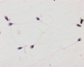
==Spermatozoa== | ==Spermatozoa== | ||
{| | {| | ||
| [[File:spermatozoa animation icon.jpg|100px]] This brief animation shows an overview of the structural components of the [[S#spermatozoa|spermatozoa]]. | | [[File:spermatozoa animation icon.jpg|100px|link=Spermatozoa Structure Movie]] This brief animation shows an overview of the structural components of the [[S#spermatozoa|spermatozoa]]. | ||
# Blue - Nucleus containing male haploid genome required to combine with oocyte haploid genome to form diploid zygote. | # Blue - Nucleus containing male haploid genome required to combine with oocyte haploid genome to form diploid zygote. | ||
| Line 78: | Line 84: | ||
* divided into head, neck and tail. | * divided into head, neck and tail. | ||
** head - (flattened, about 5 µm long and 3 µm wide) chiefly consists of the nucleus (greatly condensed chromatin!). The anterior 2/3 of the nucleus is covered by the acrosome, which contains enzymes important in the process of fertilisation. The posterior parts of the nuclear membrane forms the so-called basal plate. | ** head - (flattened, about 5 µm long and 3 µm wide) chiefly consists of the nucleus (greatly condensed chromatin!). The anterior 2/3 of the nucleus is covered by the acrosome, which contains enzymes important in the process of fertilisation. The posterior parts of the nuclear membrane forms the so-called basal plate. | ||
** neck - short (about 1 µm) and attached to the basal plate. A transversely oriented centriole is located immediately behind the basal plate. The neck also contains nine segmented columns of fibrous material, which continue as the outer dense fibres into the tail.
** tail - further divided into a middle piece, a principal piece and an end piece. The axonema ( | ** neck - short (about 1 µm) and attached to the basal plate. A transversely oriented centriole is located immediately behind the basal plate. The neck also contains nine segmented columns of fibrous material, which continue as the outer dense fibres into the tail.
| ||
| | ** tail - further divided into a middle piece, a principal piece and an end piece. The '''axonema''' (arrangement of microtubules in all cilia) begins in the middle piece. It is surrounded by nine outer dense fibres, which are not found in other cilia. In the middle piece (about 5 µm long), the axonema and dense fibres are surrounded by a sheath of mitochondria. The middle piece is terminated by a dense ring, the annulus. The principal piece is about 45 µm long. It contains a fibrous sheath, which consists of dorsal and ventral longitudinal columns interconnected by regularly spaced circumferential hoops. The fibrous sheath and the dense fibres do not extend to the tip of the tail. Along the last part (5 µm) of the tail, called the end piece, the axonema is only surrounded by a small amount of cytoplasm and the plasma membrane. | ||
| width=310px|<html5media height="240" width="300">File:Spermatozoa_animation.mp4</html5media> | |||
[[Media:Spermatozoa_animation.mp4|'''Click Here''' to play on mobile device]] | |||
|} | |} | ||
<gallery> | <gallery> | ||
| Line 145: | Line 154: | ||
[[File:Ductus_deferens_01.jpg]][[File:Ductus_deferens_02.jpg]] | [[File:Ductus_deferens_01.jpg]][[File:Ductus_deferens_02.jpg]] | ||
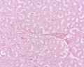
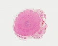
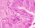
==Prostate Histology== | |||
{| | |||
| [[File:Prostate_histology_01.jpg|300px]] | |||
| [[File:Prostate_histology_02.jpg|300px]] | |||
| [[File:Prostate_histology_03.jpg|300px]] | |||
|- | |||
| '''Human prostate histology''' | |||
| '''Corpora Amylacea''' | |||
| '''Submucosal gland''' | |||
|- | |||
| (adult, low power overview) | |||
| (adult, detail) | |||
| (adult, high power detail) | |||
|} | |||
==Penis Histology== | ==Penis Histology== | ||
Latest revision as of 19:07, 27 April 2016
Introduction

|


Objectives
- Gain an overview of the microanatomy of the male reproductive system.
- Undertake a microscopical examination of the testis, epididymis, vas deferens, seminal vesicle, prostate gland, penis, and sperm. The functional significance of the various histological structures identified will be discussed.
- Links: Testis Development | Prostate Development | Histology
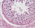
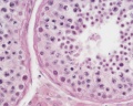
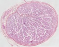
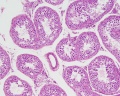
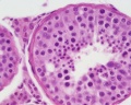
Testis Histology
Convoluted Seminiferous Tubules
Spermatogonia
** Type A spermatogonia have a rounded nucleus with very fine chromatin grains and one or two nucleoli. They are stem cells which divide to form new generations of both type A and type B spermatogonia. ** Type B spermatogonia have rounded nuclei with chromatin granules of variable size, which often attach to the nuclear membrane, and one nucleolus. Although type B spermatogonia may divide repeatedly, they do not function as stem cells and their final mitosis always results in the formation of primary spermatocytes. Primary spermatocytes
Secondary spermatocytes
Spermatids
|

|

|

|
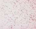
Spermatozoa
|
<html5media height="240" width="300">File:Spermatozoa_animation.mp4</html5media> |
x20
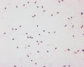
x40
x100


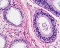
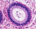
Epididymis Histology
Pseudostratified Epithelium
|

|

|

|
Human Testis (adult)
overview x2
convoluted seminiferous tubules x10
convoluted seminiferous tubules x20
convoluted seminiferous tubules x40
convoluted seminiferous tubules x40
epididymis overview x4
epididymis x10
epididymis x20
epididymis x40
ductus deferens overview x2

ductus deferens x10
ductus deferens x40


Human Testis (young)
overview Loupe
convoluted seminiferous tubules x10

convoluted seminiferous tubules x40
convoluted seminiferous tubules x40
tunica albuginea x20


Other Species
Rabbit

convoluted seminiferous tubules x20

convoluted seminiferous tubules x100


Mouse
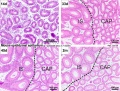
postnatal epididymis
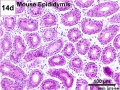
14 days postnatal
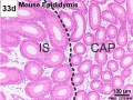
33 days postnatal
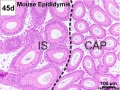
45 days postnatal
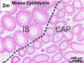
2 months postnatal


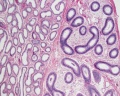
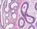
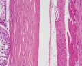
Ductus Deferens Histology


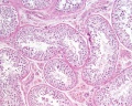
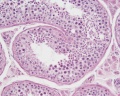
Prostate Histology

|

|

|
| Human prostate histology | Corpora Amylacea | Submucosal gland |
| (adult, low power overview) | (adult, detail) | (adult, high power detail) |
Penis Histology


Terms
- cortex - (Latin = rind, or bark) outer layer of an organ.
- hilum - or hilus (Latin,= a trifle; depression in a seed) a depression at vascular entrance/exit of a gland or organ.
- medulla - (Latin, medulla = pith, marrow) the inner portion of an organ, in contrast to cortex.
- mucosa - (Latin, = mucous membrane) thin layer which lines body cavities and passages formed by epithelium and lamina propria.
- parenchyma - (Greek," + enkeim = to pour in) the essential functional cells of an organ as opposed to its stroma.
- serosa - (Latin, serum = whey; a pale fluid) a serous membrane lining body cavities.
- stroma - (Greek, = a cover, table-cloth, bedding) term for the internal supporting frame-work of a tissue, or organ, as opposed to its parenchyma.
- tunica albuginea - a dense, white, fibrous sheath enclosing a part or organ.
- Histology Glossary: A | B | C | D | E | F | G | H | I | J | K | L | M | N | O | P | Q | R | S | T | U | V | W | X | Y | Z | ANAT2241 Support | Histology | Histology Stains | Embryology Glossary
BGDA: Lecture 1 | Lecture 2 | Practical 3 | Practical 6 | Practical 12 | Lecture Neural | Practical 14 | Histology Support - Female | Male | Tutorial
Glossary Links
- Glossary: A | B | C | D | E | F | G | H | I | J | K | L | M | N | O | P | Q | R | S | T | U | V | W | X | Y | Z | Numbers | Symbols | Term Link
Cite this page: Hill, M.A. (2024, May 6) Embryology BGDA Practical - Male Reproductive Tract Histology. Retrieved from https://embryology.med.unsw.edu.au/embryology/index.php/BGDA_Practical_-_Male_Reproductive_Tract_Histology
- © Dr Mark Hill 2024, UNSW Embryology ISBN: 978 0 7334 2609 4 - UNSW CRICOS Provider Code No. 00098G