ANAT2341 Lab 6: Difference between revisions

From Embryology
mNo edit summary
mNo edit summary
Line 49: Line 49:
{{Neural Crest Links}}


{{Sensory Links}}
[[Sensory System Development]]
|-
|-
| {{MPT2011cover_citation}}  
| {{MPT2011cover_citation}}  
Line 63: Line 63:
| Other Online Respiratory Resources
| Other Online Respiratory Resources
|   
|   
* '''Anatomy of the Human Body''' 1918 Henry Gray [[Anatomy_of_the_Human_Body_by_Henry_Gray#947_Respiratory|The Respiratory Apparatus]]
* '''Developmental Biology''' 8e Online [http://8e.devbio.com/article.php?ch=15&id=157 Lung Branching Morphogenesis]
|}
|}
{| class="wikitable mw-collapsible mw-collapsed"

Revision as of 08:07, 10 September 2014

Head Development
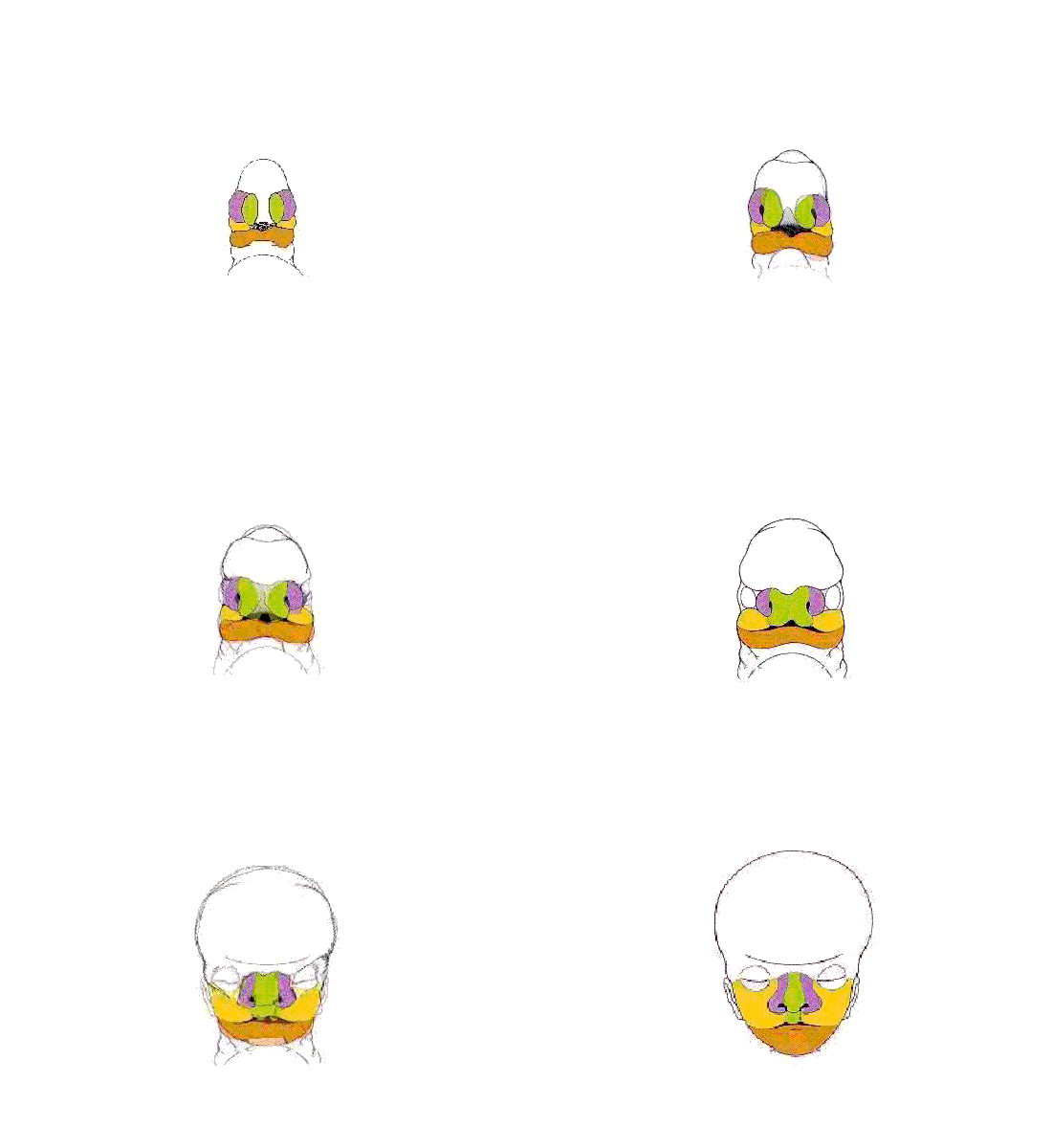
Face animation.gif

Face Development Movie | MP4 version | Quicktime version

Development of the Face

This animation shows a ventral view of development of the human face from approximately week 5 through to neonate.

The separate embryonic components that contribute to the face have been colour coded.

  • Frontonasal Prominence central portion (white)
  • Frontonasal Prominence - Lateral nasal (purple)
  • Frontonasal Prominence - Medial nasal (green)
  • Pharyngeal Arch 1 - Maxillary prominence (yellow)
  • Pharyngeal Arch 1 - Mandibular prominence (orange)
  • Stomodeum (black)

The five elements are: 1 Frontonasal Prominence, 2 Maxillary prominences and 2 Mandibular prominences.

The stomodeum is the primordial mouth region and a surface central depression lying between the forebrain bulge and the heart bulge. At the floor of the stomodeum indentation is the buccopharyngeal membrane (oral membrane).

Note the complex origin of the maxillary region (upper jaw) requiring the fusion of several embryonic elements, abnormalities of this process lead to cleft lip and cleft palate.


Links: Quicktime | Quicktime version | Flash version | Animated GIF | Lecture - Head Development | Head Development

Aim

To introduce the developmental embryology of the head and face, and their associated abnormalities.


Specific Objectives

  1. To understand the formation and contribution of the pharyngeal arches to face and neck development.
  2. To know the main structures derived from components of the pharyngeal arches (groove, pouch and arch connective tissue).
  3. To briefly understand some abnormalities associated with head development.

Textbooks

UNSW Embryology logo
Hill, M.A. (2020). UNSW Embryology (20th ed.) Retrieved May 2, 2024, from https://embryology.med.unsw.edu.au
Head Links: Introduction | Medicine Lecture | Medicine Lab | Science Lecture | Lecture Movie | Science Lab | pharyngeal arch | Craniofacial Seminar | mouth | palate | tongue | placode | skull | neural crest | Head and Face Movies | head abnormalities | Category:Head
Historic Head Embryology  
1910 Skull | 1910 Skull Images | 1912 Nasolacrimal Duct | 1921 Human Brain Vascular | 1923 Head Subcutaneous Plexus | 1919 21mm Embryo Skull | 1920 Human Embryo Head Size | 1921 43 mm Fetal Skull | Historic Disclaimer
Neural Crest Links: neural crest | Lecture - Early Neural | Lecture - Neural Crest Development | Lecture Movie | Schwann cell | adrenal | melanocyte | peripheral nervous system | enteric nervous system | cornea | cranial nerve neural crest | head | skull | cardiac neural crest | Nicole Le Douarin | Neural Crest Movies | neural crest abnormalities | Category:Neural Crest
Student Projects 2023: 1 Patterning neural border and NC | 2 NPB NEUcrest | 3 EMT and NC | 4 miRNA and NC | 5 Adrenal Gland and NC | 6 Melanocyte & Melanoma | 7 Neurocristopathies | Neural Crest
These projects are the sole work of undergraduate science students and may contain errors in fact or descriptions.


Historic Embryology - Neural Crest  
1879 Olfactory Organ | 1905 Cranial and Spinal Nerves | 1908 10 mm Peripheral | 1910 Mammal Sympathetic | 1920 Human Sympathetic | 1928 Cranial ganglia | 1939 10 Somite Embryo | 1942 Origin | 1957 Adrenal

Sensory System Development

Textbook cover The Developing Human, 9th edn.
Moore, K.L., Persaud, T.V.N. & Torchia, M.G. (2011). The developing human: clinically oriented embryology (9th ed.). Philadelphia: Saunders.
The following chapter links only work with a UNSW connection.
Textbook cover Larsen's human embryology 4th edn.
Schoenwolf, G.C., Bleyl, S.B., Brauer, P.R. & Francis-West, P.H. (2009). Larsen's human embryology (4th ed.). New York; Edinburgh: Churchill Livingstone.
The following chapter links only work with a UNSW connection.
Other Online Respiratory Resources
ECHO360 Recording
ECHO360 icon.gif

Links only work with currently enrolled UNSW students.

Lecture 9 -

Lab 6: Introduction | Trilaminar Embryo | Early Embryo | Late Embryo | Fetal | Postnatal | Abnormalities | Online Assessment
2014 Course: Week 2 Lecture 1 Lecture 2 Lab 1 | Week 3 Lecture 3 Lecture 4 Lab 2 | Week 4 Lecture 5 Lecture 6 Lab 3 | Week 5 Lecture 7 Lecture 8 Lab 4 | Week 6 Lecture 9 Lecture 10 Lab 5 | Week 7 Lecture 11 Lecture 12 Lab 6 | Week 8 Lecture 13 Lecture 14 Lab 7 | Week 9 Lecture 15 Lecture 16 Lab 8 | Week 10 Lecture 17 Lecture 18 Lab 9 | Week 11 Lecture 19 Lecture 20 Lab 10 | Week 12 Lecture 21 Lecture 22 Lab 11 | Week 13 Lecture 23 Lecture 24 Lab 12
Student Projects - Group 1 | Group 2 | Group 3 | Group 4 | Group 5 | Group 6 | Group 7 | Group 8 | Moodle