ANAT2341 Lab 10: Difference between revisions
mNo edit summary |
No edit summary |
||
| (36 intermediate revisions by 2 users not shown) | |||
| Line 1: | Line 1: | ||
= | ==Organogenesis Lab== | ||
== 1. QUIZ == | |||
== 2. Organogenesis Lab == | |||
In this lab you will dissect fertile chicken eggs and study fixed mouse embryos up to mid-gestation using dissection microscopes. You will name the embryonic anatomical structures, and describe what these will give rise to. | |||
You will | |||
[[Media:Fertile_Egg_practical_Class_2.pdf|Organogensis Lab Manual]] | |||
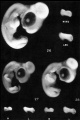
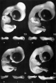
[[File:Chicken_Embryo_Hamburger_stages.jpg|600px|link=Hamburger Hamilton Stages]] | |||
''These are the Hamburger stages of chicken development'' | |||
{{ | See also the [https://www.jove.com/video/306/windowing-chicken-eggs-for-developmental-studies JoVE article on chicken egg preparation]: <pubmed>18989413</pubmed> | ||
===Additional Chicken Links=== | |||
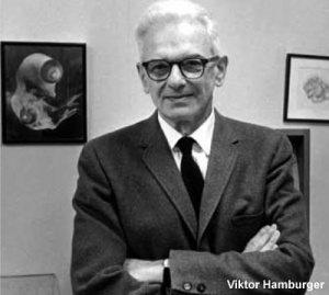
[[File:Viktor Hamburger.jpg|thumb|alt=Viktor Hamburger|link=Embryology History - Viktor Hamburger|Viktor Hamburger (1900 – 2001)]] | |||
More about chicken embryogenesis: [[Chicken Development]] | [[Hamburger Hamilton Stages]] | |||
<br> | |||
{{Chicken links}} | |||
<br> | |||
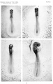
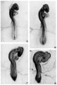
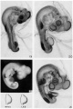
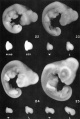
<gallery> | |||
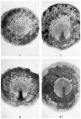
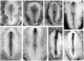
File:HHstage1-4.jpg|stage 1-4 | |||
File:HHstage5-10.jpg|stages 5-10 | |||
File:HHstage11-14.jpg|stages 11-14 | |||
File:HHstage15-18.jpg|stages 15-18 | |||
File:HHstage19-21.jpg|stages 19-21 | |||
File:HHstage22-25.jpg|stages 22-25 | |||
File:HHstage26-28.jpg|stages 26-28 | |||
File:HHstage29-32.jpg|stage 29-32 | |||
</gallery> | |||
[[File:Mouse_vs_Human_embryogenesis.jpg]] | |||
''This figure compares the human and mouse developmental stages'' | |||
More about Mouse embryogenesis: [[Mouse Timeline Detailed]] | |||
{{Chicken}} | |||
===External Links=== | |||
{{External Links}} | |||
* JOVE - [http://www.jove.com/science-education/5153/an-introduction-to-the-chick-gallus-gallus-domesticus An Introduction to the Chicken] | |||
{{2018ANAT2341}} | |||
Revision as of 08:49, 8 October 2018
Organogenesis Lab
1. QUIZ
2. Organogenesis Lab
In this lab you will dissect fertile chicken eggs and study fixed mouse embryos up to mid-gestation using dissection microscopes. You will name the embryonic anatomical structures, and describe what these will give rise to.
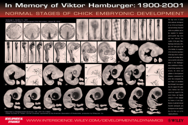
These are the Hamburger stages of chicken development
See also the JoVE article on chicken egg preparation: <pubmed>18989413</pubmed>
Additional Chicken Links
More about chicken embryogenesis: Chicken Development | Hamburger Hamilton Stages
stage 1-4
stages 5-10
stages 11-14
stages 15-18
stages 19-21
stages 22-25
stages 26-28
stage 29-32


This figure compares the human and mouse developmental stages
More about Mouse embryogenesis: Mouse Timeline Detailed
External Links
External Links Notice - The dynamic nature of the internet may mean that some of these listed links may no longer function. If the link no longer works search the web with the link text or name. Links to any external commercial sites are provided for information purposes only and should never be considered an endorsement. UNSW Embryology is provided as an educational resource with no clinical information or commercial affiliation.
| 2018 ANAT2341 - Timetable | Course Outline | Moodle | Tutorial 1 | Tutorial 2 | Tutorial 3 |
Labs: 1 Preimplantation and Implantation | 2 Reproductive Technology Revolution | 3 Group Projects | 4 GM manipulation mouse embryos | 5 Early chicken eggs | 6 Female reproductive tract | 7 Skin regeneration | 8 Vertebral development | 9 Organogenesis Lab | 10 Cardiac development | 11 Group projects | 12 Stem Cell Journal Club |
|
Lectures: 1 Introduction | 2 Fertilization | 3 Week 1/2 | 4 Week 3 | 5 Ectoderm | 6 Placenta | 7 Mesoderm | 8 Endoderm | 9 Research Technology | 10 Cardiovascular | 11 Respiratory | 12 Neural crest | 13 Head | 14 Musculoskeletal | 15 Limb | 16 Renal | 17 Genital | 18 Endocrine | 19 Sensory | 20 Fetal | 21 Integumentary | 22 Birth | 23 Stem cells | 24 Revision |
| Student Projects: Group Projects Information Project 1 | Project 3 | Project 4 | Project 5 | 2018 Test Student | Copyright |