Category:Mouse E16.5

The Embryology pages and media listed below relate to mouse embryonic day 16.5 (E16.5) of development. This staging by "days" relate to in the female presence of a vaginal plug indicating that the mating occurred, see timed pregnancy.
- Mouse Stages: E1 | E2.5 | E3.0 | E3.5 | E4.5 | E5.0 | E5.5 | E6.0 | E7.0 | E7.5 | E8.0 | E8.5 | E9.0 | E9.5 | E10 | E10.5 | E11 | E11.5 | E12 | E12.5 | E13 | E13.5 | E14 | E14.5 | E15 | E15.5 | E16 | E16.5 | E17 | E17.5 | E18 | E18.5 | E19 | E20 | Timeline | About timed pregnancy
| Carnegie | Stage | |||||||||||||||||||||||
| Human | Days | 1 | 2-3 | 4-5 | 5-6 | 7-12 | 13-15 | 15-17 | 17-19 | 20 | 22 | 24 | 28 | 30 | 33 | 36 | 40 | 42 | 44 | 48 | 52 | 54 | 55 | 58 |
| Mouse | Days | 1 | 2 | 3 | E4.5 | E5.0 | E6.0 | E7.0 | E8.0 | E9.0 | E9.5 | E10 | E10.5 | E11 | E11.5 | E12 | E12.5 | E13 | E13.5 | E14 | E14.5 | E15 | E15.5 | E16 |
| Rat | Days | 1 | 3.5 | 4-5 | 5 | 6 | 7.5 | 8.5 | 9 | 10.5 | 11 | 11.5 | 12 | 12.5 | 13 | 13.5 | 14 | 14.5 | 15 | 15.5 | 16 | 16.5 | 17 | 17.5 |
| Note these Carnegie stages are only approximate day timings for average of embryos. Links: Carnegie Stage Comparison | ||||||||||||||||||||||||
| ||||||||||||||||||||||||
| Timeline Links: human timeline | mouse timeline | mouse detailed timeline | chicken timeline | rat timeline | Medaka | Category:Timeline |
Search Pubmed: Mouse E16.5
Events
- integumentary - Mouse (129/Balb/c) crural Pacinian corpuscles develop between E16.5 and postnatal day P0 (with the delivery occurring on day E19).[1]
- integumentary - Mouse (C57BL/6J) mouse eccrine glands primordia first apparent at E16.5 and found only on the footpads (when mature resemble human eccrine glands).[2]
- musculoskeletal - Abdominal wall herniation completely reduced. Myotube unidirectional orientation in rectus, obliques, and transversus abdominis. Connective tissue layer between the rectus and panniculus carnosus is less dense than at E15.5.[3]
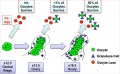
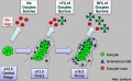
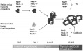
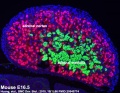
- ovary - neural crest derived neurons invade the invades the interior of the ovary around E16.5, but not the testis during mouse gonad development.[4]
References
- ↑ Sedý J, Szeder V, Walro JM, Ren ZG, Nanka O, Tessarollo L, Sieber-Blum M, Grim M & Kucera J. (2004). Pacinian corpuscle development involves multiple Trk signaling pathways. Dev. Dyn. , 231, 551-63. PMID: 15376326 DOI.
- ↑ Taylor DK, Bubier JA, Silva KA & Sundberg JP. (2012). Development, structure, and keratin expression in C57BL/6J mouse eccrine glands. Vet. Pathol. , 49, 146-54. PMID: 22135020 DOI.
- ↑ Nichol PF, Corliss RF, Yamada S, Shiota K & Saijoh Y. (2012). Muscle patterning in mouse and human abdominal wall development and omphalocele specimens of humans. Anat Rec (Hoboken) , 295, 2129-40. PMID: 22976993 DOI.
- ↑ McKey J, Bunce C, Batchvarov IS, Ornitz DM & Capel B. (2019). Neural crest-derived neurons invade the ovary but not the testis during mouse gonad development. Proc. Natl. Acad. Sci. U.S.A. , 116, 5570-5575. PMID: 30819894 DOI.
Search Pubmed: Mouse E16.5
Pages in category 'Mouse E16.5'
The following 2 pages are in this category, out of 2 total.
Media in category 'Mouse E16.5'
The following 21 files are in this category, out of 21 total.
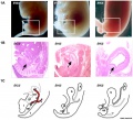 Mouse bladder development E12.5-E16.5.jpg 1,105 × 1,000; 202 KB
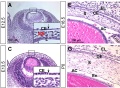 Mouse cornea development 01.jpg 1,200 × 880; 325 KB
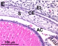 Mouse cornea E16.5.jpg 701 × 562; 113 KB
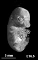 Mouse CT E16.5.jpg 221 × 344; 9 KB
 Mouse gonad Gcnf expression 01.jpg 1,947 × 843; 304 KB
 Mouse gonad Gcnf expression E16.5.jpg 325 × 786; 40 KB
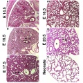
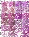 Mouse lung development 01.jpg 1,000 × 1,254; 791 KB
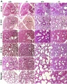 Mouse lung development 01a.jpg 800 × 1,003; 495 KB
 Mouse lung development 02.jpg 922 × 922; 239 KB
 Mouse lung development 03.jpg 540 × 1,200; 349 KB
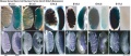
 Mouse melanoblast distribution 01.jpg 697 × 1,000; 192 KB
 Mouse melanoblast distribution 06.jpg 761 × 524; 74 KB
 Mouse model of ovarian cord formation 01.jpg 800 × 491; 85 KB
 Mouse model of ovarian cord formation.jpg 800 × 491; 85 KB
 Mouse placenta 01.jpg 429 × 463; 63 KB
 Mouse posterior neuropore Axd mutant.jpg 475 × 1,074; 95 KB
 Mouse thyroid Hes1 model.jpg 600 × 364; 33 KB
 Mouse tongue Pax9 expression 01.jpg 1,200 × 687; 259 KB
 Mouse tongue Pax9 expression 02.jpg 1,200 × 833; 299 KB
 Mouse ventral body wall development 01.jpg 1,200 × 635; 130 KB
 Mouse-adrenal gland E16.5.jpg 600 × 463; 93 KB


