Category:Human
From Embryology
This Embryology category shows pages and media related to human development.
Subcategories
This category has the following 72 subcategories, out of 72 total.
C
- Carnegie Embryo
- Carnegie Embryo 1
- Carnegie Embryo 112
- Carnegie Embryo 1134B
- Carnegie Embryo 116
- Carnegie Embryo 1266
- Carnegie Embryo 1455
- Carnegie Embryo 148
- Carnegie Embryo 172
- Carnegie Embryo 19
- Carnegie Embryo 239
- Carnegie Embryo 2393
- Carnegie Embryo 240
- Carnegie Embryo 248
- Carnegie Embryo 256
- Carnegie Embryo 296
- Carnegie Embryo 3527
- Carnegie Embryo 3956
- Carnegie Embryo 4046
- Carnegie Embryo 4059
- Carnegie Embryo 407
- Carnegie Embryo 4148
- Carnegie Embryo 43
- Carnegie Embryo 4405
- Carnegie Embryo 460
- Carnegie Embryo 463
- Carnegie Embryo 523
- Carnegie Embryo 5541
- Carnegie Embryo 5609
- Carnegie Embryo 5652
- Carnegie Embryo 5682
- Carnegie Embryo 5874
- Carnegie Embryo 6032
- Carnegie Embryo 625
- Carnegie Embryo 6426
- Carnegie Embryo 6581
- Carnegie Embryo 7618
- Carnegie Embryo 7669
- Carnegie Embryo 786
- Carnegie Embryo 808
- Carnegie Embryo 8147
- Carnegie Embryo 8239
- Carnegie Embryo 8370
- Carnegie Embryo 858
- Carnegie Embryo 8630
- Carnegie Embryo 8967
- Carnegie Embryo 9296
- Carnegie Embryo 96
- Carnegie Embryo 963
- Carnegie Embryo 966
- Carnegie Embryo 9697
D
F
H
Pages in category 'Human'
The following 200 pages are in this category, out of 332 total.
(previous page) (next page)A
B
- Template:B050966
- Template:B100658
- Template:B220849
- Template:Bardeen1906 figures
- Template:Barniville1914 figures
- Template:Bartelmez1922 figures
- Template:BaxterBoyd1939 figures
- BGDA Practical 7 - Week 6
- Birth MRI Movie
- Book - Contributions to Embryology Carnegie Institution No.10
- Book - Contributions to Embryology Carnegie Institution No.112
- Book - Contributions to Embryology Carnegie Institution No.131
- Book - Contributions to Embryology Carnegie Institution No.159
- Book - Contributions to Embryology Carnegie Institution No.21
- Book - Contributions to Embryology Carnegie Institution No.22
- Book - Contributions to Embryology Carnegie Institution No.27
- Book - Contributions to Embryology Carnegie Institution No.29
- Book - Contributions to Embryology Carnegie Institution No.30
- Book - Contributions to Embryology Carnegie Institution No.32
- Book - Contributions to Embryology Carnegie Institution No.33
- Book - Contributions to Embryology Carnegie Institution No.34
- Book - Contributions to Embryology Carnegie Institution No.35
- Book - Contributions to Embryology Carnegie Institution No.38
- Book - Contributions to Embryology Carnegie Institution No.40
- Book - Contributions to Embryology Carnegie Institution No.42
- Book - Contributions to Embryology Carnegie Institution No.43
- Book - Contributions to Embryology Carnegie Institution No.44
- Book - Contributions to Embryology Carnegie Institution No.46
- Book - Contributions to Embryology Carnegie Institution No.47
- Book - Contributions to Embryology Carnegie Institution No.48
- Book - Contributions to Embryology Carnegie Institution No.52
- Book - Contributions to Embryology Carnegie Institution No.55
- Book - Contributions to Embryology Carnegie Institution No.59
- Book - Contributions to Embryology Carnegie Institution No.61
- Book - Contributions to Embryology Carnegie Institution No.65
- Book - Contributions to Embryology Carnegie Institution No.69
- Book - Contributions to Embryology Carnegie Institution No.72
- Book - Contributions to the Development of the Human Brain (1919)
- History:Book - Contributions to the Development of the Human Brain (1919)
- Book - Human embryos of different ages examined in median sections - a contribution to the mechanics of development
- Template:Braune 1877 Plate 2
C
E
F
H
- Template:Hamilton1944 figures
- Harvard Collection
- Template:Hearing EAM timeline
- Hela Apoptosis Movie
- Template:HillH12inks
- Template:HillH13 links
- Template:HillH145 links
- Template:HillH159 links
- Template:HillH202 links
- Template:HillH257 links
- Template:HillH4 links
- Template:HillH5 links
- Template:HillH52
- Template:HillH58 links
- Template:HillH6 links
- Template:HillH8 links
- Template:Hochstadter plates
- Template:Huber1905 table1
- Template:Human 7.5mm Embryo links
- Human Adult Brain Movie
- Human Development Timeline Movie
- Human Embryo - Scanning electron microscopy
- Template:Human embryo neck links
- Human Embryo SEM
- Human Fertilization Detail Movie
- Human Fertilization Movie
- Template:Human Fertilization Movie 1 frame table
- Template:Human Fertilization Movie 1 frames
- Template:Human Fertilization Movie 2 frame table
- Template:Human follicles lm and em links
- Template:Human ovary - corpus luteum links
- Template:Human Spermatozoa Statistics collapse table
- Template:Human Spermatozoa Statistics table
- Human Sylvian Fissure Movie
- Template:Human timeline
- Hutchinson-Gilford Progeria Syndrome
J
K
L
M
- Template:Macklin1921 figures
- Template:Mall1912 figures
- Template:Mall1916 figures
- Template:Mall1917 figures
- Menstrual Cycle - Histology
- Model Embryo 1.6mm Movie 1
- Model Embryo 10mm Movie 1
- Model Embryo 3.1mm Movie 1
- Model Embryo 7.5mm Movie 1
- Monosomic Embryo Movie 1
- Template:Morton1949 figures
- Template:Mouse Human lung table
- Movie - Neural Sylvian Fissure
N
P
- Palate Development
- Paper - A human embryo before the appearance of the myotomes (1918)
- Paper - A Human Embryo of Twenty-five Somites
- Paper - A Human Embryo of Twenty-seven Pairs of Somites, Embedded in Decidua
- Paper - A human embryo with head-process and commencing arch enteric canal
- Paper - A Human Embryo with Seven Pairs of Somites Measuring about 2 mm in Length
- Paper - A human embryo with seventeen pairs of somites (1930)
- Paper - A morphological study of testicular descent
- Paper - A Note on the Development of the Septum Transversum and the Liver
- Paper - A presomite human embryo (Shaw) - the implantation
- Paper - A presomite human embryo (Shaw) - the implantation (1942)
- Paper - A presomite human embryo showing a yolk-sac duct
- Paper - A presomite human embryo showing an early stage of the primitive streak
- Paper - A presomite human embryo with a neurenteric canal (embryo R.S.)
- Paper - A study of a 7 mm human embryo with special reference to its peculiar spirally twisted form, and its large aortic cell-clusters
- Paper - A study of the development of certain features of the cerebellum (1920)
- Paper - A very Young Human Embryo found embedded in a "Decidual Cast" of the Uterus
- Paper - A well-preserved human embryo of 10 somites (1929)
- Paper - A Young Human Embryo (Embryo Dobbin) with Head-Process and Prochordal Plate
- Paper - An early human embryo (no. 1285, Manchester Collection) with capsular attachment of the connecting stalk (1935)
- Paper - An Early Human Embryo (No. 1285, Manchester Collection), with Capsular Attachment of the Connecting Stalk
- Paper - An Early Human Embryo, with 0.55 mm long Embryonic Shield
- Paper - An Early Human Ovum (Thomson) in situ
- Paper - An iconometrographic representation of the growth of the central nervous system in man
- Paper - Breech fused twin monster (1934)
- Paper - Changes in fetuses due to formalin preservation
- Special:Badtitle/NS501:Paper - Description of a Human Embryo of 13-14 Mesodermic Somites
- Paper - Description of a Human Embryo of Twenty-three Paired Somites
- History:Paper - Description of a Human Embryo of Twenty-two paired Somites
- Paper - Description of a reconstruction of the head of a thirty-millimetre embryo (1910)
- Paper - Development and variation of the nerves and the musculature of the inferior extremity and of the neighboring regions of the trunk in man
- Paper - Development of the human heart from its earliest appearance to the stage found in embryos of twenty paired somites (1927)
- Paper - Developmental Changes in the Pericardium, the Mesocardia, and the Pleural Sacs in the Human Embryo
- Paper - Early development of the cervical vertebrae and the base of the occipital bone in man
- Paper - Early Stages of Human Development
- Paper - Factors Involved In The Formation Of The Filum Terminale
- Paper - Fine structure of the human ovum in the pronuclear stage
- Paper - Further note on the pro-chordal plate in man
- Paper - History of the development of the human ovum (1834)
Media in category 'Human'
The following 200 files are in this category, out of 2,421 total.
(previous page) (next page) Bartelmez1922-fig10.jpg 1,000 × 1,136; 209 KB
 Basic Heart Development Timeline.jpg 1,658 × 556; 73 KB
 BaxterBoyd1939-fig01.jpg 361 × 736; 43 KB
 BaxterBoyd1939-fig02.jpg 459 × 851; 65 KB
 BaxterBoyd1939-fig03.jpg 732 × 1,000; 170 KB
 BaxterBoyd1939-fig04.jpg 642 × 615; 109 KB
 BaxterBoyd1939-fig05.jpg 1,000 × 864; 263 KB
 BaxterBoyd1939-fig06.jpg 489 × 917; 141 KB
 BaxterBoyd1939-fig07.jpg 795 × 917; 242 KB
 BaxterBoyd1939-plate01.jpg 1,680 × 2,400; 786 KB
 BaxterBoyd1939-plate02.jpg 1,681 × 2,400; 860 KB
 BaxterBoyd1939-text-fig01.jpg 1,283 × 1,000; 138 KB
 BaxterBoyd1939-text-fig02.jpg 1,200 × 861; 133 KB
 Bedford01.jpg 734 × 1,000; 82 KB
 Bedford02.jpg 1,069 × 911; 266 KB
 Bicornuate uterus01.jpg 1,452 × 1,691; 209 KB
 Blood test result for glucose and iron.jpg 879 × 345; 54 KB
 Bone histology 014.jpg 1,280 × 1,024; 541 KB
 Bone histology 015.jpg 1,280 × 1,024; 519 KB
 Bone histology 016.jpg 1,280 × 1,024; 379 KB
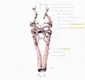
 Bone-femur.jpg 798 × 1,000; 150 KB
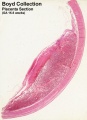 Boyd collection icon.jpg 400 × 554; 56 KB
 Brain growth and birth size.jpg 800 × 492; 70 KB
 Braune 1877 plate 2 fig1.jpg 960 × 1,000; 181 KB
 Braune 1877 plate 2 fig2.jpg 806 × 1,000; 200 KB
 Braune 1877 plate 2 fig3.jpg 893 × 1,000; 208 KB
 Braune 1877 plate 2 fig4.jpg 809 × 1,000; 213 KB
 Braune 1877 plate 2 fig5.jpg 771 × 1,000; 169 KB
 Braune 1877 plate 2 fig6.jpg 690 × 1,000; 144 KB
 Braune 1877 plate 2 fig7.jpg 893 × 1,000; 241 KB
 Braune 1877 plate 2A.jpg 780 × 1,200; 263 KB
 Braune 1877 plate 2B.jpg 801 × 1,200; 264 KB
 Bremer1914 plate04.jpg 643 × 1,000; 127 KB
 Brown015.jpg 800 × 654; 72 KB
 Brown021.jpg 800 × 783; 94 KB
 Brown024.jpg 558 × 800; 52 KB
 Brown025.jpg 600 × 521; 43 KB
 Brown026.jpg 457 × 423; 25 KB
 Brown028.jpg 700 × 730; 52 KB
 Bryce1908 fig01.jpg 500 × 500; 20 KB
 Bryce1908 fig02.jpg 828 × 1,000; 110 KB
 Bryce1908 fig03.jpg 801 × 500; 40 KB
 Bryce1908 fig04.jpg 600 × 400; 28 KB
 Bryce1908 fig05.jpg 700 × 536; 0 bytes
 Bryce1908 fig06.jpg 600 × 500; 39 KB
 Bryce1908 table01.jpg 837 × 1,000; 141 KB
 Bryce1908 table02.jpg 702 × 1,000; 68 KB
 Bryce1908 table03.jpg 731 × 1,000; 78 KB
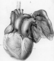
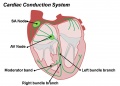 Cardiac Conduction System.jpg 1,201 × 862; 81 KB
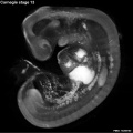
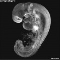 Carnegie stage 12 OPT.jpg 800 × 801; 46 KB
 Carnegie stage 13 OPT.jpg 800 × 801; 53 KB
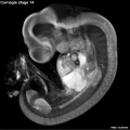 Carnegie stage 14 OPT.jpg 800 × 801; 55 KB
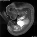 Carnegie stage 15 OPT.jpg 800 × 801; 56 KB
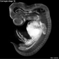 Carnegie stage 16 OPT.jpg 800 × 801; 51 KB
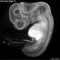 Carnegie stage 17 OPT.jpg 800 × 801; 57 KB
 Carnegie stage 18 OPT.jpg 800 × 801; 43 KB
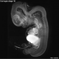
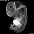 Carnegie stage 19 OPT.jpg 800 × 801; 46 KB
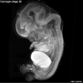 Carnegie stage 20 OPT.jpg 800 × 801; 47 KB
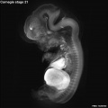 Carnegie stage 21 OPT.jpg 800 × 801; 39 KB
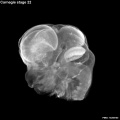 Carnegie stage 22 OPT.jpg 800 × 801; 38 KB
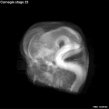 Carnegie stage 23 OPT.jpg 800 × 801; 36 KB
 Caudal duplication syndrome.jpg 700 × 599; 47 KB
 Cerebral blood supply development 01.jpg 1,200 × 460; 67 KB
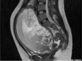
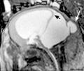
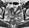
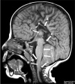 Chiari II malformation MRI01.jpg 723 × 800; 88 KB
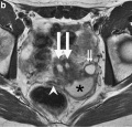
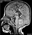 Chiari II malformation MRI02.jpg 723 × 800; 101 KB
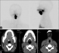
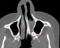 Choanal atresia computed tomography 01.jpg 598 × 477; 35 KB
 Chromosome telomeres.jpg 600 × 471; 33 KB
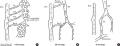
 Classification of Oesophageal atresia.jpg 513 × 726; 55 KB
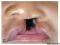
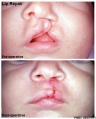
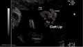
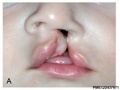 Cleft lip 001.jpg 400 × 300; 16 KB
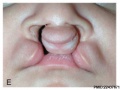
 Cleft lip 002.jpg 400 × 300; 22 KB
 Cleft lip 003.jpg 400 × 300; 19 KB
 Cleft lip 004.jpg 400 × 300; 21 KB
 Cleft lip 005.jpg 400 × 300; 0 bytes
 Cleft lip 006.jpg 400 × 300; 22 KB
 Cleft lip 007.jpg 405 × 504; 38 KB
 Cleft lip 01.jpg 585 × 438; 34 KB
 Cleft lip 02.jpg 641 × 362; 22 KB
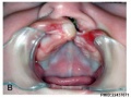
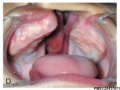
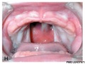
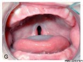 Cleft palate 001.jpg 400 × 300; 22 KB
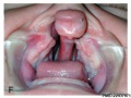
 Cleft palate 002.jpg 400 × 300; 20 KB
 Cleft palate 003.jpg 407 × 600; 46 KB
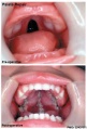
 Cleft palate feeder.jpg 276 × 600; 19 KB
 Cockle01.jpg 881 × 1,000; 158 KB
 Cockle02.jpg 1,459 × 929; 266 KB
 Cockle03.jpg 810 × 1,062; 158 KB
 Cockle04.jpg 740 × 1,065; 135 KB
 Common bile duct in duodenal bulb.jpg 367 × 364; 38 KB
 Complete hydatidiform mole 01.jpg 748 × 560; 50 KB
 Complete hydatidiform mole 02.jpg 748 × 560; 60 KB
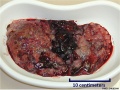 Complete hydatidiform mole 03.jpg 748 × 560; 103 KB
 Complete hydatidiform mole 04.jpg 748 × 560; 124 KB
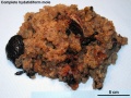 Complete hydatidiform mole 05.jpg 1,280 × 960; 324 KB
 Complete hydatidiform mole 06.jpg 1,280 × 960; 553 KB
 Congdon-table01.jpg 764 × 1,000; 167 KB
 Congdon1922-1-16.jpg 980 × 1,000; 157 KB
 Congdon1922-17.jpg 1,000 × 411; 55 KB
 Congdon1922-18-25.jpg 1,200 × 795; 179 KB
 Congdon1922-18.jpg 494 × 506; 29 KB
 Congdon1922-19.jpg 653 × 471; 31 KB
 Congdon1922-20.jpg 794 × 446; 43 KB
 Congdon1922-21.jpg 578 × 407; 27 KB
 Congdon1922-22.jpg 511 × 489; 27 KB
 Congdon1922-23.jpg 519 × 412; 26 KB
 Congdon1922-24.jpg 790 × 482; 40 KB
 Congdon1922-25.jpg 509 × 358; 25 KB
 Congdon1922-26.jpg 746 × 726; 54 KB
 Congdon1922-27-28.jpg 997 × 612; 68 KB
 Congdon1922-29.jpg 976 × 1,000; 81 KB
 Congdon1922-30.jpg 1,133 × 1,000; 176 KB
 Congdon1922-31.jpg 1,063 × 1,000; 93 KB
 Congdon1922-32.jpg 1,133 × 1,000; 132 KB
 Congdon1922-33.jpg 920 × 1,000; 107 KB
 Congdon1922-34.jpg 920 × 1,000; 122 KB
 Congdon1922-35.jpg 920 × 1,000; 97 KB
 Congdon1922-36.jpg 920 × 1,000; 113 KB
 Congdon1922-37.jpg 1,200 × 838; 163 KB
 Congdon1922-38.jpg 1,187 × 1,000; 165 KB
 Congdon1922-39.jpg 1,200 × 756; 138 KB
 Congdon1922-40.jpg 1,013 × 1,000; 110 KB
 Congdon1922-plate01.jpg 877 × 1,200; 145 KB
 Congdon1922-plate02.jpg 877 × 1,200; 191 KB
 Congdon1922-plate03.jpg 1,200 × 885; 182 KB
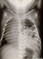
 Congenital diaphragmatic hernia 01.jpg 1,000 × 494; 108 KB
 Congenital diaphragmatic hernia 02.jpg 578 × 800; 48 KB
 Congenital diaphragmatic hernia 03.jpg 611 × 800; 82 KB
 Congenital diaphragmatic hernia 04.jpg 637 × 600; 66 KB
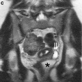
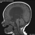 Congenital hydrocephalus MRI01.jpg 595 × 600; 43 KB
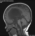 Congenital hydrocephalus MRI02.jpg 595 × 600; 42 KB
 Cord blood induced stem cells 01.jpg 800 × 787; 157 KB
 Cord blood induced stem cells 02.jpg 700 × 966; 189 KB
 Cranial neural crest skeletal fate 01.jpg 800 × 633; 59 KB
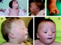
 Craniofrontonasal syndrome.jpg 1,280 × 543; 130 KB
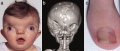
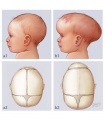 Craniosynostosis .jpg 375 × 430; 85 KB
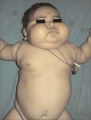 Cushing's syndrome.jpg 450 × 592; 62 KB
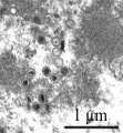
 Cytomegalovirus infected spermatozoa EM01.jpg 990 × 991; 204 KB
 Cytomegalovirus infected spermatozoa.jpg 1,000 × 1,260; 324 KB
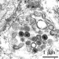
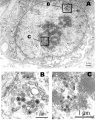
 Cytomegalovirus virions EM.jpg 911 × 987; 212 KB
 Dandy Walker malformation MRI 01.jpg 600 × 506; 37 KB
 Dandy1910-plate01.jpg 1,738 × 2,359; 541 KB
 Dandy1910-plate02.jpg 1,754 × 2,400; 951 KB
 Dandy1910-plate03.jpg 1,000 × 1,617; 224 KB
 Dandy1910-plate04.jpg 1,000 × 1,875; 174 KB
 Dandy1910-plate05.jpg 1,000 × 2,034; 238 KB
 Dandy1910-plate06.jpg 1,000 × 2,166; 265 KB
 Davis1927 plate01.jpg 1,357 × 1,500; 501 KB
 Davis1927 plate02.jpg 1,357 × 1,500; 795 KB
 Developing human cerebellum 01.jpg 1,009 × 1,200; 494 KB
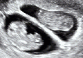 Dichorionic twins ultrasound 01.gif 401 × 282; 2.51 MB
 Differentially expressed RefSeq genes in human trisomy 21.jpg 661 × 847; 171 KB
 Double meningomyelocele.jpg 504 × 800; 49 KB
 Double tetrasomy 18 mosaicism.jpg 600 × 447; 78 KB
 Duodenal atresia.jpg 600 × 873; 63 KB
 Early human telomere length.jpg 1,800 × 1,034; 70 KB
 Early human telomeres.jpg 1,280 × 1,006; 194 KB
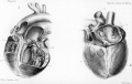
 Ectopia cordis.jpg 800 × 603; 37 KB
 Ectopic molar pregnancy 01.jpg 700 × 535; 60 KB
 Ectopic pregnancy 01.jpg 735 × 596; 45 KB
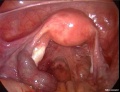
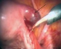
 Ectopic thyroid - lingual 01.jpg 600 × 525; 34 KB
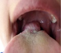
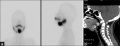 Ectopic thyroid - sublingual and suprahyoid.jpg 1,000 × 383; 42 KB
 Ectopic thyroid - sublingual, suprahyoid and subhyoid.jpg 800 × 721; 59 KB
 Embryo 7.5mm model 01.gif 448 × 600; 903 KB
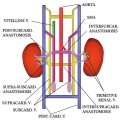 Embryo renal venous cartoon.jpg 600 × 600; 68 KB
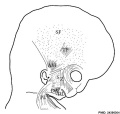 Embryonic neck muscle cartoon.jpg 600 × 570; 45 KB
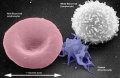
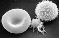 Erythrocyte and lymphocyte SEM01.jpg 800 × 522; 74 KB
 Erythrocyte and lymphocyte SEM02.jpg 800 × 522; 78 KB
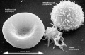 Erythrocyte and lymphocyte SEM03.jpg 800 × 522; 80 KB
 Extravillous trophoblasts week 5.5.jpg 1,280 × 900; 421 KB
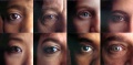 Eye collage 2.jpg 848 × 417; 112 KB
 Fawcett1910 fig01.jpg 891 × 639; 148 KB
 Fawcett1910 fig02.jpg 1,035 × 949; 265 KB
 Fawcett1910 fig03.jpg 905 × 978; 160 KB
 Fawcett1910 fig04.jpg 969 × 1,110; 115 KB
 Fawcett1913 fig01.jpg 793 × 573; 67 KB
 Fawcett1913 fig02.jpg 679 × 579; 42 KB
 Fawcett1913 fig03.jpg 687 × 581; 33 KB
 Fawcett1913 fig04.jpg 928 × 664; 122 KB
 Fawcett1913 fig05.jpg 979 × 671; 98 KB
 Fawcett1913 fig06.jpg 899 × 675; 80 KB
 Fawcett1913 fig07.jpg 1,113 × 763; 97 KB
 Fawcett1913 fig08.jpg 867 × 397; 28 KB
 Female genital and ureter abnormality 01.jpg 766 × 732; 86 KB
 Female genital and ureter abnormality 02.jpg 766 × 733; 78 KB
 Female genital and ureter abnormality 03.jpg 766 × 762; 79 KB
 Fetal blood flow 01.jpg 1,000 × 599; 75 KB
 Fetal blood flow 02.jpg 491 × 599; 38 KB
 Fetal blood flow 03.jpg 500 × 599; 43 KB
 Fetal blood flow 04.jpg 506 × 599; 50 KB
 Fetal blood flow liver and brain.jpg 677 × 790; 69 KB
 Fetal cardiac state diagram 01.jpg 1,200 × 321; 29 KB
 Fetal cardiac state diagram 02.jpg 1,200 × 458; 27 KB
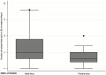 Fetal cells maternal blood graph.jpg 600 × 426; 18 KB
 Fetal ductus venosus pressure wave 01.jpg 706 × 755; 52 KB
 Fetal ductus venosus ultrasound 01.jpg 783 × 1,000; 68 KB
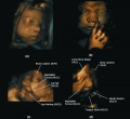
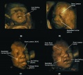 Fetal facial expression 01.jpg 1,200 × 1,094; 122 KB
 Fetal facial expression 02.jpg 1,914 × 1,762; 205 KB
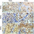 Fetal gonad retinoid receptor expression 01.jpg 1,004 × 1,000; 226 KB
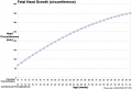 Fetal head growth circumference graph01.jpg 905 × 613; 58 KB
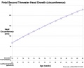 Fetal head growth circumference graph02.jpg 800 × 650; 44 KB
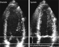 Fetal heart atrioventricular plane displacement 01.jpg 915 × 733; 55 KB
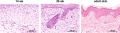 Fetal integumentary histology 01.jpg 800 × 219; 74 KB
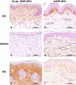 Fetal integumentary histology 02.jpg 600 × 664; 145 KB


