Category:Histology
From Embryology
Introduction
This Embryology category lists files and media related to histology. Most links are to histological images that relate to tissue structure and development. Many images are sourced from the original UNSW Anatomy Histology slide set and UWA Blue Histology online images.
Subcategories
This category has the following 15 subcategories, out of 15 total.
Pages in category 'Histology'
The following 134 pages are in this category, out of 334 total.
(previous page) (next page)P
- Paper - A morphological study of the development of the human liver 1
- Paper - A morphological study of the development of the human liver 2
- Paper - A note on the post-natal growth of the kidney, thyroid gland and liver (1924)
- Paper - A quantitative study of the fetal growth changes in the parts of the human stomach wall
- Paper - A Study of the Structural Unit of the Liver
- Paper - A subject with complete transposition of viscera (1917)
- Paper - Appendix vermiformis duplex (1936)
- Paper - Case of abnormal duodenum (1924)
- Paper - Changes in fetuses due to formalin preservation
- Paper - Chiefly concerning the genito-mesenteric fold of peritoneum
- Paper - Congenital absence of the appendix of the caecum (1915)
- Paper - Congenital anomalies of the duodenum (1940)
- Paper - Congenital Anomalies of the Liver (1929)
- Paper - Congenital atresia of the oesophagus
- Paper - Congenital hernia into the umbilical cord - two cases, one associated with persistent cloaca
- Paper - Congenital malformations of the oesophagus
- Paper - Cytogenesis of the human fetal pancreas (1962)
- Paper - Cytological studies of Langerhans's islets, with special reference to the problem of their relation to the pancreatic acinus tissue (1920)
- Paper - Early differentiation of the foregut in the dog
- Paper - Imperfect torsion of the intestinal loop
- Paper - Normal development of the trachea and esophagus in man
- Paper - Notes on the origin of the liver (1891)
- Paper - Obstructions about the mesentery in infants (1936)
- Paper - On abnormalities of the caecum and colon with reference to development
- Paper - On the development of the villi of the human intestine
- Paper - On the developmental topography of the thoracic and abdominal viscera (1909)
- Paper - On the factors concerned in causing rotation of the intestine in man
- Paper - On the histogenesis of gastric glands
- Paper - On the relation of the liver cells to the blood-vessels and lymphatics
- Paper - On the so-called ultimobranchial body of the mammalian embryo (1915)
- Paper - Retrogressive Changes in the Fetal Vessels and the Suspensory Ligament of the Liver
- Paper - Sequential innervation of the intestinal loop in the human embryo
- Paper - Some factors influencing the position of the small intestine (1915)
- Paper - Studies of the intestine and peritoneum in the human foetus - part 1
- Paper - Studies of the intestine and peritoneum in the human foetus - part 2
- Paper - Studies of the intestine and peritoneum in the human foetus - part 3
- Paper - Studies of the intestine and peritoneum in the human foetus - part 4
- Paper - Studies of the intestine and peritoneum in the human foetus - part 5
- Paper - Studies of the intestine and peritoneum in the human foetus - part 6
- Paper - The angiology, angiogenesis, and organogenesis of the submaxillary gland
- Paper - The application of trichrome staining methods to embryological technique (1940)
- Paper - The bi-lobed form of the ventral pancreas in mammals
- Paper - The comparative anatomy of the lips and labial villi of vertebrates
- Paper - The critical period in the development of the intestines (1914)
- Paper - The development of the form of the gastrointestinal canal in humans 1
- Paper - The development of the form of the gastrointestinal canal in humans 2
- Paper - The development of the great omentum and transverse mesocolon
- Paper - The development of the human pharynx
- Paper - The development of the lobule of the pig's liver (1919)
- Paper - The development of the lobus quadratus of the liver with special reference to an unusual anomaly of this lobe in the adult (1914)
- Paper - The development of the mucous membrane oesophagus stomach and small intestine in human embryo
- Paper - The development of the mucous membrane of the large intestine and vermiform process in the human embryo
- Paper - The development of the rectum in the human embryo
- Paper - The development of the serous glands (von Ebner's) of the vallate papillae in man (1917)
- Paper - The development of the spiral coil in the large intestine of the pig
- Paper - The early looping of the alimentary canal in the mammalian and human foetus and the mechanisms assumed to be active in this process
- Paper - The early stages of the development of the ileo-colic sphincter (1924)
- Paper - The embryogenesis of human bile capillaries and ducts
- Paper - The form of the stomach in human embryos with notes upon the nomenclature of the stomach
- Paper - The formation of the duodenal curve
- Paper - The formation of the duodenal curve (1919)
- Paper - The gall bladder and the extrahepatic biliary passages in late embryonic and early fetal life
- Paper - The genesis of Jackson's membrane (1914)
- Paper - The genesis of Jackson's membrane: notes on the genito-mesenteric fold of peritoneum and the supra-adhesion foramen
- Paper - The lachrymal gland (1916)
- Paper - The morphology and development of intestinal folds and villi in vertebrates
- Paper - The nature of the malformations of the rectum and urogenital passages
- Paper - The origin of blood cells (1916)
- Paper - The regular occurrence of intestinal diverticula in embryos of the pig, rabbit and man
- Paper - The regular occurrence of intestinal diverticula in embryos of the pig, rabbit, and man
- Paper - The relations of endogenous and exogenous factors in bone and tooth development (1937)
- Paper - The relative frequency of the various positions of the vermiform appendix (1924)
- Paper - The role of the primitive mesothelium in the development of the mammalian spleen (1936)
- Paper - The shrinkage of embryos in the processes preparatory to sectioning
- Paper - Transposition of Abdominal Viscera (1926)
- Paper - V. Meckel's diverticulum patent at the navel (1902)
- Template:Peripheral Nerve Histology
- Placenta - Histology
- Template:Placenta Cord Histology
- Template:Placenta histology
R
- Template:Ref-Arey1917
- Template:Ref-BacsichSmout1938
- Template:Ref-Badertscher1915b
- Template:Ref-Baxter1940
- Template:Ref-Bloom1931
- Template:Ref-BöhmDavidoffHuber1910
- Template:Ref-Cooper1938
- Template:Ref-Danchakoff1916b
- Template:Ref-Danchakoff1916c
- Template:Ref-Danchakoff1918
- Template:Ref-HallpikePeet1939
- Template:Ref-Hassall1849
- Template:Ref-Herring1908a
- Template:Ref-Ingalls1915
- Template:Ref-Lewis1906
- Template:Ref-Nonidez1941
- Template:Ref-Orban1944
- Template:Ref-PattenPhilpott1921
- Template:Ref-ScharpenbergWindle1938
- Template:Ref-Schultz1919
- Template:Ref-ThielDowney1921
- Template:Ref-WheatersHistology2006
- Template:Ref-WislockiDempsey1945
- Template:Renal Histology
- Template:Renal histology
- Renal System Histology
- Respiratory System - Histology
S
- Salivary Gland Development
- Sertoli cell
- SH Lecture - Lymphatic Structure and Organs
- SH Practical - Lymphatic Structure and Organs
- SH Practical - Respiratory
- Site Map
- Talk:Site Map
- Skeletal Muscle Histology
- Template:SmMhistolinks
- Smooth Muscle Development
- Smooth Muscle Histology
- Template:Spalteholz method
- Template:Spleen Histology
- Template talk:Spleen Histology
- Template:Spleen Histology gallery
- Template:Stage 22 histology gallery
- Template:Stage 22 histology gallery table
- Template:Stains
- Template:Stomach Histology
T
Media in category 'Histology'
The following 114 files are in this category, out of 718 total.
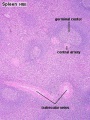
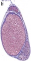
(previous page) (next page) Spleen histology 01.jpg 450 × 600; 133 KB
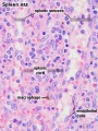
 Spleen histology 02.jpg 455 × 606; 142 KB
 Spleen histology 03.jpg 450 × 600; 83 KB
 Spleen histology 04.jpg 450 × 600; 108 KB
 Spleen histology 05.jpg 450 × 600; 170 KB
 Spleen histology 06.jpg 1,000 × 800; 408 KB
 Spleen histology 07.jpg 1,000 × 800; 251 KB
 Spleen histology 08.jpg 1,000 × 800; 304 KB
 Spleen histology 09.jpg 1,280 × 1,024; 692 KB
 Spleen histology 10.jpg 1,280 × 1,024; 444 KB
 Spleen histology 11.jpg 1,280 × 1,024; 410 KB
 Spleen histology 12.jpg 600 × 450; 93 KB
 Spleen histology 13.jpg 600 × 450; 80 KB
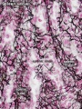
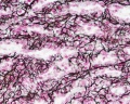
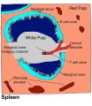
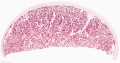
 Spleen structure 01.jpg 1,200 × 463; 211 KB
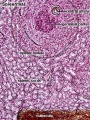
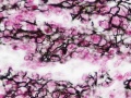
 Spleen structure 02.jpg 401 × 463; 46 KB
 Stage11 histology-neural tube roof plate 1.jpg 800 × 594; 134 KB
 Stage11 histology-neural tube roof plate 2.jpg 800 × 552; 145 KB
 Stage11 histology-optic pit.jpg 800 × 510; 157 KB
 Stage11 histology-optic vesicle-hindbrain.jpg 800 × 536; 158 KB
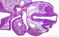
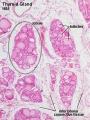
 Stage13 and 22 thyroid development a.jpg 800 × 640; 94 KB
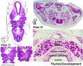
 Stage13 and 22 thyroid development.jpg 1,000 × 800; 283 KB
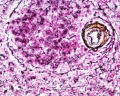
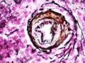
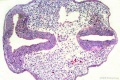
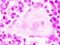
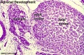 Stage22 adrenal.jpg 596 × 392; 65 KB
 Stage22 HPA1L.jpg 640 × 433; 65 KB
 Stage22 pancreas a.jpg 800 × 640; 99 KB
 Stage22 pancreas b.jpg 600 × 480; 63 KB
 Stage22 pancreas c.jpg 400 × 320; 38 KB
 Stage22 pancreas.jpg 1,000 × 800; 264 KB
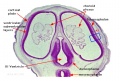
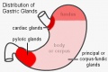 Stomach gastric gland distribution.jpg 500 × 334; 28 KB
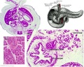
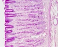
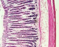
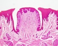
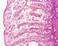
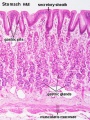 Stomach histology 001.jpg 400 × 533; 87 KB
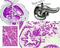
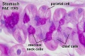
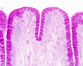
 Stomach histology 002.jpg 400 × 532; 53 KB
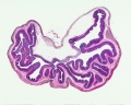
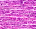
 Stomach histology 003.jpg 599 × 400; 53 KB
 Stomach histology 004.jpg 1,280 × 1,024; 435 KB
 Stomach histology 005.jpg 1,280 × 1,024; 432 KB
 Stomach histology 006.jpg 1,280 × 1,024; 227 KB
 Stomach histology 007.jpg 1,280 × 1,024; 308 KB
 Stomach histology 008.jpg 1,280 × 1,024; 452 KB
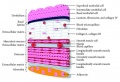
 Stratified epithelia cartoon.jpg 696 × 1,000; 166 KB
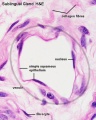
 Sublingual gland histology 02.jpg 500 × 500; 23 KB
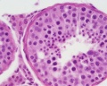
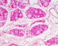
 Testis histology 001.jpg 1,280 × 1,024; 574 KB
 Testis histology 002.jpg 1,280 × 1,024; 599 KB
 Testis histology 003.jpg 1,280 × 1,024; 183 KB
 Testis histology 004.jpg 1,280 × 1,024; 396 KB
 Testis histology 005.jpg 1,280 × 1,024; 266 KB
 Testis histology 006.jpg 1,280 × 1,024; 251 KB
 Testis histology 007.jpg 1,280 × 1,024; 256 KB
 Testis histology 008.jpg 1,280 × 1,024; 454 KB
 Testis histology 009.jpg 1,280 × 1,024; 339 KB
 Testis histology 010.jpg 1,280 × 1,024; 422 KB
 Testis histology 011.jpg 1,280 × 1,024; 245 KB
 Testis histology 012.jpg 1,280 × 1,024; 266 KB
 Testis histology 013.jpg 1,280 × 1,024; 418 KB
 Testis histology 014.jpg 1,280 × 1,024; 352 KB
 Testis histology 015.jpg 1,280 × 1,024; 281 KB
 Testis histology 016.jpg 1,280 × 1,024; 322 KB
 Testis histology 017.jpg 1,280 × 1,024; 283 KB
 Testis histology 018.jpg 1,280 × 1,024; 350 KB
 Testis histology 019.jpg 1,280 × 1,024; 239 KB
 Testis histology 02.jpg 246 × 481; 49 KB
 Testis histology 020.jpg 1,300 × 685; 334 KB
 Testis histology 021.jpg 1,200 × 962; 312 KB
 Testis histology 022.jpg 1,229 × 966; 311 KB
 Testis histology 023.jpg 600 × 375; 35 KB
 Testis histology 1.jpg 400 × 500; 113 KB
 Testis histology 2.jpg 400 × 500; 32 KB
 Testis histology.jpg 400 × 500; 54 KB
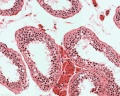
 Testis, young H&E reproductive system, male, convoluted seminiferous tubules x10.jpg 1,280 × 1,024; 396 KB
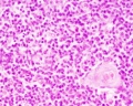
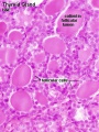
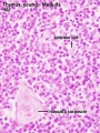 Thymus - young 01.jpg 450 × 600; 93 KB
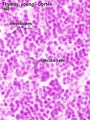 Thymus - young 02.jpg 450 × 600; 91 KB
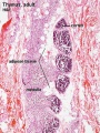 Thymus adult.jpg 450 × 600; 138 KB
 Thymus histology 01.jpg 1,280 × 1,024; 723 KB
 Thymus histology 02.jpg 1,280 × 1,024; 287 KB
 Thymus histology 03.jpg 1,280 × 1,024; 325 KB
 Thymus histology 04.jpg 1,280 × 1,024; 511 KB
 Thymus histology 05.jpg 513 × 385; 41 KB
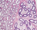
 Thyroid histology 001.jpg 450 × 600; 96 KB
 Thyroid histology 002.jpg 450 × 600; 85 KB
 Thyroid histology 003.jpg 1,280 × 1,024; 209 KB
 Thyroid histology 004.jpg 1,280 × 1,024; 351 KB
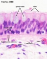
 Tongue histology 05.jpg 1,280 × 1,024; 418 KB
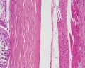
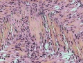 Tongue-muscle.jpg 300 × 228; 43 KB
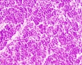
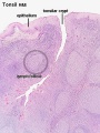 Tonsil histology 01.jpg 450 × 600; 106 KB
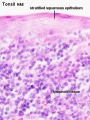 Tonsil histology 02.jpg 450 × 600; 62 KB
 Trachea histology 01.jpg 480 × 600; 47 KB
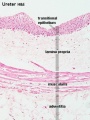 Ureter histology 001.jpg 375 × 500; 50 KB
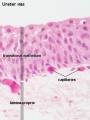 Ureter histology 002.jpg 375 × 500; 34 KB
 Urinary Bladder Histology.jpg 581 × 399; 42 KB
 Uterine gland proliferative phase.jpg 400 × 533; 51 KB
 Uterine gland secretory phase.jpg 400 × 533; 49 KB
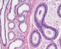
 Uterine tube histology 02.jpg 400 × 533; 55 KB
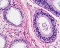
 Uterine tube histology 03.jpg 400 × 533; 34 KB
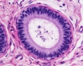
 Uterine tube histology 04.jpg 1,280 × 1,024; 253 KB
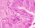
 Uterine tube histology 05.jpg 1,063 × 1,063; 508 KB
 Uterine tube histology.jpg 1,280 × 1,024; 568 KB
 Uterus proliferative phase.jpg 400 × 533; 52 KB
 Uterus secretory phase 01.jpg 1,280 × 1,024; 318 KB
 Uterus secretory phase 02.jpg 1,280 × 1,024; 359 KB
 Uterus secretory phase.jpg 400 × 533; 68 KB
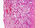
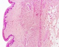
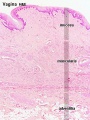 Vagina histology 01.jpg 450 × 600; 92 KB
 Vagina histology 02.jpg 1,280 × 1,024; 555 KB
 Vein histology 01.jpg 480 × 600; 57 KB
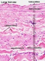 Vein histology 02.jpg 400 × 533; 76 KB
 Vein histology 03.jpg 400 × 533; 76 KB
 Vein valve animation.gif 300 × 200; 54 KB
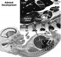 Week10 adrenal.jpg 600 × 564; 48 KB
 Wheater’s Functional Histology.jpg 425 × 600; 66 KB
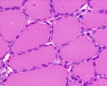
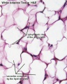 White adipose 01.jpg 500 × 625; 65 KB
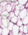 White adipose 02.jpg 500 × 625; 61 KB
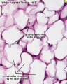 White adipose histology.jpg 400 × 500; 57 KB
 William Bowman.jpg 600 × 665; 49 KB
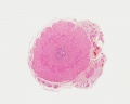
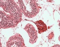
 Wilms tumor.jpg 776 × 512; 310 KB
 XMRV-infected prostate cells.png 485 × 599; 528 KB
 Zorn2008 fig01.jpg 1,200 × 1,158; 110 KB


