File:Stage12 bf3.jpg: Difference between revisions
(Stage12day26somite21-dorsal-bf3.jpg) |
No edit summary |
||
| Line 1: | Line 1: | ||
Stage12day26somite21-dorsal-bf3.jpg | Human Embryo | ||
Carnegie Stage 12 | |||
Facts: Week 4, 26 days, 5 mm, Somite Number 21 | |||
View: Dorsal view, day 26, 21 somites, amniotic membrane removed, scale background | |||
Features: day 26, 27 somites, forebrain, site of lens placode, site of otic placode , stomodeum, 1st pharyngeal arch, 2nd pharyngeal arch, 3rdpharyngeal arch, heart prominence, somite | |||
Identify: forebrain, site of lens placode, site of otic placode, 1st pharyngeal arch, 2nd pharyngeal arch, 3rd pharyngeal arch, heart prominence, somite | |||
[[:File:Stage12_sem1.jpg|Scanning EM image version]] of this image also available. | |||
Original File Name: Stage12day26somite21-dorsal-bf3.jpg | |||
'''Image version links:''' [[:File:Stage12 bf1.jpg|Large 1000px]] | [[:File:Stage12 bf1a.jpg| 800px]] | | |||
[[:File:Stage12 bf1b.jpg|Medium 600px]] | [[:File:Stage12 bf1c.jpg|Small 400px]] | |||
'''Related Images:''' [[:File:Stage12 bf2.jpg|Bright Field 2]] | [[:File:Stage12 bf3.jpg|Bright Field 3]] | [[:File:Stage12_sem1.jpg|Scanning EM image version]] | |||
{{Template:SEM}} | |||
{{Template:Carnegie_stages}} | |||
{{Template:Footer}} | |||
[[Category:Carnegie Stage 12]] | |||
Revision as of 12:28, 7 September 2009
Human Embryo
Carnegie Stage 12
Facts: Week 4, 26 days, 5 mm, Somite Number 21
View: Dorsal view, day 26, 21 somites, amniotic membrane removed, scale background
Features: day 26, 27 somites, forebrain, site of lens placode, site of otic placode , stomodeum, 1st pharyngeal arch, 2nd pharyngeal arch, 3rdpharyngeal arch, heart prominence, somite
Identify: forebrain, site of lens placode, site of otic placode, 1st pharyngeal arch, 2nd pharyngeal arch, 3rd pharyngeal arch, heart prominence, somite
Scanning EM image version of this image also available.
Original File Name: Stage12day26somite21-dorsal-bf3.jpg
Image version links: Large 1000px | 800px |
Medium 600px | Small 400px
Related Images: Bright Field 2 | Bright Field 3 | Scanning EM image version
Image Source: Scanning electron micrographs of the Carnegie stages of the early human embryos are reproduced with the permission of Prof Kathy Sulik, from embryos collected by Dr. Vekemans and Tania Attié-Bitach. Images are for educational purposes only and cannot be reproduced electronically or in writing without permission.
- Carnegie Stages: 1 | 2 | 3 | 4 | 5 | 6 | 7 | 8 | 9 | 10 | 11 | 12 | 13 | 14 | 15 | 16 | 17 | 18 | 19 | 20 | 21 | 22 | 23 | About Stages | Timeline
Cite this page: Hill, M.A. (2024, June 26) Embryology Stage12 bf3.jpg. Retrieved from https://embryology.med.unsw.edu.au/embryology/index.php/File:Stage12_bf3.jpg
- © Dr Mark Hill 2024, UNSW Embryology ISBN: 978 0 7334 2609 4 - UNSW CRICOS Provider Code No. 00098G
File history
Yi efo/eka'e gwa ebo wo le nyangagi wuncin ye kamina wunga tinya nan
| Gwalagizhi | Nyangagi | Dimensions | User | Comment | |
|---|---|---|---|---|---|
| current | 12:26, 7 September 2009 | 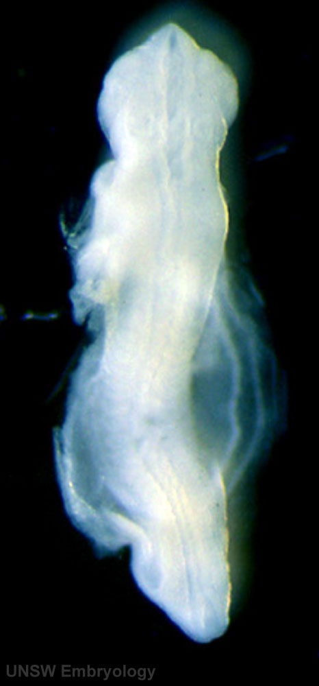 | 466 × 1,000 (43 KB) | S8600021 (talk | contribs) | Stage12day26somite21-dorsal-bf3.jpg |
You cannot overwrite this file.
File usage
The following 3 pages use this file: