Gastrointestinal Tract - Stomach Development: Difference between revisions
mNo edit summary |
mNo edit summary |
||
| Line 218: | Line 218: | ||
{{Ref-GrosserLewisMcmurrich1912}} | {{Ref-GrosserLewisMcmurrich1912}} | ||
{{Ref-Bardeen1914}} | {{Ref-Bardeen1914}} | ||
Revision as of 20:06, 18 April 2017
| Embryology - 15 Jun 2024 |
|---|
| Google Translate - select your language from the list shown below (this will open a new external page) |
|
العربية | català | 中文 | 中國傳統的 | français | Deutsche | עִברִית | हिंदी | bahasa Indonesia | italiano | 日本語 | 한국어 | မြန်မာ | Pilipino | Polskie | português | ਪੰਜਾਬੀ ਦੇ | Română | русский | Español | Swahili | Svensk | ไทย | Türkçe | اردو | ייִדיש | Tiếng Việt These external translations are automated and may not be accurate. (More? About Translations) |


Introduction
This section of notes gives an overview of how the stomach and duodenum develops. The GIT is best imagined as a simple tube, the upper part being the foregut diverticulum, which is further divided into oesophagus and stomach.
During week 4 at the level where the stomach will form the tube begins to dilate, forming an enlarged lumen. The dorsal border grows more rapidly than ventral, which establishes the greater curvature of the stomach.[1] A second rotation (of 90 degrees) occurs on the longitudinal axis establishing the adult orientation of the stomach.
Some Recent Findings
|
| More recent papers |
|---|
 This table allows an automated computer search of the external PubMed database using the listed "Search term" text link.
More? References | Discussion Page | Journal Searches | 2019 References | 2020 References Search term: Stomach Embryology <pubmed limit=5>Stomach Embryology</pubmed> |
Components of Stomach Formation
primitive endoderm
- foregut diverticulum (pocket)
- pharyngeal region of foregut
- laryngo-tracheal groove (see respiratory tract)
- oesophageal region of foregut
- oesophagus
- stomach
- glandular/proventricular/pyloric stenosis
- fundus/pyloric antrum
- pyloric sphincter
- fundus/pyloric antrum
- dorsal mesogastrium
- lieno-renal ligament
- splenic primordium
- spleen
- gastro-splenic ligament
- duodenum (rostral half)
- splenic primordium
- lieno-renal ligament
- glandular/proventricular/pyloric stenosis
- stomach
- oesophagus
- pharyngeal region of foregut
- foregut-midgut junction
- midgut region
- hindgut diverticulum (pocket)
Modified from [4]
Movies
|
|
|
|
|
|
Stage 13

The images below link to larger cross-sections of the mid-embryonic period (end week 4) stage 13 embryo starting just above the level of the stomach and then in sequence through the stomach to the level of the duodenum.

|
|

|
| D2 Cardio-oesophageal junction | D3 Stomach body | D4 Stomach body |

|

|

|
| D5 Stomach body | D6 Pyloric junction | D7 Duodenum |
|
This is an animation based on a reconstruction of the above embryo entire stage 13 gastrointestinal tract. |
Stage 22
The images below link to larger cross-sections of the end of the embryonic period (week 8) stage 22 embryo starting just above the level of the stomach and then in sequence through the stomach to the level of the duodenum. Note how the stomach is "embedded" within the large developing liver.


|

|

|
|
| E5 Oesophagus | E6 Cardio-oesophageal junction | E7 Stomach body Pyloric junction | F1 Stomach body Pylorus |

|

|

|

|
| F2 Stomach body | F3 Stomach body Duodenum | F4 Duodenal-Jejunal junction | F5 Duodenum Jejunum |
|
This is an animation based on a reconstruction of the above embryo entire stage 22 gastrointestinal tract. |
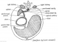
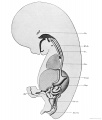
Stomach Mesentery
In the second trimester, the ventral and dorsal mesenteries associated with the stomach are still anatomically different from the newborn. The figure shows a lateral view of this process comparing the early second trimester arrangement with the newborn structure.
Ventral Mesogastrium
Attached to the superior end of the stomach will form the lesser omentum. This structure will connect the lesser curvature of the stomach to the liver as a ligamentous structure.
Dorsal Mesogastrium
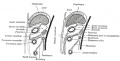
Attached to the inferior end of the stomach initially as an extended fold, this later fuses as a single "apron-like" structure, the greater omentum. Fusion will also incorporate the transverse colon part of the large intestine. This will also contribute the gastrosplenic ligament (gastrolienal ligament).

|
The greater omentum hangs like an apron over the small intestine and transverse colon. It begins attacted to the inferior end of the stomach as a fold of the dorsal mesogastrium which later fuses to form the structure we recognise anatomically. The figure below shows a lateral view of this process comparing the early second trimester arrangement with the newborn structure. |
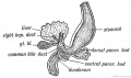
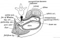
Duodenum/Pancreas Rotation

|
After the stomach the initial portion of the gastrointestinal tract tube is the duodenum which initially lies in the midline within the peritoneal cavity.
This region, along with the attached pancreas, undergoes rotation to become a retroperitoneal structure. This diagram shows the rotation with spinal cord at the top, vertebral body then dorsal aorta then pertioneal wall and cavity. |
Stomach Hormonal Development
The gastrointestinal tract has its own complex entero-endocrine system (enterohormones) that regulates many regional tract functions.
Cells within the stomach express a range of peptide hormones known to regulate a range of gastric functions including secretion of digestive enzymes, mucous and the movement of the luminal contents. The list below shows the earliest detectible presence of specific hormone-containing cells in regions of the developing human stomach.
Hormonal Timecourse
8 weeks - Gastrin containing cells in stomach antrum. Somatostatin cells in both the antrum and the fundus.
10 weeks - Glucagon containing cells in stomach fundus.
11 weeks - Serotonin containing cells in both the antrum and the fundus.
Expression data based upon[5]
Other Gut Peptides
- Cholecystokinin (CCK), pancreatic polypeptide, peptide YY, glucagon-like peptide-1 (GLP-1), oxyntomodulin (increase satiety and decrease food intake) and ghrelin
Mouse
The images below show differential gene expression of some selected markers during development (E10.5 and E13.5) of the mouse gastrointestinal tract.[6]


- Links: Mouse Development | Full original figure | E10.5 | E13.5
References
Reviews
<pubmed>26884394</pubmed> <pubmed>19575677</pubmed> <pubmed>16109035</pubmed> <pubmed>7526882</pubmed> <pubmed>3922287</pubmed> <pubmed>4923462</pubmed>
Articles
<pubmed>16616737</pubmed> <pubmed>11329933</pubmed>
Search Pubmed
Search Bookshelf: Stomach Development
Search Pubmed Now: Stomach Development
Historic References
Template:Ref-GrosserLewisMcmurrich1912
Bardeen CR. The critical period in the development of the intestines. (1914) Amer. J Anat. 16: 427 – 445.
Images
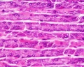
- Stomach Histology
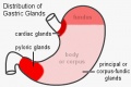
adult stomach gastric gland distribution
rat stomach overview
stomach labeled overview
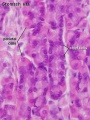
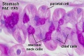
parietal cells - chief cells
mucus neck - parietal cells - chief cells
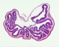
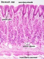
stomach overview
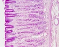
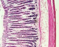
stomach mucosa
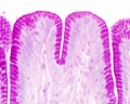
mucosa - secretory epithelial sheath - goblet cell
gastric glands - parietal cells - chief cells


Historic Images
| Historic Disclaimer - information about historic embryology pages |
|---|
|
1902 transverse section mesogastrium
1902 4th week human embryo
1902 lesser sac of peritoneum
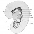
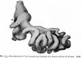
1912 human embryo 4.9 mm
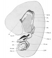
1912 human embryo 7.5 mm
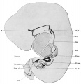
1912 human embryo 9.4 mm
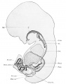
1912 human embryo 22.8 mm
1912 human embryo 42 mm
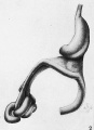
1914 lateral view human embryo 27 mm
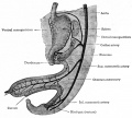
1918 greater omentum
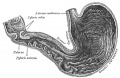
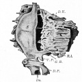
1918 interior adult stomach
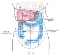
1918 adult stomach position
1921 human embryo 8 mm
1921 human embryo 6 weeks
1921 human embryo 28 mm


Glossary Links
- Glossary: A | B | C | D | E | F | G | H | I | J | K | L | M | N | O | P | Q | R | S | T | U | V | W | X | Y | Z | Numbers | Symbols | Term Link
Cite this page: Hill, M.A. (2024, June 15) Embryology Gastrointestinal Tract - Stomach Development. Retrieved from https://embryology.med.unsw.edu.au/embryology/index.php/Gastrointestinal_Tract_-_Stomach_Development
- © Dr Mark Hill 2024, UNSW Embryology ISBN: 978 0 7334 2609 4 - UNSW CRICOS Provider Code No. 00098G