Sensory - Hearing Abnormalities: Difference between revisions
| Line 58: | Line 58: | ||
[[File:Microtia.jpg|thumb|Microtia]] | [[File:Microtia.jpg|thumb|Microtia]] | ||
[[File:Preauricular sinus.jpg|thumb|Preauricular sinus]] | [[File:Preauricular sinus.jpg|thumb|Preauricular sinus]] | ||
Several genetic effects and syndromes | Several genetic effects and syndromes can include impacts on developmental of the external ear either directly or by altering development of the skull or face. Several developmental environment effects can be indicated by changes in the relative position or appearance of the external ear at birth. (More? [../Defect/maternal.htm Abnormal Development - Environment] | (Fetal Alcohol Syndrome). | ||
* Microtia - abnormally small external ear | * Microtia - abnormally small external ear | ||
* Preauricular sinus - occurs in 0.25% births, bilateral (hereditary) 25-50%, unilateral (mainly the left), duct runs inward can extend into the parotid gland, Postnatally sites for infection | * Preauricular sinus - occurs in 0.25% births, bilateral (hereditary) 25-50%, unilateral (mainly the left), duct runs inward can extend into the parotid gland, Postnatally sites for infection | ||
=== Microtia === | |||
[[ | The condition in humans of an abnormally small external ear is called Microtia. This is generally surgically repaired by use of rib cartilage to reconstruct the external ear. A recent study has identified a mouse model for this condition with the knockout of the Pact gene. | ||
<center>[[Image:microtia.jpg]] </center> | |||
<center>Newborn Microtia (Image: NZ National Women's Health [http://www.adhb.govt.nz/newborn/TeachingResources/Dermatology/EarAnomalies.htm Ear Anomalies]) </center> | |||
'''OMIM''' Database Search: "[http://www.ncbi.nlm.nih.gov:80/entrez/query.fcgi?db=omim&cmd=search&doptcmdl=DocSum&term=Microtia Microtia]" (2006 - 25 search results) | |||
'''Search PubMed''' May 2006 "Microtia" '''449''' reference articles of which '''37''' were reviews. | |||
'''Search PubMed:''' [http://www.ncbi.nlm.nih.gov/entrez/query.fcgi?db=pubmed&cmd=search&term=Microtia Microtia] | [http://www.ncbi.nlm.nih.gov/entrez/query.fcgi?db=pubmed&cmd=search&term=external+ear+defects external ear defects] | |||
'''References''' | |||
[http://www.ncbi.nlm.nih.gov:80/entrez/query.fcgi?cmd=Retrieve&db=PubMed&list_uids=16571658&dopt=Abstract Rowe TM, Rizzi M, Hirose K, Peters GA, Sen GC.] A role of the double-stranded RNA-binding protein PACT in mouse ear development and hearing. Proc Natl Acad Sci U S A. 2006 Mar 29 ".. Pact(-/-) mouse were reduced size and severe microtia. As a result of the congenital abnormality of both outer and middle ears, these mice were hearing impaired. Our study demonstrated an essential role of PACT in mammalian ear development and produced the first animal model for studying human microtia." | |||
[http://www.ncbi.nlm.nih.gov:80/entrez/query.fcgi?cmd=Retrieve&db=PubMed&list_uids=14515077&dopt=Abstract Zim SA.] Microtia reconstruction: an update. Curr Opin Otolaryngol Head Neck Surg. 2003 Aug;11(4):275-81. Review. "...autogenous rib cartilage continues to be the gold standard for microtia repair. Numerous refinements and modifications in the original technique described by Tanzer have paved the way for exceptional results in experienced hands. However, ideal results are not always achieved, and there continue to be drawbacks with the standard approach to reconstruction with autogenous rib cartilage. In an attempt to circumvent these shortcomings, surgeons have developed alternative or adjuvant techniques to repair the microtic ear, including the use of tissue expansion, alloplastic implants, and osseointegrated prostheses. Finally, greater emphasis is being placed on early atresia repair in appropriate candidates." | |||
=== Preauricular Sinus === | |||
[[Image:preauricular_sinus_sm.jpg]] | |||
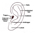
Preauricular sinus in ascending limb of the helix | |||
Preauricular sinus occurs in 0.25% births, is bilateral (hereditary) in 25-50% of cases and unilateral (mainly the left). They are developmental and generally occur on the surface in anterior margin of the ascending limb of the helix, and the duct runs inward to the perichondrium of the auricular cartilage and in some cases extend into the parotid gland. Postnatally they are a site for infection. | |||
'''Search PubMed:''' [http://www.ncbi.nlm.nih.gov/entrez/query.fcgi?db=pubmed&cmd=search&term=Preauricular%20Sinus Preauricular Sinus] | |||
'''Links:''' [http://www.nlm.nih.gov/medlineplus/ency/article/003304.htm Medline Plus - Preauricular tag or pit] | |||
=== Preauricular Tag === | |||
[[Image:preauricular_tag1.jpg]] [[Image:preauricular_tag2.jpg]] | |||
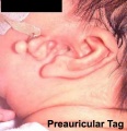
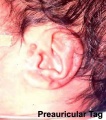
Skin tags in front of the external ear opening are common in neonates and in most cases are normal, though in some cases are indicative of other associated abnormalities. | |||
'''Search PubMed:''' [http://www.ncbi.nlm.nih.gov/entrez/query.fcgi?db=pubmed&cmd=search&term=Preauricular%20Tag Preauricular Tag] | |||
'''Links:''' [http://www.nlm.nih.gov/medlineplus/ency/article/003304.htm Medline Plus - Preauricular tag or pit] | |||
=== External Meatus Stenosis === | |||
Stenosis (narrowing) of the external auditory meatus is uncommon and can be due to chronic otitis externa or acquired atresia. The condition can be treated surgically by meatoplasty (reconstructive surgery of the canal) alone, though acquired atresia requires removal of the soft tissue plug and a split skin graft. | |||
'''Search PubMed:''' [http://www.ncbi.nlm.nih.gov/entrez/query.fcgi?db=pubmed&cmd=search&term=external%20meatus%20stenosis external meatus stenosis] | |||
== Congenital Deafness == | == Congenital Deafness == | ||
Revision as of 17:39, 5 June 2010

Introduction

How and why do things go wrong in development? Developing of hearing requires a complex origin, organisation, and timecourse means that abnormal development of any one system can impact upon the development of hearing. There are many different abnormalities of hearing development that can result in hearing loss and can broadly be divided into either conductive or sensorineural loss. These abnormalities can have genetic, environmental or unknown origins. In addition, abnormalities of the external ear (position and structure) is used as a clinical diagnostic tool for developmental abnormalities in other systems.
In Australia, there is now an early postnatal screening of neonatal hearing as part of a NSW State Wide Infant Screening Hearing (SWISH) Program using Automated Auditory Brainstem Response (AABR).
| Abnormality Links: abnormal development | abnormal genetic | abnormal environmental | Unknown | teratogens | ectopic pregnancy | cardiovascular abnormalities | coelom abnormalities | endocrine abnormalities | gastrointestinal abnormalities | genital abnormalities | head abnormalities | integumentary abnormalities | musculoskeletal abnormalities | limb abnormalities | neural abnormalities | neural crest abnormalities | placenta abnormalities | renal abnormalities | respiratory abnormalities | hearing abnormalities | vision abnormalities | twinning | Developmental Origins of Health and Disease | ICD-11 | ||
|
Some Recent Findings
- Review - The etiology of otosclerosis[1] "Otosclerosis is a common form of hearing loss characterized by abnormal bone remodeling in the otic capsule. It is a complex genetic disease, caused by a combination of genetic and environmental factors. During the past decade, several attempts have been made to identify factors for otosclerosis."
Inner Ear Abnormalities
Common cavity, severe cochlear hypoplasia
Cholesteatoma
Epithelium trapped within skull base in development, erosion of bones: temporal bone, middle ear, mastoid
Middle Ear Abnormalities
Rare and can be part of first arch syndrome.
Fixation of the middle ear ossicles Malleus, Incus and Stapes Middle ear abnormalities (ossicular anomalies) are rare and can be part of first arch syndrome.
- familial expansile osteolysis
- malleus/incus fixation
- absence of the long process of the incus
- congenital fixation of stapes (stapes anchored to oval window)
- failure of annular ligament development
- cholesteatoma
Familial Expansile Osteolysis (FEO)
A rare congenital (autosomal dominant, 18q21.1-q22) disorder similar to Paget’s disease of bone. Osteolytic lesions occur in all bones (mainly long bones) causing medullar expansions and lead eventually to middle ear and jaw abnormalities.
Daneshi A, Shafeghati Y, Karimi-Nejad MH, Khosravi A, Farhang F. Hereditary bilateral conductive hearing loss caused by total loss of ossicles: a report of familial expansile osteolysis. Otol Neurotol. 2005 Mar;26(2):237-40.
Malleus/Incus Fixation
Wehrs RE. Congenital absence of the long process of the incus. Laryngoscope. 1999 Feb;109(2 Pt 1):192-7.
Congenital Fixation of Stapes
In this condition the stapes is anchored to oval window often by growth of bone around the stapes (otosclerosis). Surgicallly treated by stapedectomy, where the bone and stapes is removed and replaced by a prosthesis.
Seidman MD, Babu S. A new approach for malleus/incus fixation: no prosthesis necessary. Otol Neurotol. 2004 Sep;25(5):669-73.
Cholesteatoma
Squamous epithelium that has been trapped within the skull base during development (congenital) and also occurs in an acquired form. The presence of this abnormality leads to erosion of the bones (temporal bone, middle ear, or mastoid) in which the epithelium is embedded.
Outer Ear Abnormalities

Several genetic effects and syndromes can include impacts on developmental of the external ear either directly or by altering development of the skull or face. Several developmental environment effects can be indicated by changes in the relative position or appearance of the external ear at birth. (More? [../Defect/maternal.htm Abnormal Development - Environment] | (Fetal Alcohol Syndrome).
- Microtia - abnormally small external ear
- Preauricular sinus - occurs in 0.25% births, bilateral (hereditary) 25-50%, unilateral (mainly the left), duct runs inward can extend into the parotid gland, Postnatally sites for infection
Microtia
The condition in humans of an abnormally small external ear is called Microtia. This is generally surgically repaired by use of rib cartilage to reconstruct the external ear. A recent study has identified a mouse model for this condition with the knockout of the Pact gene.

OMIM Database Search: "Microtia" (2006 - 25 search results)
Search PubMed May 2006 "Microtia" 449 reference articles of which 37 were reviews.
Search PubMed: Microtia | external ear defects
References
Rowe TM, Rizzi M, Hirose K, Peters GA, Sen GC. A role of the double-stranded RNA-binding protein PACT in mouse ear development and hearing. Proc Natl Acad Sci U S A. 2006 Mar 29 ".. Pact(-/-) mouse were reduced size and severe microtia. As a result of the congenital abnormality of both outer and middle ears, these mice were hearing impaired. Our study demonstrated an essential role of PACT in mammalian ear development and produced the first animal model for studying human microtia."
Zim SA. Microtia reconstruction: an update. Curr Opin Otolaryngol Head Neck Surg. 2003 Aug;11(4):275-81. Review. "...autogenous rib cartilage continues to be the gold standard for microtia repair. Numerous refinements and modifications in the original technique described by Tanzer have paved the way for exceptional results in experienced hands. However, ideal results are not always achieved, and there continue to be drawbacks with the standard approach to reconstruction with autogenous rib cartilage. In an attempt to circumvent these shortcomings, surgeons have developed alternative or adjuvant techniques to repair the microtic ear, including the use of tissue expansion, alloplastic implants, and osseointegrated prostheses. Finally, greater emphasis is being placed on early atresia repair in appropriate candidates."
Preauricular Sinus
File:Preauricular sinus sm.jpg
Preauricular sinus in ascending limb of the helix
Preauricular sinus occurs in 0.25% births, is bilateral (hereditary) in 25-50% of cases and unilateral (mainly the left). They are developmental and generally occur on the surface in anterior margin of the ascending limb of the helix, and the duct runs inward to the perichondrium of the auricular cartilage and in some cases extend into the parotid gland. Postnatally they are a site for infection.
Search PubMed: Preauricular Sinus
Links: Medline Plus - Preauricular tag or pit
Preauricular Tag
File:Preauricular tag1.jpg File:Preauricular tag2.jpg
Skin tags in front of the external ear opening are common in neonates and in most cases are normal, though in some cases are indicative of other associated abnormalities.
Search PubMed: Preauricular Tag
Links: Medline Plus - Preauricular tag or pit
External Meatus Stenosis
Stenosis (narrowing) of the external auditory meatus is uncommon and can be due to chronic otitis externa or acquired atresia. The condition can be treated surgically by meatoplasty (reconstructive surgery of the canal) alone, though acquired atresia requires removal of the soft tissue plug and a split skin graft.
Search PubMed: external meatus stenosis
Congenital Deafness
Sensorineural - cochlear or central auditory pathway Conductive - disease of outer and middle ear
Sensorineural
Cochlear or central auditory pathway
- Hereditary
- recessive- severe
- dominant- mild
- can be associated with abnormal pigmentation (hair and irises)
- Acquired
- rubella (German measles), maternal infection during 2nd month of pregnancy, vaccination of young girls
- cytomegalovirus [2]
- streptomycin
- antibiotic
- thalidomide
Conductive
Disease of outer and middle ear
- produced by otitis media with effusion, is widespread in young children.
- temporary blockage of outer or middle ear
Newborn Hearing Screening
In Australia, there is now an early postnatal screening of neonatal hearing as part of a NSW State Wide Infant Screening Hearing (SWISH) Program using Automated Auditory Brainstem Response (AABR).
- Very low birthweight infants and universal newborn hearing screening in a developing country[3]
Links: NSW Statewide Infant Screening - Hearing (SWISH) Program
Bionic Ear
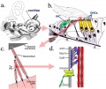
The "Cochlear Implant" was pioneered in development by Professor Graeme Clark (1960s, Australia).[4] It consists of an array of electrodes implanted within cochlea, that directly electrically stimulate the auditory nerve fibres.
- Young children with cochlear implants compared with children with normal hearing.[5]
References
Online Textbooks
- Clinical Methods 63. Cranial Nerves IX and X: The Glossopharyngeal and Vagus Nerves | The Tongue | 126. The Ear and Auditory System | An Overview of the Head and Neck - Ears and Hearing | Audiometry
- Health Services/Technology Assessment Text (HSTAT) Bethesda (MD): National Library of Medicine (US), 2003 Oct. Developmental Disorders Associated with Failure to Thrive
- Search Bookshelf hearing development
Reviews
- The International Journal of Developmental Biology Vol. 51 Nos. 6/7 (2007) Ear Development
Articles
Search Pubmed
Search Pubmed: Abnormalities | Middle ear ossicular anomalies | familial expansile osteolysis | cholesteatoma |
Additional Images
Inner ear hair cells
External ear anatomy
Preauricular tag
Preauricular tag


External Links
- Embryo Images - Hearing
- NIDCD - Balance Disorders
- NSW Health - NSW Statewide Infant Screening - Hearing (SWISH) Program
- American Academy of Audiology - American Academy of Audiology | In Memoriam: Judy Gravel
Glossary Links
- Glossary: A | B | C | D | E | F | G | H | I | J | K | L | M | N | O | P | Q | R | S | T | U | V | W | X | Y | Z | Numbers | Symbols | Term Link
Cite this page: Hill, M.A. (2024, June 15) Embryology Sensory - Hearing Abnormalities. Retrieved from https://embryology.med.unsw.edu.au/embryology/index.php/Sensory_-_Hearing_Abnormalities
- © Dr Mark Hill 2024, UNSW Embryology ISBN: 978 0 7334 2609 4 - UNSW CRICOS Provider Code No. 00098G