Musculoskeletal System - Joint Development: Difference between revisions
mNo edit summary |
mNo edit summary |
||
| Line 20: | Line 20: | ||
|-bgcolor="F5FAFF" | |-bgcolor="F5FAFF" | ||
| | | | ||
* '''Precise spatial restriction of BMP signaling in developing joints is perturbed upon loss of embryo movement'''{{#pmid:29467244|PMID29467244}} "Dynamic mechanical loading of synovial joints is necessary for normal joint development, as evidenced in certain clinical conditions, congenital disorders and animal models where dynamic muscle contractions are reduced or absent. Although the importance of mechanical forces on joint development is unequivocal, little is known about the molecular mechanisms involved. Here, using chick and mouse embryos, we observed that molecular changes in expression of multiple genes analyzed in the absence of mechanical stimulation are consistent across species. Our results suggest that abnormal joint development in immobilized embryos involves inappropriate regulation of Wnt and BMP signaling during definition of the emerging joint territories, i.e. reduced β-catenin activation and concomitant upregulation of pSMAD1/5/8 signaling. Moreover, dynamic mechanical loading of the developing knee joint activates Smurf1 expression; our data suggest that Smurf1 insulates the joint region from pSMAD1/5/8 signaling and is essential for maintenance of joint progenitor cell fate." | * '''Precise spatial restriction of BMP signaling in developing joints is perturbed upon loss of embryo movement'''{{#pmid:29467244|PMID29467244}} "Dynamic mechanical loading of synovial joints is necessary for normal joint development, as evidenced in certain clinical conditions, congenital disorders and animal models where dynamic muscle contractions are reduced or absent. Although the importance of mechanical forces on joint development is unequivocal, little is known about the molecular mechanisms involved. Here, using chick and mouse embryos, we observed that molecular changes in expression of multiple genes analyzed in the absence of mechanical stimulation are consistent across species. Our results suggest that abnormal joint development in immobilized embryos involves inappropriate regulation of {{Wnt}} and {{BMP}} signaling during definition of the emerging joint territories, i.e. reduced β-catenin activation and concomitant upregulation of pSMAD1/5/8 signaling. Moreover, dynamic mechanical loading of the developing knee joint activates Smurf1 expression; our data suggest that Smurf1 insulates the joint region from pSMAD1/5/8 signaling and is essential for maintenance of joint progenitor cell fate." | ||
* '''Development of the human shoulder joint during the embryonic and early fetal stages'''{{#pmid:29193070|PMID29193070}} | * '''Development of the human shoulder joint during the embryonic and early fetal stages'''{{#pmid:29193070|PMID29193070}} | ||
Revision as of 12:17, 11 April 2018
| Embryology - 16 Jun 2024 |
|---|
| Google Translate - select your language from the list shown below (this will open a new external page) |
|
العربية | català | 中文 | 中國傳統的 | français | Deutsche | עִברִית | हिंदी | bahasa Indonesia | italiano | 日本語 | 한국어 | မြန်မာ | Pilipino | Polskie | português | ਪੰਜਾਬੀ ਦੇ | Română | русский | Español | Swahili | Svensk | ไทย | Türkçe | اردو | ייִדיש | Tiếng Việt These external translations are automated and may not be accurate. (More? About Translations) |
Introduction

In the adult, the region where two skeletal bones meet and articulate is called a "joint", that are classified based upon their: anatomical structure, mobility and shape.
In the embryo, the majority of the vertebrate skeleton is initially formed as a cartilage template, that is later replaced by bone except at the interface between two adjacent bones, leaving in the adult a layer of cartilage in this region. The musculoskeletal system consists of skeletal muscle, bone, and cartilage and is mainly mesoderm in origin with some neural crest contribution.
| Joint Links: joint | synovial joint | temporomandibular joint | musculoskeletal | cartilage | Category:Joint | ||
|
Historic Embryology:
- Whillis J. The development of synovial joints. (1940) J Anat. 74(Pt 2): 277-283. PMID: 17104813
- Symons NB. The development of the human mandibular joint. (1952) J Anat. 86(3):326-32. PMID 12980883
Some Recent Findings
"In our study, serial sections of 32 human embryos (Carnegie stages 16-23) and 26 fetuses (9-13 weeks) were analyzed. The chondrogenic anlagen of the humerus and the medial border of the scapula can be observed from as early as Carnegie stage 17, whereas that of the rest of the scapula appears at stage 18. The osteogenic process begins in week 10 for the humeral head and week 11 for the scapula. At stage 19 the interzone becomes apparent, which will form the glenohumeral joint. In the next stage the glenohumeral joint will begin delaminating and exhibiting a looser central band. Denser lateral bands will join the humeral head (caput humeri) and the margins of the articular surface of the scapula, thus forming the glenoid labrum, which can be fully appreciated by stage 22. In 24-mm embryos (stage 21) we can observe, for the first time, the long head of the biceps tendon (which is already inserted in the glenoid labrum by week 9), and the intertubercular sulcus, whose depth is apparent since week 12. Regarding ligamentous structures, the coracohumeral ligament is observed at the end of Carnegie stage 23, whereas the primitive glenohumeral ligament already appeared in week 10." Shoulder Development | Musculoskeletal - Limb Development
|
| More recent papers |
|---|
 This table allows an automated computer search of the external PubMed database using the listed "Search term" text link.
More? References | Discussion Page | Journal Searches | 2019 References | 2020 References Search term: Joint Development <pubmed limit=5>Joint Development</pubmed> |
Joint Types

Classification
- Fibrous (synarthrodial) - immoveable joints found in cranial vault and teeth
- Cartilagenous (synchondroses and sympheses) - partially moveable joints
- Synovial (diarthrosis) - freely moveable joints are the most common found in the skeleton
Movement

- Hinge - (elbow and knee) Flexion/Extension
- Pivot - (neck, atlas and axis bones) Rotation of one bone around another
- Ball and Socket - (shoulder and hip)
- Saddle - (thumb)
- Condyloid - (wrist joints)
- Gliding - (intercarpal joints) Gliding movements
Synovial Joint Development
| Skeletal joint cavity development (cavitation) occurs along planes of the future articular surfaces of synovial joints. A number of different markers have been shown to be present in the interzone at the time of cavitation (hyaluronan and hyaluronan synthase, but not chondroitin sulphates).
Fibroblast-like cells (and/or adjacent chondrocytes) with uridine-diphospho glucose dehydrogenase (UDPGD) activity contribute to glycosaminoglycan levels (increases in hyaluronan). These cells are located on the intimal surface of the synovial lining and have been suggested as the possible cavitation mechanism, switching from cellular cohesion to dissociation.[6] |

|
Shoulder
This timeline data comes from a recent study of 32 human embryos (Carnegie stages 16-23) and 26 fetuses (9-13 weeks.[2]
| Carnegie Stage | Event |
|---|---|
| 17 | chondrogenic progenitor of the humerus and the medial border of the scapula can be observed. |
| 18 | chondrogenic progenitor for rest of the scapula appears. |
| 19 | glenohumeral joint will begin delaminating and showing a looser central band (interzone). Denser lateral bands will join the humeral head (caput humeri) and the margins of the articular surface of the scapula, thus forming the glenoid labrum (glenoid ligament). |
| 21 | long head of the biceps tendon present |
| 22 | glenoid labrum (glenoid ligament) present |
| 23 | coracohumeral ligament present |
| Week | |
| Fetal Week 10 | osteogenic process begins in the humeral head. Primitive glenohumeral ligament present |
| Fetal Week 11 | osteogenic process begins in the scapula |
| Links: shoulder | joint | limb | timeline Data from human histological study.[2] | |
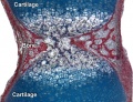
Knee
In human embryo at week 7 the femur and tibia cartilage template is present (stage 18), by week 8 the posterior cruciate ligament appears (stage 21), and by stage 23 the knee cavity and the anterior cruciate ligament are both also present.[7]

|
Knee Morphogenesis
Figure from recent BMP review.[8] |
Joint Abnormalities
FGFR-Related Craniosynostosis Syndromes
Pfeiffer syndrome, Apert syndrome, Crouzon syndrome, Beare-Stevenson syndrome, FGFR2-related isolated coronal synostosis, Jackson-Weiss syndrome, Crouzon syndrome with acanthosis nigricans (AN), and Muenke syndrome
Multiple Epiphyseal Dysplasia
Arthrogryposis
Arthrogryposis (arthrogryposis multiplex congenital, AMC) is a congenital joint contracture occurring in two or more body regions.
Large range of causes including:
- single-gene disorders autosomal recessive, autosomal dominant or X-linked traits.
- part of chromosomal disorders (Trisomy 18, many microdeletions and micro duplications)
- connective tissue disorders
Temporomandibular Disorders
Osteoarthritis
Clutton's joints
Historic clinical term for a symmetrical joint swelling occurring in patients of both sexes between 5 to 20 years of age with congenital syphilis. Joint swelling is usually in the knees, but can also affect the ankles, elbows, wrists and fingers. Named after Henry Hugh Clutton who first described the condition in 1886.
- Links: Abnormal Development - Syphilis)
References
- ↑ Singh PNP, Shea CA, Sonker SK, Rolfe RA, Ray A, Kumar S, Gupta P, Murphy P & Bandyopadhyay A. (2018). Precise spatial restriction of BMP signaling in developing joints is perturbed upon loss of embryo movement. Development , 145, . PMID: 29467244 DOI.
- ↑ 2.0 2.1 2.2 Hita-Contreras F, Sánchez-Montesinos I, Martínez-Amat A, Cruz-Díaz D, Barranco RJ & Roda O. (2018). Development of the human shoulder joint during the embryonic and early fetal stages: anatomical considerations for clinical practice. J. Anat. , 232, 422-430. PMID: 29193070 DOI.
- ↑ Giorgi M, Carriero A, Shefelbine SJ & Nowlan NC. (2014). Mechanobiological simulations of prenatal joint morphogenesis. J Biomech , 47, 989-95. PMID: 24529755 DOI.
- ↑ Khan IM, Redman SN, Williams R, Dowthwaite GP, Oldfield SF & Archer CW. (2007). The development of synovial joints. Curr. Top. Dev. Biol. , 79, 1-36. PMID: 17498545 DOI.
- ↑ Iwamoto M, Tamamura Y, Koyama E, Komori T, Takeshita N, Williams JA, Nakamura T, Enomoto-Iwamoto M & Pacifici M. (2007). Transcription factor ERG and joint and articular cartilage formation during mouse limb and spine skeletogenesis. Dev. Biol. , 305, 40-51. PMID: 17336282 DOI.
- ↑ <pubmed>7525525</pubmed>
- ↑ <pubmed>9185992</pubmed>
- ↑ <pubmed>26893264</pubmed>
Online Textbooks
Developmental Biology Gilbert, Scott F. Sunderland (MA): Sinauer Associates, Inc. ; c2000 Forming the joints
Reviews
Articles
<pubmed>15492776</pubmed> <pubmed>10645964</pubmed> <pubmed>7544653</pubmed> <pubmed>7525525</pubmed>
Search PubMed
Search July 2010 "Joint Development" All (19900) Review (3137) Free Full Text (3325)
Search Pubmed: Joint Development
Additional Images

Adult axial skeleton

Adult appendicular skeleton

Bone structure
Developing vertebra

Endochondral bone

Fetal head lateral (12 weeks)

Fetal head medial (12 weeks)

Fetal head section (12 weeks)
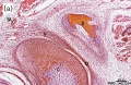
Fetal temporomandibular joint 10 weeks
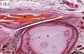
Fetal temporomandibular joint 12 weeks


External Links
External Links Notice - The dynamic nature of the internet may mean that some of these listed links may no longer function. If the link no longer works search the web with the link text or name. Links to any external commercial sites are provided for information purposes only and should never be considered an endorsement. UNSW Embryology is provided as an educational resource with no clinical information or commercial affiliation.
Glossary Links
- Glossary: A | B | C | D | E | F | G | H | I | J | K | L | M | N | O | P | Q | R | S | T | U | V | W | X | Y | Z | Numbers | Symbols | Term Link
Cite this page: Hill, M.A. (2024, June 16) Embryology Musculoskeletal System - Joint Development. Retrieved from https://embryology.med.unsw.edu.au/embryology/index.php/Musculoskeletal_System_-_Joint_Development
- © Dr Mark Hill 2024, UNSW Embryology ISBN: 978 0 7334 2609 4 - UNSW CRICOS Provider Code No. 00098G