File:B220849-02.jpg: Difference between revisions
From Embryology
mNo edit summary |
|||
| Line 7: | Line 7: | ||
** Chorionic plate and villi forming the early embryonic portion of the placenta. | ** Chorionic plate and villi forming the early embryonic portion of the placenta. | ||
** Yolk sac is not visible in this section. The yolk sac is seen in a [[:File:B220849-03.jpg|more lateral section]] from this embryo. | ** Yolk sac is not visible in this section. The yolk sac is seen in a [[:File:B220849-03.jpg|more lateral section]] from this embryo. | ||
* '''Central nervous system''' - Primary brain vesicles formed by large ventricular cavity with a thin wall that will later develop the central nervous system. | * '''Central nervous system''' - Primary brain vesicles formed by large ventricular cavity with a thin wall that will later develop the central nervous system. Beneath the hindbrain the neural tube narrows to the spinal cord, not visible in this section, and the section now passes through the lateral wall of the tube. | ||
* | * | ||
{{B220849}} | {{B220849}} | ||
Revision as of 21:05, 8 December 2013
Human Embryo (CRL 4.2 mm)
Sagittal section through embryo week 4 approximately Carnegie stage 11 to 12.
- Extra-embryonic coeloms - spaces and membranes that lie outside the embryo.
- Amniotic cavity surrounding the embryo.
- Chorionic plate and villi forming the early embryonic portion of the placenta.
- Yolk sac is not visible in this section. The yolk sac is seen in a more lateral section from this embryo.
- Central nervous system - Primary brain vesicles formed by large ventricular cavity with a thin wall that will later develop the central nervous system. Beneath the hindbrain the neural tube narrows to the spinal cord, not visible in this section, and the section now passes through the lateral wall of the tube.
- Embryo Image Links: labeled embryo | unlabeled embryo | yolk sac | Carnegie stage 11 | Carnegie stage 12
| Week: | 1 | 2 | 3 | 4 | 5 | 6 | 7 | 8 |
| Carnegie stage: | 1 2 3 4 | 5 6 | 7 8 9 | 10 11 12 13 | 14 15 | 16 17 | 18 19 | 20 21 22 23 |
Image source: The Blechschmidt Collection images are reproduced with the permission of Prof. Christoph Viebahn, director of the Institute of Anatomy and Embryology, , University Medical Center Göttingen. Images are for educational purposes only and cannot be reproduced electronically or in writing without permission.
Cite this page: Hill, M.A. (2024, June 1) Embryology B220849-02.jpg. Retrieved from https://embryology.med.unsw.edu.au/embryology/index.php/File:B220849-02.jpg
- © Dr Mark Hill 2024, UNSW Embryology ISBN: 978 0 7334 2609 4 - UNSW CRICOS Provider Code No. 00098G
File history
Click on a date/time to view the file as it appeared at that time.
| Date/Time | Thumbnail | Dimensions | User | Comment | |
|---|---|---|---|---|---|
| current | 02:06, 8 December 2013 | 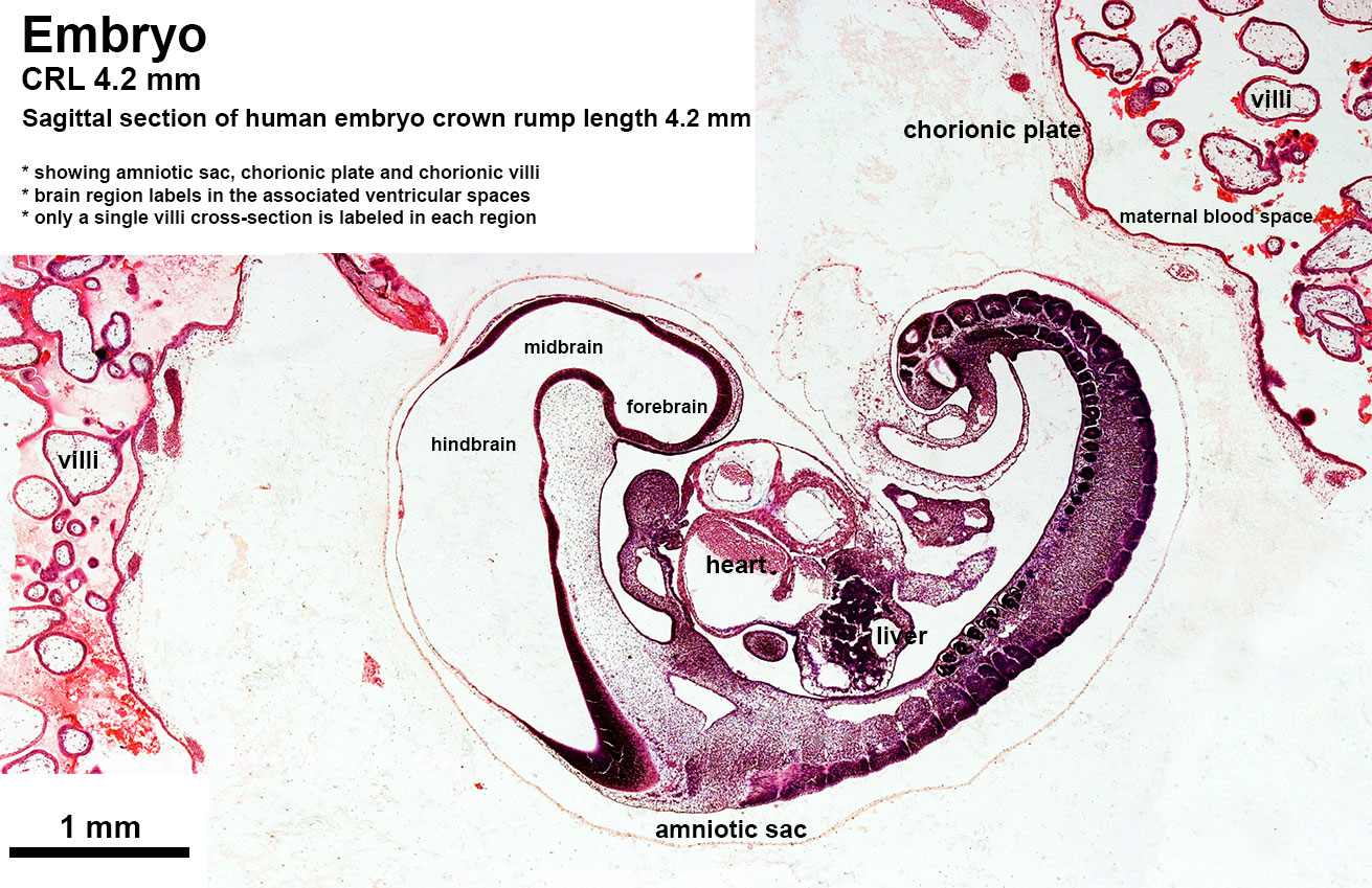 | 1,311 × 849 (358 KB) | Z8600021 (talk | contribs) | ==Human Embryo (CRL 4.2 mm)== Sagittal section through embryo week 4 approximately Carnegie stage 11 to 12. {{Blechschmidt collection}} Category:HumanCategory:Week 4 |
You cannot overwrite this file.
File usage
The following page uses this file: