BGDA Practical 3 - Gametogenesis: Difference between revisions
mNo edit summary |
|||
| (85 intermediate revisions by 3 users not shown) | |||
| Line 1: | Line 1: | ||
{{ | {{BGDALab3}} | ||
== Female Gametogenesis == | == Female Gametogenesis == | ||
[[File:Ovary_histology_061.jpg|thumb|Oocyte and support cells in an astral follicle]] | |||
In females, the total number of eggs ever to be produced are present in the newborn female. | In females, the total number of eggs ever to be produced are present in the newborn female. | ||
| Line 8: | Line 9: | ||
# The secondary oocyte then commences '''meiosis 2''' which arrests at metaphase and will not continue without fertilization. | # The secondary oocyte then commences '''meiosis 2''' which arrests at metaphase and will not continue without fertilization. | ||
# At fertilization '''meiosis 2''' completes, forming a second polar body. Note that the first polar body may also undergo this process forming a third polar body. | # At fertilization '''meiosis 2''' completes, forming a second polar body. Note that the first polar body may also undergo this process forming a third polar body. | ||
<br><br> | |||
{| | |||
| valign="bottom"|{{Meiosis movie 1}} | |||
| A mouse oocyte undergoing meiosis spindle migration followed by first polar body extrusion and MII spindle positioning.{{#pmid:23439682|PMID23439682}} | |||
* <font color=blue>'''blue'''</font> - Hoechst staining of chromosomes. | |||
* <font color=green>'''green'''</font> - UtrCH-GFP was used to label cortical changes during spindle migration. | |||
The video shows that cytoplasmic streaming continues to the MII arrest stage to maintain the oocyte set of chromosomes/MII spindle in place close to the cortex. Frames are 11 min apart, and video length is 840 min. Bar, 20 µm. | |||
|} | |||
<br><br> | |||
[[File:Female_gametogenesis.jpg|600px|Female gametogenesis]] | |||
[[File: | <br> | ||
[[File:Oogenesis and meiosis cartoon.jpg|600px]] | |||
Meiosis and Oogenesis{{#pmid:22705668|PMID22705668}} | |||
<br> | |||
{| | |||
|-bgcolor="CEDFF2" | |||
! Meiosis | |||
! Oogenesis | |||
|- | |||
| Divided into 3 temporally distinct phases. | |||
# Prophase - after DNA replication, homologous chromosomes (shown in red and blue) undergo pairing, synapsis and recombination, and arrest at the diplotene (dictyate) stage. | |||
# Dictyate arrest - oocytes remain in meiotic arrest until the female reaches maturity and the oocyte has completed an extensive period of growth following follicle formation. | |||
# Divisions - luteinizing hormone (LH) surge that triggers ovulation also causes resumption and completion of the first meiotic division in the periovulatory oocyte. The ovulated egg is arrested at second meiotic metaphase, and anaphase onset and completion of meiosis II only occur if the egg is fertilized. | |||
| Complex involving 4 distinct phases. | |||
# Commitment to meiosis and meiotic initiation - occurs at {{GA}} 8–10 weeks in humans. | |||
# Follicle formation - occurs during the [[Second Trimester|second trimester]] in humans. | |||
# Oocyte growth - occurs in the sexually mature female under the control of paracrine and endocrine signals. Oocyte growth is thought to take approximately 85+ days in humans and typically culminates in the ovulation of a single egg. | |||
# Fertilization - of the ovulated egg results in the completion of the second meiotic division. | |||
|} | |||
===Female Abnormalities=== | ===Female Abnormalities=== | ||
Meiotic non-disjunction resulting in aneuploidy, most are embryonic lethal and not seen. The most common non-lethal abnormality is [[trisomy 21|Trisomy 21 or Down syndrome]]. The potential for this and most genetic abnormalities [[Genetic_risk_maternal_age|increase with maternal age]]. | |||
Meiotic non-disjunction resulting in aneuploidy, most are embryonic lethal and not seen. The potential for genetic abnormalities [[Genetic_risk_maternal_age|increase with maternal age]]. | |||
{| | |||
| [[File:Trisomy21female.jpg|400px]] | |||
| [[File:Turner syndrome karyotype.jpg|400px]] | |||
|- | |||
| Trisomy 21 female karyotype | |||
| Turner's Syndrome karyotype | |||
|} | |||
<br> | |||
{{Chromosome Aneuploidy table}} | |||
== Male Gametogenesis == | == Male Gametogenesis == | ||
In males, sperm continues to be generated throughout life from a stem cell population in the testis. Spermatozoa maturation involves two processes meiosis and spermiogenesis | [[File:Seminiferous-tubule-HEx40.jpg|thumb|Adult seminiferous tubule showing spermatozoa developmental stages]] | ||
The histology will be covered in a separate Male Histology Practical ([[BGDA_Practical_-_Male_Reproductive_Tract_Histology|support page]]). | |||
In males, sperm continues to be generated throughout life from a stem cell population in the testis. Spermatozoa maturation involves two processes meiosis and spermiogenesis. | |||
{{Seminiferous Tubule puberty table}} | |||
[[File:Male_gametogenesis.jpg|600px]] | [[File:Male_gametogenesis.jpg|600px]] | ||
| Line 33: | Line 67: | ||
<gallery> | <gallery> | ||
File:Historic-testis.jpg|Historic testis drawing | File:Historic-testis.jpg|Historic testis drawing | ||
File: | File:Testis_histology_006.jpg|Immature (child) seminiferous tubule showing no spermatozoa | ||
File:Testis_histology_2.jpg|Seminiferous tubule cross-section and supporting cells | File:Testis_histology_2.jpg|Seminiferous tubule cross-section and supporting cells | ||
File:Human-spermatozoa.jpg|Human spermatozoa | File:Human-spermatozoa.jpg|Human spermatozoa | ||
| Line 40: | Line 74: | ||
===Human Spermatozoa Development=== | ===Human Spermatozoa Development=== | ||
* Spermatogenesis process of spermatagonia mature into | * Spermatogenesis process of spermatagonia mature into spermatozoa (sperm). | ||
* Continuously throughout life occurs in the seminiferous tubules in the male gonad- testis (plural testes). | * Continuously throughout life occurs in the seminiferous tubules in the male gonad- testis (plural testes). | ||
* At puberty spermatagonia activate and proliferate (mitosis). | * At puberty spermatagonia activate and proliferate (mitosis). | ||
| Line 47: | Line 81: | ||
* follicle stimulating hormone (FSH) - stimulates the spermatogenic epithelium | * follicle stimulating hormone (FSH) - stimulates the spermatogenic epithelium | ||
* luteinizing-hormone (LH) - stimulates testosterone production by Leydig cells | * luteinizing-hormone (LH) - stimulates testosterone production by Leydig cells | ||
===Spermiogenesis=== | |||
[[File:Human_spermatozoa_acrosomal_protein_SP-10.jpg|600px]] | |||
[[File:Human-spermatozoa_EM01.jpg|600px]] | [[File:Human-spermatozoa_EM01.jpg|600px]] | ||
| Line 65: | Line 102: | ||
|} | |} | ||
{{Spermatozoa Terms collapse table}} | |||
:'''Links:''' [[Spermatozoa Development]] | [http://www.ncbi.nlm.nih.gov/bookshelf/br.fcgi?book=mboc4&part=A3729 MBoC - Sperm] | [http://www.ncbi.nlm.nih.gov/bookshelf/br.fcgi?book=mboc4&part=A3729&rendertype=figure&id=A3735 MBoC - Highly simplified drawing of a cross-section of a seminiferous tubule in a mammalian testis] | [http://www.ncbi.nlm.nih.gov/bookshelf/br.fcgi?book=mboc4&part=A3729&rendertype=figure&id=A3736 MBoC - Cytoplasmic bridges in developing sperm cells and their precursors] | |||
===Puberty=== | ===Puberty=== | ||
* In humans at puberty, hormonal and morphological changes occur within the gonad and other systems (secondary sex characteristics). | * In humans at puberty, hormonal and morphological changes occur within the gonad and other systems (secondary sex characteristics). | ||
* Within the testis the immature Sertoli cells cease to proliferate and differentiate. | * Within the testis the immature Sertoli cells cease to proliferate and differentiate. | ||
* Spermatogonium proliferate and spermatogenesis begins. | * Spermatogonium (plural, spermatogonia) proliferate and spermatogenesis begins. | ||
* It takes about 70 days for cells to mature from the diploid spermatogonium to a primary spermatocyte. | * It takes about 70 days for cells to mature from the diploid spermatogonium to a primary spermatocyte. | ||
* This maturation occurs in waves along the seminiferous tubules. | * This maturation occurs in waves along the seminiferous tubules. | ||
=== | ===Ejaculate=== | ||
[[File:Azoospermia.jpg|thumb|Azoospermia - Non-obstructive azoospermia (NOA) and Obstructive azoospermia (OA)]] | [[File:Azoospermia.jpg|thumb|Azoospermia - Non-obstructive azoospermia (NOA) and Obstructive azoospermia (OA)]] | ||
*release of spermatozoa and accessory gland secretions from the male genital tract (3.5 ml) | *release of spermatozoa and accessory gland secretions from the male genital tract (3.5 ml) | ||
| Line 82: | Line 122: | ||
===Male Abnormalities=== | ===Male Abnormalities=== | ||
Clinically abnormality studies relate mainly to infertility and [[#Genetics|inherited genetic disorders]]. | |||
* '''Oligospermia''' - (Low Sperm Count) less than 20 million sperm after 72 hour abstinence from sex | * '''Oligospermia''' - (Low Sperm Count) less than 20 million sperm after 72 hour abstinence from sex | ||
* '''Azoospermia''' - (Absent Sperm) blockage of duct network | * '''Azoospermia''' - (Absent Sperm) blockage of duct network | ||
* '''Immotile Cilia Syndrome''' - lack of sperm motility | * '''Immotile Cilia Syndrome''' - lack of sperm motility | ||
For spermatozoa genetic abnormalities see [https://www.ncbi.nlm.nih.gov/books/NBK6064/|Cytogenetics of Human Sperm]. | |||
== Differences in Mammalian Meioses == | == Differences in Mammalian Meioses == | ||
| Line 121: | Line 162: | ||
|} | |} | ||
{{BGDA Practical 3 - Gametogenesis Interactive}} | |||
{{ | {{BGDALab3}} | ||
==Additional Information== | ==Additional Information== | ||
{{Med Prac additional Information}} | |||
=== | <br> | ||
'''Links:''' | [[File:Chromosome- structure.jpg|thumb|Chromosome structure]] | ||
* [[BGDA_Practical_-_Male_Reproductive_Tract_Histology|Male Histology]] - covered in another practical class. From today's class you should have a basic understanding of seminiferous tubule structure in relation to spermatozoa development. (More? {{spermatozoa}}, spermatogenesis review{{#pmid:26537427|PMID26537427}}) | |||
* {{genetics}} - covered elsewhere in your course. | |||
* {{mitosis}} and {{meiosis}} - Cell Biology of mitosis and meiosis also covered in Foundations. | |||
Martin RH. Cytogenetics of Human Sperm. In: Madame Curie Bioscience Database [Internet]. Austin (TX): Landes Bioscience; 2000-2013. Available from: https://www.ncbi.nlm.nih.gov/books/NBK6064/ | |||
{{#pmid:19934213}} | |||
===Mitosis and Meiosis=== | |||
[[File:Mitosis_and_meiosis.jpg|600px]] | |||
{| | |||
! Chromosome connections in meiosis{{#pmid:20231873|PMID20231873}} | |||
|- | |||
| [[File:Chromosome connections in meiosis.jpg|alt=Chromosome connections in meiosis|500px]] | |||
| Kinetochores attach homologous chromosomes to opposite halves of the spindle. | |||
Homologs are held together by chiasmata, in which recombinant chromatids cross each other. | |||
Sisters are held together by cohesins and possibly by catenation of centromeric DNA threads, which have been observed in human mitosis. | |||
Cohesion is released in two steps: | |||
# on chromosome arms to resolve chiasmata and separate homologs in the first meiotic division. | |||
# around centromeres to separate sisters in the second meiotic division. | |||
|} | |||
:'''Links:''' {{mitosis}} | {{meiosis}} | |||
===Aneuploidy and Fertility=== | |||
{| | |||
| valign=top|Homologous Recombination {{meiosis}} - In the two sexes genetic recombination between homologous chromosomes proceeds analogously through most stages. | |||
<br> | |||
'''Female''' | |||
* In female, but not male, it has been shown that about 25% of the intermediates that should mature into crossover products actually fail, called "female-specific crossover maturation inefficiency".{{#pmid:28262352|PMID28262352}} | |||
* Some contributory factors include: cohesion deterioration, uncoordinated sister kinetochore behaviour, erroneous microtubule attachments, spindle instability and structural chromosomal defects that impact centromeres and telomeres. | |||
* Mouse model shows that in oocytes both cohesin and centromere-specific histones are long-lived proteins, though without obvious renewal pathways, "their deterioration with age provides an appealing explanation for at least some of the problems in older oocytes."{{#pmid:29244163|PMID29244163}} | |||
'''Male''' | |||
* In male infertility (non-obstructive azoospermia '''NOA''' and obstructive azoospermia '''OA'''), spermatozoa development has been shown to have both an altered crossover distribution and frequency.{{#pmid:27273078|PMID27273078}} | |||
:'''Links:''' {{meiosis}} | {{genetic abnormalities}} | {{trisomy 21}}) | |||
| [[File:Meiotic chromosome crossovers 01.jpg|300px]] | |||
Meiotic chromosome crossovers | |||
|} | |||
<br> | |||
{{Human Spermatozoa Statistics collapse table}} | |||
===Genetics=== | ===Genetics=== | ||
Where genes are located and how they are inherited relate to the chromosome that the gene is located upon, the parental origin of the gene and the dominant/recessive nature of the gene. | |||
<gallery> | <gallery> | ||
File:Autosomal_dominant_inheritance.jpg|Autosomal dominant inheritance | File:Autosomal_dominant_inheritance.jpg|Autosomal dominant inheritance | ||
| Line 149: | Line 246: | ||
</gallery> | </gallery> | ||
{{ | {{GHR Inheritance}} | ||
== | ==Terms== | ||
''' | * '''autosomal inheritance''' - some hereditary diseases are described as autosomal which means that the disease is due to a DNA error in one of the 22 pairs that are not sex chromosomes. Both boys and girls can then inherit this error. If the error is in a sex chromosome, the inheritance is said to be sex-linked. | ||
* '''cascade testing''' - Clinical genetic term for the testing of genetic relatives for a mutation that has been identified in the first affected family member. [[Abnormal Development - Genetic]] | |||
* '''gene''' - a sequence of DNA that encodes an individual protein. | |||
* '''genome''' - the complete genetic information in the form of DNA available to a specific species. | |||
* '''Johnsen score''' - a clinical score (1-10) for assessing spermatogenesis in a human testicular biopsy. Named after the author of the original article. [https://www.ncbi.nlm.nih.gov/pubmed/5527187 PMID 5527187] | |||
* '''sperm''' - ({{spermatozoa}}) The male haploid reproductive cell, often used generically (and incorrectly) to describe these cells and the fluid of the ejaculate. Term is a shortened form of scientifically correct term {{spermatozoa}}. | |||
* '''sperm''' - | |||
* '''sperm annulus''' - (Jensen's ring; Latin, ''annulus'' = ring) A region of the mammalian sperm flagellum connecting the midpiece and the principal piece. The annulus is a septin-based structure formed from SEPT1, 4, 6, 7 and 12. Septins are polymerizing GTPases that can act as a scaffold forming hetero-oligomeric filaments required for cytokinesis and other cell cycle roles. | * '''sperm annulus''' - (Jensen's ring; Latin, ''annulus'' = ring) A region of the mammalian sperm flagellum connecting the midpiece and the principal piece. The annulus is a septin-based structure formed from SEPT1, 4, 6, 7 and 12. Septins are polymerizing GTPases that can act as a scaffold forming hetero-oligomeric filaments required for cytokinesis and other cell cycle roles. | ||
* '''spermatid''' - Intermediate cell in spermatozoa development, within the testis seminiferous tubule they lie in the luminal cell layer to the [[S#secondary spermatocyte|secondary spermatocyte]]. These small cells are haploid and in [[S#spermiogenesis|spermiogenesis]] change their cellular structure and shape to form [[S#spermatozoa|spermatozoa]]. | * '''spermatid''' - Intermediate cell in spermatozoa development, within the testis seminiferous tubule they lie in the luminal cell layer to the [[S#secondary spermatocyte|secondary spermatocyte]]. These small cells are haploid and in [[S#spermiogenesis|spermiogenesis]] change their cellular structure and shape to form [[S#spermatozoa|spermatozoa]]. (More? {{spermatozoa}} | {{testis}} | {{fertilization}} | [[Lecture - Fertilization]]) | ||
* '''{{spermatogenesis}}''' - (Greek, ''genesis'' = origin, creation, generation) The term used to describe the process of diploid spermatagonia division and differentiation to form haploid spermatozoa within the testis (male gonad). The process includes the following cellular changes: meiosis, reoorganization of DNA, reduction in DNA content, reorganization of cellular organelles, morphological changes (cell shape). The final process of change in cell shape is also called [[S#spermiogenesis|spermiogenesis]]. | |||
* '''spermatogenesis''' - (Greek, ''genesis'' = origin, creation, generation) The term used to describe the process of diploid spermatagonia division and differentiation to form haploid | |||
* '''spermiogenesis''' - (Greek, ''genesis'' = origin, creation, generation) The maturation process of the already haploid [[S#spermatid|spermatids]] into the mature [[S#spermatozoa|spermatozoa]] shape and organization. This process involves reorganization of cellular organelles ([[E#endoplasmic reticulum|endoplasmic reticulum]], [[G#golgi apparatus|Golgi apparatus]], [[M#mitochondria|mitochondria]]), cytoskeletal changes (microtubule organization) and morphological changes (cell shape, acrosome and tail formation). The process of maturation of the [[S#spermatid|spermatids]] into spermatozoa: chromatin condenses, nucleus becomes smaller, the [[G#golgi apparatus|Golgi apparatus]] is modified to form the [[A#acrosome|acrosome]], microtubules are reorganised to form the tail, mitochondria are relocated to the initial segment of the tail and the majority of cell cytoplasm is discarded. | * '''spermiogenesis''' - (Greek, ''genesis'' = origin, creation, generation) The maturation process of the already haploid [[S#spermatid|spermatids]] into the mature [[S#spermatozoa|spermatozoa]] shape and organization. This process involves reorganization of cellular organelles ([[E#endoplasmic reticulum|endoplasmic reticulum]], [[G#golgi apparatus|Golgi apparatus]], [[M#mitochondria|mitochondria]]), cytoskeletal changes (microtubule organization) and morphological changes (cell shape, acrosome and tail formation). The process of maturation of the [[S#spermatid|spermatids]] into spermatozoa: chromatin condenses, nucleus becomes smaller, the [[G#golgi apparatus|Golgi apparatus]] is modified to form the [[A#acrosome|acrosome]], microtubules are reorganised to form the tail, mitochondria are relocated to the initial segment of the tail and the majority of cell cytoplasm is discarded. | ||
* '''spermatogonia''' - These cells form in the embryo from the [[P#primordial germ cell|primordial germ cell]] and are located in the seminiferous tubule adjacent to the basal membrane. The cells can either divide and separate to renew the stem cell population, or they divide and stay together as a pair ([[A#Apr spermatogonia|Apr spermatogonia]]) connected by an intercellular cytoplasmic bridge to begin to differentiate and eventually form | * '''{{spermatogonia}}''' - These cells form in the embryo from the [[P#primordial germ cell|primordial germ cell]] and are located in the seminiferous tubule adjacent to the basal membrane. The cells can either divide and separate to renew the stem cell population, or they divide and stay together as a pair ([[A#Apr spermatogonia|Apr spermatogonia]]) connected by an intercellular cytoplasmic bridge to begin to differentiate and eventually form {{spermatozoa}}. | ||
* '''spermatozoa''' - (spermatozoon, singular term) The male haploid gamete cell produced by meiosis in the testis (male gonad) seminiferous tubule. In humans, produced from puberty onwards and develop from the diploid stem cell the [[S#spermatogonia|spermatogonia]]. The developmental meiosis is called | * '''{{spermatozoa}}''' - (spermatozoon, singular term) The male haploid gamete cell produced by meiosis in the testis (male gonad) seminiferous tubule. In humans, produced from puberty onwards and develop from the diploid stem cell the [[S#spermatogonia|spermatogonia]]. The developmental meiosis is called {{spermatogenesis}} and the final morphologiccal (shape) change is called {{spermiogenesis}}. The mature human spermatozoon formed from the [[S#spermatid|spermatid]] has a head, neck and tail and is about 60 µm long. At ejaculation these cells undergo [[C#capacitation|capacitation]] are activated and become motile. | ||
* '''spermatozoa head''' - Following | * '''spermatozoa head''' - Following {{spermiogenesis}}, the first region of the [[S#spermatozoa|spermatozoa]] containing the haploid nucleus and acrosome. In humans, it is a flattened structure (5 µm long by 3 µm wide) with the posterior part of nuclear membrane forming the basal plate region. The human [[S#spermatozoa|spermatozoa]] is about 60 µm long, actively motile and divided into 3 main regions ([[S#spermatozoa head|head]], [[S#spermatozoa neck|neck]] and [[S#spermatozoa tail|tail]]). | ||
* '''spermatozoa neck''' - Following [[S#spermiogenesis|spermiogenesis]], the second region of the [[S#spermatozoa|spermatozoa]] attached to basal plate, transverse oriented centriole, contains nine segmented columns of fibrous material, continue as outer dense fibres in tail. In humans, it forms a short structure (1 µm). The human [[S#spermatozoa|spermatozoa]] is about 60 µm long, actively motile and divided into 3 main regions ([[S#spermatozoa head|head]], [[S#spermatozoa neck|neck]] and [[S#spermatozoa tail|tail]]). | * '''spermatozoa neck''' - Following [[S#spermiogenesis|spermiogenesis]], the second region of the [[S#spermatozoa|spermatozoa]] attached to basal plate, transverse oriented centriole, contains nine segmented columns of fibrous material, continue as outer dense fibres in tail. In humans, it forms a short structure (1 µm). The human [[S#spermatozoa|spermatozoa]] is about 60 µm long, actively motile and divided into 3 main regions ([[S#spermatozoa head|head]], [[S#spermatozoa neck|neck]] and [[S#spermatozoa tail|tail]]). | ||
| Line 188: | Line 280: | ||
* '''spermatozoa tail''' - Following [[S#spermiogenesis|spermiogenesis]], the third region of the [[S#spermatozoa|spermatozoa]] that has a ([[S#spermatozoa head|head]], [[S#spermatozoa neck|neck]] and [[S#spermatozoa tail|tail]]). The tail is also divided into 3 structural regions a middle piece, a principal piece and an end piece. In humans: the middle piece (5 µm long) is formed by [[A#axonema|axonema]] and dense fibres surrounded by mitochondria; the principal piece (45 µm long) fibrous sheath interconnected by regularly spaced circumferential hoops; the final end piece (5 µm long) has an [[A#axonema|axonema]] surrounded by small amount of cytoplasm and plasma membrane. | * '''spermatozoa tail''' - Following [[S#spermiogenesis|spermiogenesis]], the third region of the [[S#spermatozoa|spermatozoa]] that has a ([[S#spermatozoa head|head]], [[S#spermatozoa neck|neck]] and [[S#spermatozoa tail|tail]]). The tail is also divided into 3 structural regions a middle piece, a principal piece and an end piece. In humans: the middle piece (5 µm long) is formed by [[A#axonema|axonema]] and dense fibres surrounded by mitochondria; the principal piece (45 µm long) fibrous sheath interconnected by regularly spaced circumferential hoops; the final end piece (5 µm long) has an [[A#axonema|axonema]] surrounded by small amount of cytoplasm and plasma membrane. | ||
* '''spermatogonial stem cells''' - (SSCs) The [[S#spermatagonia|spermatagonia cells]] located beside the seminiferous tubule basal membrane that either divide and separate to renew the stem cell population, or they divide and stay together as a pair ([[A#Apr spermatogonia|Apr spermatogonia]]) connected by an intercellular cytoplasmic bridge to differentiate and eventually form | * '''spermatogonial stem cells''' - (SSCs) The [[S#spermatagonia|spermatagonia cells]] located beside the seminiferous tubule basal membrane that either divide and separate to renew the stem cell population, or they divide and stay together as a pair ([[A#Apr spermatogonia|Apr spermatogonia]]) connected by an intercellular cytoplasmic bridge to differentiate and eventually form spermatozoa}}. | ||
* '''sperm protein 56''' - A component of the spermatozoa acrosomal matrix released to the sperm surface during [[C#capacitation|capacitation]]. | * '''sperm protein 56''' - A component of the spermatozoa acrosomal matrix released to the sperm surface during [[C#capacitation|capacitation]]. | ||
<br><br> | |||
{{Spermatozoa Terms collapse table}} | |||
<br> | |||
{{Cell Division terms}} | |||
<br> | |||
{{Oocyte terms collapse table}} | |||
<br> | |||
{{Glossary}} | |||
<br> | |||
==References== | |||
<references/> | |||
<br> | |||
{{BGDALab3}} | |||
{{BGDAFooter}} | |||
[[Category:Meiosis]] [[Category:Gametogenesis]] | [[Category:Meiosis]] [[Category:Gametogenesis]] | ||
Latest revision as of 10:59, 8 May 2019
Female Gametogenesis

In females, the total number of eggs ever to be produced are present in the newborn female.
- All eggs are arrested at an early stage of the first meiotic division as a primary oocyte (primordial follicle). Following purberty, during each menstrual cycle, pituitary gonadotrophin stimulates completion of meiosis 1 the day before ovulation.
- In meiosis 1, a diploid cell becomes 2 haploid (23 chromosomes) daughter cells, each chromosome has two chromatids. One cell becomes the secondary oocyte the other cell forms the first polar body.
- The secondary oocyte then commences meiosis 2 which arrests at metaphase and will not continue without fertilization.
- At fertilization meiosis 2 completes, forming a second polar body. Note that the first polar body may also undergo this process forming a third polar body.
|
A mouse oocyte undergoing meiosis spindle migration followed by first polar body extrusion and MII spindle positioning.[1]
The video shows that cytoplasmic streaming continues to the MII arrest stage to maintain the oocyte set of chromosomes/MII spindle in place close to the cortex. Frames are 11 min apart, and video length is 840 min. Bar, 20 µm. |


Meiosis and Oogenesis[2]
| Meiosis | Oogenesis |
|---|---|
Divided into 3 temporally distinct phases.
|
Complex involving 4 distinct phases.
|
Female Abnormalities
Meiotic non-disjunction resulting in aneuploidy, most are embryonic lethal and not seen. The most common non-lethal abnormality is Trisomy 21 or Down syndrome. The potential for this and most genetic abnormalities increase with maternal age.

|

|
| Trisomy 21 female karyotype | Turner's Syndrome karyotype |
| Autosomal | Sex chromosome |
|---|---|
Male Gametogenesis

The histology will be covered in a separate Male Histology Practical (support page). In males, sperm continues to be generated throughout life from a stem cell population in the testis. Spermatozoa maturation involves two processes meiosis and spermiogenesis.
| Testis - Seminiferous Tubule | |
|---|---|
| Pre-puberty | Post-puberty |

|

|
| Cross-sectional view of the seminiferous tubule histology before and after puberty. | |

The above figure compares meiosis to the female (the polar bodies have been removed and labelling updated).
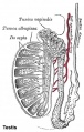
Historic testis drawing

Immature (child) seminiferous tubule showing no spermatozoa

Seminiferous tubule cross-section and supporting cells
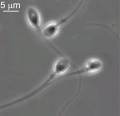
Human spermatozoa


Human Spermatozoa Development
- Spermatogenesis process of spermatagonia mature into spermatozoa (sperm).
- Continuously throughout life occurs in the seminiferous tubules in the male gonad- testis (plural testes).
- At puberty spermatagonia activate and proliferate (mitosis).
- about 48 days from entering meiosis until morphologically mature spermatozoa
- about 64 days to complete spermatogenesis, depending reproduction time of spermatogonia
- follicle stimulating hormone (FSH) - stimulates the spermatogenic epithelium
- luteinizing-hormone (LH) - stimulates testosterone production by Leydig cells
Spermiogenesis


Mature human spermatozoa
|
| Spermatozoa Development (expand to see terms) | ||
|---|---|---|
|
Note there are additional glossaries associated with genital, spermatozoa, oocyte and renal.
See also: Spermatozoa Terms collapse table
|

- Links: Spermatozoa Development | MBoC - Sperm | MBoC - Highly simplified drawing of a cross-section of a seminiferous tubule in a mammalian testis | MBoC - Cytoplasmic bridges in developing sperm cells and their precursors
Puberty
- In humans at puberty, hormonal and morphological changes occur within the gonad and other systems (secondary sex characteristics).
- Within the testis the immature Sertoli cells cease to proliferate and differentiate.
- Spermatogonium (plural, spermatogonia) proliferate and spermatogenesis begins.
- It takes about 70 days for cells to mature from the diploid spermatogonium to a primary spermatocyte.
- This maturation occurs in waves along the seminiferous tubules.
Ejaculate

- release of spermatozoa and accessory gland secretions from the male genital tract (3.5 ml)
- 200-600 million sperm, by volume less than 10 % spermatozoa
- Accessory Gland secretions - 60 % seminal vesicle, 30 % prostate and 10 % bulbourethral
Male Abnormalities
Clinically abnormality studies relate mainly to infertility and inherited genetic disorders.
- Oligospermia - (Low Sperm Count) less than 20 million sperm after 72 hour abstinence from sex
- Azoospermia - (Absent Sperm) blockage of duct network
- Immotile Cilia Syndrome - lack of sperm motility
For spermatozoa genetic abnormalities see of Human Sperm.
Differences in Mammalian Meioses
| Female Oogenesis | Male Spermatogenesis | |
| Meiosis initiated | once in a finite population of cells | continuously in mitotically dividing stem cell population |
| Gametes produced | 1 / meiosis | 4 / meiosis |
| Meiosis completed | delayed for months or years | completed in days or weeks |
| Meiosis Arrest | arrest at 1st meiotic prophase | no arrest differentiation proceed continuously |
| Chromosome Equivalence | All chromosomes exhibit equivalent transcription and recombination during meiotic prophase | Sex chromosomes excluded from recombination and transcription during first meiotic prophase |
| Gamete Differentiation | occurs while diploid (in first meiotic prophase) | occurs while haploid (after meiosis ends) |
Gametogenesis Interactive Component
| Attempt the Quiz - Gametogenesis |
|---|

Here are a few simple Quiz questions that relate to Gametogenesis from the lecture and practical. See your Quiz Result - Answer all the questions, then click "submit" to complete. The page will reload and you can then reopen this table to see your result and feedback.
|
Additional Information
| Additional Information - Content shown under this heading is not part of the material covered in this class. It is provided for those students who would like to know about some concepts or current research in topics related to the current class page. |

- Male Histology - covered in another practical class. From today's class you should have a basic understanding of seminiferous tubule structure in relation to spermatozoa development. (More? spermatozoa, spermatogenesis review[3])
- genetics - covered elsewhere in your course.
- mitosis and meiosis - Cell Biology of mitosis and meiosis also covered in Foundations.
Martin RH. Cytogenetics of Human Sperm. In: Madame Curie Bioscience Database [Internet]. Austin (TX): Landes Bioscience; 2000-2013. Available from: https://www.ncbi.nlm.nih.gov/books/NBK6064/
Cooper TG, Noonan E, von Eckardstein S, Auger J, Baker HW, Behre HM, Haugen TB, Kruger T, Wang C, Mbizvo MT & Vogelsong KM. (2010). World Health Organization reference values for human semen characteristics. Hum. Reprod. Update , 16, 231-45. PMID: 19934213 DOI.
Mitosis and Meiosis

| Chromosome connections in meiosis[4] | |
|---|---|

|
Kinetochores attach homologous chromosomes to opposite halves of the spindle.
Homologs are held together by chiasmata, in which recombinant chromatids cross each other. Sisters are held together by cohesins and possibly by catenation of centromeric DNA threads, which have been observed in human mitosis. Cohesion is released in two steps:
|
Aneuploidy and Fertility
| Homologous Recombination meiosis - In the two sexes genetic recombination between homologous chromosomes proceeds analogously through most stages.
Male
|

Meiotic chromosome crossovers |
| Human Spermatozoa Statistics | ||||||||||||||||||||||||||||
|---|---|---|---|---|---|---|---|---|---|---|---|---|---|---|---|---|---|---|---|---|---|---|---|---|---|---|---|---|
| ||||||||||||||||||||||||||||
Genetics
Where genes are located and how they are inherited relate to the chromosome that the gene is located upon, the parental origin of the gene and the dominant/recessive nature of the gene.
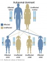
Autosomal dominant inheritance
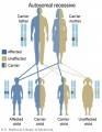
Autosomal recessive inheritance
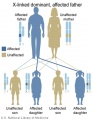
X-Linked dominant (affected father)
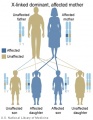
X-Linked dominant (affected mother)
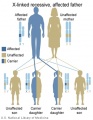
X-Linked recessive (affected father)
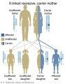
X-Linked recessive (carrier mother)
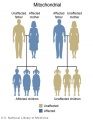
Mitochondrial genome inheritance
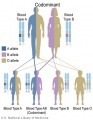
Codominant inheritance


.jpg)
.jpg)
.jpg)
.jpg)


- Inheritance Pattern images: Genetic Abnormalities | autosomal dominant | autosomal recessive | X-linked dominant (affected father) | X-Linked dominant (affected mother) | X-Linked recessive (affected father) | X-Linked recessive (carrier mother) | mitochondrial inheritance | Codominant inheritance | Genogram symbols | Genetics
Terms
- autosomal inheritance - some hereditary diseases are described as autosomal which means that the disease is due to a DNA error in one of the 22 pairs that are not sex chromosomes. Both boys and girls can then inherit this error. If the error is in a sex chromosome, the inheritance is said to be sex-linked.
- cascade testing - Clinical genetic term for the testing of genetic relatives for a mutation that has been identified in the first affected family member. Abnormal Development - Genetic
- gene - a sequence of DNA that encodes an individual protein.
- genome - the complete genetic information in the form of DNA available to a specific species.
- Johnsen score - a clinical score (1-10) for assessing spermatogenesis in a human testicular biopsy. Named after the author of the original article. PMID 5527187
- sperm - (spermatozoa) The male haploid reproductive cell, often used generically (and incorrectly) to describe these cells and the fluid of the ejaculate. Term is a shortened form of scientifically correct term spermatozoa.
- sperm annulus - (Jensen's ring; Latin, annulus = ring) A region of the mammalian sperm flagellum connecting the midpiece and the principal piece. The annulus is a septin-based structure formed from SEPT1, 4, 6, 7 and 12. Septins are polymerizing GTPases that can act as a scaffold forming hetero-oligomeric filaments required for cytokinesis and other cell cycle roles.
- spermatid - Intermediate cell in spermatozoa development, within the testis seminiferous tubule they lie in the luminal cell layer to the secondary spermatocyte. These small cells are haploid and in spermiogenesis change their cellular structure and shape to form spermatozoa. (More? spermatozoa | testis | fertilization | Lecture - Fertilization)
- spermatogenesis - (Greek, genesis = origin, creation, generation) The term used to describe the process of diploid spermatagonia division and differentiation to form haploid spermatozoa within the testis (male gonad). The process includes the following cellular changes: meiosis, reoorganization of DNA, reduction in DNA content, reorganization of cellular organelles, morphological changes (cell shape). The final process of change in cell shape is also called spermiogenesis.
- spermiogenesis - (Greek, genesis = origin, creation, generation) The maturation process of the already haploid spermatids into the mature spermatozoa shape and organization. This process involves reorganization of cellular organelles (endoplasmic reticulum, Golgi apparatus, mitochondria), cytoskeletal changes (microtubule organization) and morphological changes (cell shape, acrosome and tail formation). The process of maturation of the spermatids into spermatozoa: chromatin condenses, nucleus becomes smaller, the Golgi apparatus is modified to form the acrosome, microtubules are reorganised to form the tail, mitochondria are relocated to the initial segment of the tail and the majority of cell cytoplasm is discarded.
- spermatogonia - These cells form in the embryo from the primordial germ cell and are located in the seminiferous tubule adjacent to the basal membrane. The cells can either divide and separate to renew the stem cell population, or they divide and stay together as a pair (Apr spermatogonia) connected by an intercellular cytoplasmic bridge to begin to differentiate and eventually form spermatozoa.
- spermatozoa - (spermatozoon, singular term) The male haploid gamete cell produced by meiosis in the testis (male gonad) seminiferous tubule. In humans, produced from puberty onwards and develop from the diploid stem cell the spermatogonia. The developmental meiosis is called spermatogenesis and the final morphologiccal (shape) change is called spermiogenesis. The mature human spermatozoon formed from the spermatid has a head, neck and tail and is about 60 µm long. At ejaculation these cells undergo capacitation are activated and become motile.
- spermatozoa head - Following spermiogenesis, the first region of the spermatozoa containing the haploid nucleus and acrosome. In humans, it is a flattened structure (5 µm long by 3 µm wide) with the posterior part of nuclear membrane forming the basal plate region. The human spermatozoa is about 60 µm long, actively motile and divided into 3 main regions (head, neck and tail).
- spermatozoa neck - Following spermiogenesis, the second region of the spermatozoa attached to basal plate, transverse oriented centriole, contains nine segmented columns of fibrous material, continue as outer dense fibres in tail. In humans, it forms a short structure (1 µm). The human spermatozoa is about 60 µm long, actively motile and divided into 3 main regions (head, neck and tail).
- spermatozoa tail - Following spermiogenesis, the third region of the spermatozoa that has a (head, neck and tail). The tail is also divided into 3 structural regions a middle piece, a principal piece and an end piece. In humans: the middle piece (5 µm long) is formed by axonema and dense fibres surrounded by mitochondria; the principal piece (45 µm long) fibrous sheath interconnected by regularly spaced circumferential hoops; the final end piece (5 µm long) has an axonema surrounded by small amount of cytoplasm and plasma membrane.
- spermatogonial stem cells - (SSCs) The spermatagonia cells located beside the seminiferous tubule basal membrane that either divide and separate to renew the stem cell population, or they divide and stay together as a pair (Apr spermatogonia) connected by an intercellular cytoplasmic bridge to differentiate and eventually form spermatozoa}}.
- sperm protein 56 - A component of the spermatozoa acrosomal matrix released to the sperm surface during capacitation.
| Spermatozoa Development (expand to see terms) | ||
|---|---|---|
|
Note there are additional glossaries associated with genital, spermatozoa, oocyte and renal.
See also: Spermatozoa Terms collapse table
|
| Cell Division Terms (expand to view) | ||
|---|---|---|
meiosis | mitosis
| ||
|
| Oocyte Development (expand to see terms) | ||
|---|---|---|
|
Note there are additional specific term glossaries available listed at bottom of this table.
|
Glossary Links
- Glossary: A | B | C | D | E | F | G | H | I | J | K | L | M | N | O | P | Q | R | S | T | U | V | W | X | Y | Z | Numbers | Symbols | Term Link
References
- ↑ Yi K, Rubinstein B, Unruh JR, Guo F, Slaughter BD & Li R. (2013). Sequential actin-based pushing forces drive meiosis I chromosome migration and symmetry breaking in oocytes. J. Cell Biol. , 200, 567-76. PMID: 23439682 DOI.
- ↑ Nagaoka SI, Hassold TJ & Hunt PA. (2012). Human aneuploidy: mechanisms and new insights into an age-old problem. Nat. Rev. Genet. , 13, 493-504. PMID: 22705668 DOI.
- ↑ Griswold MD. (2016). Spermatogenesis: The Commitment to Meiosis. Physiol. Rev. , 96, 1-17. PMID: 26537427 DOI.
- ↑ Talbert PB & Henikoff S. (2010). Centromeres convert but don't cross. PLoS Biol. , 8, e1000326. PMID: 20231873 DOI.
- ↑ Wang S, Hassold T, Hunt P, White MA, Zickler D, Kleckner N & Zhang L. (2017). Inefficient Crossover Maturation Underlies Elevated Aneuploidy in Human Female Meiosis. Cell , 168, 977-989.e17. PMID: 28262352 DOI.
- ↑ Greaney J, Wei Z & Homer H. (2017). Regulation of chromosome segregation in oocytes and the cellular basis for female meiotic errors. Hum. Reprod. Update , , . PMID: 29244163 DOI.
- ↑ Ren H, Ferguson K, Kirkpatrick G, Vinning T, Chow V & Ma S. (2016). Altered Crossover Distribution and Frequency in Spermatocytes of Infertile Men with Azoospermia. PLoS ONE , 11, e0156817. PMID: 27273078 DOI.
- ↑ Amann RP & Howards SS. (1980). Daily spermatozoal production and epididymal spermatozoal reserves of the human male. J. Urol. , 124, 211-5. PMID: 6772801
- ↑ Cooper TG, Noonan E, von Eckardstein S, Auger J, Baker HW, Behre HM, Haugen TB, Kruger T, Wang C, Mbizvo MT & Vogelsong KM. (2010). World Health Organization reference values for human semen characteristics. Hum. Reprod. Update , 16, 231-45. PMID: 19934213 DOI.
BGDA: Lecture 1 | Lecture 2 | Practical 3 | Practical 6 | Practical 12 | Lecture Neural | Practical 14 | Histology Support - Female | Male | Tutorial
Glossary Links
- Glossary: A | B | C | D | E | F | G | H | I | J | K | L | M | N | O | P | Q | R | S | T | U | V | W | X | Y | Z | Numbers | Symbols | Term Link
Cite this page: Hill, M.A. (2024, June 10) Embryology BGDA Practical 3 - Gametogenesis. Retrieved from https://embryology.med.unsw.edu.au/embryology/index.php/BGDA_Practical_3_-_Gametogenesis
- © Dr Mark Hill 2024, UNSW Embryology ISBN: 978 0 7334 2609 4 - UNSW CRICOS Provider Code No. 00098G