File:Stage19 em01.jpg: Difference between revisions
mNo edit summary |
|||
| (5 intermediate revisions by the same user not shown) | |||
| Line 1: | Line 1: | ||
==Human Embryo Carnegie Stage | ==Human Embryo Carnegie Stage 19== | ||
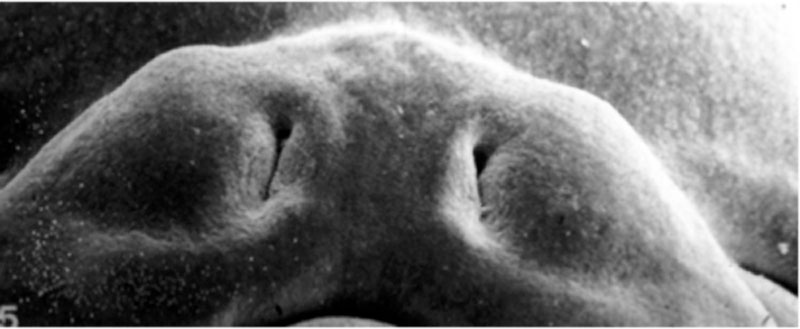
Scanning election micrograph (SEM). View: Ventral view head. Amniotic membrane removed. ([[Week 7]], [[Carnegie stage 19]], 48 - 51 days, CRL 16 - 18 mm. ([[:File:Stage19 em11.jpg|see labeled image]]) | |||
View: Ventral view head. Amniotic membrane removed. | |||
This electron micrograph shows a detailed view of the maxilla and upper lip development during embryonic week 7 ({{GA}} 9). This is the last in a series (stage 16 to 19) of EM images focussing on this developmentally important region and time for embryonic cleft lip and palate. | This electron micrograph shows a detailed view of the maxilla and upper lip development during embryonic week 7 ({{GA}} 9). This is the last in a series (stage 16 to 19) of EM images focussing on this developmentally important region and time for embryonic cleft lip and palate. | ||
Note: | |||
* completely fused upper lip, with indentation still present at philtrum. | |||
* narrowing nasal openings | |||
{{Lip EM images 16-19}} | {{Lip EM images 16-19}} | ||
Latest revision as of 08:17, 23 February 2014
Human Embryo Carnegie Stage 19
Scanning election micrograph (SEM). View: Ventral view head. Amniotic membrane removed. (Week 7, Carnegie stage 19, 48 - 51 days, CRL 16 - 18 mm. (see labeled image)
This electron micrograph shows a detailed view of the maxilla and upper lip development during embryonic week 7 (GA 9). This is the last in a series (stage 16 to 19) of EM images focussing on this developmentally important region and time for embryonic cleft lip and palate.
Note:
- completely fused upper lip, with indentation still present at philtrum.
- narrowing nasal openings
- EM Links: Image - stage 16 | Labeled Image - stage 16 | Image - stage 17 | labeled Image - stage 17 | Image - stage 18 | Labeled Image - stage 18 | Image - stage 19 | Labeled Image - stage 19 | Palate Development
SEM Image Source: Prof Virginia Diewert
- Carnegie Stages: 1 | 2 | 3 | 4 | 5 | 6 | 7 | 8 | 9 | 10 | 11 | 12 | 13 | 14 | 15 | 16 | 17 | 18 | 19 | 20 | 21 | 22 | 23 | About Stages | Timeline
Cite this page: Hill, M.A. (2024, June 26) Embryology Stage19 em01.jpg. Retrieved from https://embryology.med.unsw.edu.au/embryology/index.php/File:Stage19_em01.jpg
- © Dr Mark Hill 2024, UNSW Embryology ISBN: 978 0 7334 2609 4 - UNSW CRICOS Provider Code No. 00098G
File history
Yi efo/eka'e gwa ebo wo le nyangagi wuncin ye kamina wunga tinya nan
| Gwalagizhi | Nyangagi | Dimensions | User | Comment | |
|---|---|---|---|---|---|
| current | 16:19, 15 May 2013 | 800 × 329 (37 KB) | Z8600021 (talk | contribs) | ==Human Embryo Carnegie Stage 18== View: Ventral view head. Amniotic membrane removed. {{Template:Carnegie_stages}} {{Virginia Diewert images}} {{Template:Footer}} Category:Carnegie Stage 18 Category:Week 6 |
You cannot overwrite this file.
File usage
The following 9 pages use this file: