Hearing - Outer Ear Development: Difference between revisions
mNo edit summary |
|||
| (31 intermediate revisions by the same user not shown) | |||
| Line 1: | Line 1: | ||
{{Header}} | |||
==Introduction== | ==Introduction== | ||
[[File:Adult_hearing_embryonic_origins.jpg|thumb|400px|Adult hearing embryonic origins.]] | [[File:Adult_hearing_embryonic_origins.jpg|thumb|400px|Adult hearing embryonic origins.]] | ||
[[File: | [[File:Streeter1922-fig02.jpg|thumb|300px|Adult external ear]] | ||
The external ear is derived from 6 surface hillocks (auricular hillocks), three on each of pharyngeal arch 1 and 2. | The {{outer ear}} or external ear is derived from 6 surface hillocks (auricular hillocks), three on each of pharyngeal arch 1 and 2. | ||
The external auditory meatus is derived from the 1st pharyngeal cleft. | The external auditory meatus is derived from the 1st pharyngeal cleft. | ||
The postnatal human outer ear structure also selectively boosts frequencies around 3 kHz, by a sound pressure level of 30 to 100-fold, that correspond to frequencies associated with speech. The anatomical position, on either side of the head, also allows exquisite localization of sounds in space by neural comparison of signals reaching each ear. | |||
{{Hearing Links}} | |||
== Some Recent Findings == | == Some Recent Findings == | ||
| Line 15: | Line 18: | ||
|-bgcolor="F5FAFF" | |-bgcolor="F5FAFF" | ||
| | | | ||
* '''Movement of the external ear in human embryo''' | * '''Movement of the external ear in human embryo'''{{#pmid:22296782|PMID22296782}} "In all, 171 samples between Carnegie stage (CS) 17 and CS 23 were selected from MR image datasets of human embryos obtained from the Kyoto Collection of Human Embryos. The three-dimensional absolute position of 13 representative anatomical landmarks, including external and internal ears, from MRI data was traced to evaluate the movement between the different stages with identical magnification. Two different sets of reference axes were selected for evaluation and comparison of the movements. ...The results indicate that movement of all anatomical landmarks, including external and internal ears, can be explained by differential growth. Also, when the external ear is recognized as one of the facial landmarks and having a relative position to other landmarks such as the eyes and mouth, the external ears seem to move cranially." | ||
* '''Age- and sex-related changes in the normal human ear''' | |||
* '''Age- and sex-related changes in the normal human ear'''{{#pmid:19356871|PMID19356871}} "All ear dimensions were significantly larger in men than in women (p<0.001). A significant effect of age was found (p<0.001), with larger values in older individuals. The ear width-to-length ratio and the sagittal angle of the auricle significantly decreased as a function of age (p<0.001) but without sex-related differences. On average, the three-dimensional position of ears was symmetric, with symmetry coefficients ranging between 92% and 96%. Asymmetry was found in the sagittal angle of the auricle (both sexes), in the ear width-to-length ratio and ear width (men only)." | |||
|} | |} | ||
{| class="wikitable mw-collapsible mw-collapsed" | |||
! More recent papers | |||
|- | |||
| [[File:Mark_Hill.jpg|90px|left]] {{Most_Recent_Refs}} | |||
Search term: [http://www.ncbi.nlm.nih.gov/pubmed/?term=Outer+Ear+Development ''Outer Ear Development''] | |||
<pubmed limit=5>Outer Ear Development</pubmed> | |||
|} | |||
==Pinna- Auricle== | ==Pinna- Auricle== | ||
| Line 30: | Line 43: | ||
* develops from six aural hillocks: 3 on first pharyngeal arch and 3 on the second pharyngeal arch. | * develops from six aural hillocks: 3 on first pharyngeal arch and 3 on the second pharyngeal arch. | ||
* originally on neck, moves cranially during mandible development | * originally on neck, moves cranially during mandible development | ||
<gallery mode="packed-hover"> | |||
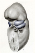
File:Streeter1922-fig09.jpg|Embryo 6mm | |||
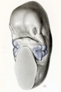
File:Streeter1922-fig10.jpg|Embryo 12 mm | |||
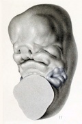
File:Streeter1922-fig11.jpg|Embryo 14mm | |||
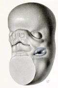
File:Streeter1922-fig12.jpg|Embryo 18mm | |||
</gallery> | |||
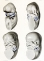
[[File:Human embryo head week 6 to 8.jpg|400px]] | [[File:Human embryo head week 6 to 8.jpg|400px]] | ||
'''Movement of the external ear in human embryo''' (week 6 to 8) | '''Movement of the external ear in human embryo''' (week 6 to 8){{#pmid:22296782|PMID22296782}} | ||
===Pharyngeal Contributions=== | ===Pharyngeal Contributions=== | ||
[[File:External ear anatomy.jpg|thumb|External ear anatomy]] | [[File:External ear anatomy.jpg|thumb|External ear simplified anatomy]] | ||
{| class="prettytable" | {| class="prettytable" | ||
|-bgcolor="CEDFF2" | |-bgcolor="CEDFF2" | ||
| Line 80: | Line 100: | ||
* ectodermal diverticulum | * ectodermal diverticulum | ||
* week 5 - extends inwards to pharynx | * week 5 - extends inwards to pharynx | ||
* until week 18 has ectodermal plug - plug forms stratified squamous epithelia of canal and outer eardrum | * until week 18 has ectodermal plug - plug forms stratified squamous epithelia of canal and outer eardrum | ||
===Human Timeline=== | ===Human Timeline=== | ||
| Line 105: | Line 125: | ||
|} | |} | ||
Based on data from | Based on data from{{#pmid:1441991|PMID1441991}} | ||
==Auricular Cartilage== | ==Auricular Cartilage== | ||
{| | {| | ||
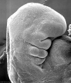
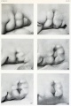
| [[File:Streeter1922-06-07.jpg|500px]] | | [[File:Streeter1922-06-07.jpg|500px]] | ||
| | | Image shows the embryonic and fetal growth of the auricular cartilage within the pinna.<ref name=Streeter1922>{{Ref-Streeter1922}}</ref> | ||
'''Fig. 6.''' Lateral views of left auricular cartilage, taken from reconstructions of human embryos of the Carnegie Collection: No. 460 (21 mm.), No. 417 (32 mm.), No. 886 (43 mm.). X14. | '''Fig. 6.''' Lateral views of left auricular cartilage, taken from reconstructions of human embryos of the Carnegie Collection: No. 460 (21 mm.), No. 417 (32 mm.), No. 886 (43 mm.). X14. | ||
| Line 131: | Line 154: | ||
==External Auditory Meatus== | ==External Auditory Meatus== | ||
{| | |||
|+ '''External Auditory Meatus''' | |||
|-bgcolor="CEDFF2" | |||
! At Birth | |||
! Adult | |||
|- | |||
| [[File:Keith1902 fig036b.jpg|400px]] | |||
| [[File:Keith1902 fig036a.jpg|400px]] | |||
|} | |||
Development of the human external auditory meatus (EAM) begins in the late embryo and continues through the fetal second trimester. The period the "metal plug" is present has been variously described. The best EAM developmental time course is described in two studies.{{#pmid:2756906|PMID2756906}}{{#pmid:1441991|PMID1441991}} | |||
{{External Auditory Meatus Timeline table}} | |||
'''Fetal epithelium'''{{#pmid:2756906|PMID2756906}} | |||
# originates as a tube derived from the epithelium of the fundus of the primary external canal | |||
# composed of a thin, flat epithelium on the medial side | |||
# continuous with a thicker epithelium on the lateral side | |||
# then merges with the external epithelium of the primary external canal | |||
Epithelium cornification begins in the second trimester, at week 16 ({{GA}} week 18), and is followed by clearing of keratinous debris to the exterior. | |||
The adult stratified squamous epithelium lines the external auditory meatus and covers tympanic membrane. | |||
==Innervation== | ==Innervation== | ||
| Line 142: | Line 186: | ||
==Postnatal Growth== | ==Postnatal Growth== | ||
Postnatally, human ears continue to grow throughout the entire lifetime and have a sexually dimorphic pattern, described in a large study. | Postnatally, human ears continue to grow throughout the entire lifetime and have a sexually dimorphic pattern, described in a large study.{{#pmid:18196763|PMID18196763}} Three anatomical features of the ear were found to not grow at all after birth; Concha auriculae width, Incisura intertragica width, and the helical brim diameter of the auricle. | ||
* birth - external ear bigger than the large head in proportion to the body | * birth - external ear bigger than the large head in proportion to the body | ||
| Line 149: | Line 193: | ||
{| | {| | ||
|+ '''Ear Length''' (mm +/-SD) Data | |+ '''Ear Length''' (mm +/-SD) Data{{#pmid:18196763|PMID18196763}} | ||
|-bgcolor="CEDFF2" | |-bgcolor="CEDFF2" | ||
| width=120px|'''Age''' | | width=120px|'''Age''' | ||
| Line 172: | Line 216: | ||
* '''Darwin's tubercle''' - (Woolnerian tip) is a tubercle is seen along the upper, posterior portion of the helix (upper and middle thirds). | * '''Darwin's tubercle''' - (Woolnerian tip) is a tubercle is seen along the upper, posterior portion of the helix (upper and middle thirds). | ||
* "railroad track" - associated with fatal alcohol syndrome, the curve at top part of outer ear is underdeveloped and folded over parallel to curve beneath. | * "railroad track" - associated with fatal alcohol syndrome, the curve at top part of outer ear is underdeveloped and folded over parallel to curve beneath. | ||
==Lobe Attachment== | |||
In the normal population, free earlobes have been described as dominant.{{#pmid:14277139|PMID14277139}} With some researchers suggesting that "attached" would be better described as "lobeless". There have been several historic studies identifying attached ear lobes in some population groups (Indian{{#pmid:17585565|PMID17585565}}, Malaysian). There are a number of syndromes and genetic disorders associated with variation in lobe attachment. | |||
:'''Links:''' [http://omim.org/entry/128900 OMIM 128900] | PMID 14277139 | |||
==Molecular== | ==Molecular== | ||
| Line 178: | Line 228: | ||
* controlled by genes that regulate arch 1 and 2 development | * controlled by genes that regulate arch 1 and 2 development | ||
* related to hindbrain segmentation (rhombomere 4) | * related to hindbrain segmentation (rhombomere 4) | ||
* Mouse - Hox a1/ | * Mouse - {{Hox}}a1/{{Hox}}b1, goosecoid, Endothelin1, dHAND | ||
==Abnormalities== | ==Abnormalities== | ||
[[File:FASface.jpg|thumb|Facial appearance of fetal alcohol syndrome. Ear curve at top part of outer ear is underdeveloped and folded over parallel to curve beneath and gives the appearance of a "railroad track"]] | [[File:FASface.jpg|thumb|Facial appearance of fetal alcohol syndrome. Ear curve at top part of outer ear is underdeveloped and folded over parallel to curve beneath and gives the appearance of a "railroad track"]] | ||
There are a range of external ear abnormalities relate to final structure, size and position. In some cases these abnormalities relate directly to pharyngeal arch development or may be part of a wider spectrum of abnormalities associated with a genetic or environmental (fetal alcohol syndrome) disorders. Some known abnormalities include: anotia, microtia, prominent ear, lop ear, cup ear, cryptotia and Stahl's ear. Other associated external ear abnormalities include the formation of the external auditory meatus (canal) and pre-auricular fistulae (pits) and appendages. Finally, a range of abnormalities can be found associated with the overlying skin of both the external ear and the ear canal. | There are a range of external ear abnormalities relate to final structure, size and position. In some cases these abnormalities relate directly to pharyngeal arch development or may be part of a wider spectrum of abnormalities associated with a genetic or environmental (fetal alcohol syndrome) disorders. Some known abnormalities include: anotia, microtia, prominent ear, lop ear, cup ear, cryptotia and Stahl's ear. Other associated external ear abnormalities include the formation of the external auditory meatus (canal) and pre-auricular fistulae (pits) and appendages. Finally, a range of abnormalities can be found associated with the overlying skin of both the external ear and the ear canal.{#pmid:18261212|PMID18261212}} | ||
Minor structural anomalies have been shown to be corrected by appropriate splinting in the early neonatal period. | Minor structural anomalies have been shown to be corrected by appropriate splinting in the early neonatal period.{#pmid:18490209|PMID18490209}} | ||
:'''Links:''' [[Sensory - Hearing Abnormalities]] | :'''Links:''' [[Sensory - Hearing Abnormalities]] | ||
| Line 198: | Line 248: | ||
[[File:Microtia.jpg|300px]] | [[File:Microtia.jpg|300px]] | ||
'''Microtia (autosomal-recessive)''' - A mutation in HOXA2 | '''Microtia (autosomal-recessive)''' - A mutation has been identified in {{Hox}}A2 (https://www.omim.org/entry/604685 HOXA2] {{Chr7}}p15.2){#pmid:18394579|PMID18394579}} | ||
:Links: {{Hox}} | |||
===Cleft Lobule=== | ===Cleft Lobule=== | ||
'''Oculo-auricular syndrome''' - A mutation in the NKX5-3 | '''Oculo-auricular syndrome''' - A mutation in the NKX5-3 ([https://www.omim.org/entry/613380 HMX3] {{Chr10}}q26.13) human homeobox gene.{#pmid:18423520|PMID18423520}} | ||
===Stahl's Ear=== | ===Stahl's Ear=== | ||
| Line 211: | Line 264: | ||
The external auditory meatus (canal) can also fail to canalise leading to a range of malformation including membranous and/or bony atresia and stenosis. | The external auditory meatus (canal) can also fail to canalise leading to a range of malformation including membranous and/or bony atresia and stenosis. | ||
'''External Auditory Meatus Stenosis''' | '''External Auditory Meatus Stenosis'''{{#pmid:18054456|PMID18054456}} | ||
* Type A - a marked narrowing of the canal with an intact skin layer. | * Type A - a marked narrowing of the canal with an intact skin layer. | ||
* Type B - a partial development of the canal with an atresia plate at the medial part. | * Type B - a partial development of the canal with an atresia plate at the medial part. | ||
| Line 233: | Line 286: | ||
:'''Links:''' [[Sensory - Hearing Abnormalities]] | :'''Links:''' [[Sensory - Hearing Abnormalities]] | ||
==Additional Images== | |||
===Historical Images=== | |||
<gallery> | |||
File:Keith1902 fig035.jpg|Fig. 35. Diagrammatic Section through the Cephalic region of an embryo, showing the origin of the Auditory System. | |||
File:Keith1902 fig036a.jpg|Fig. 36 A. Section of the External Auditory Meatus of the Adult. | |||
File:Keith1902 fig036b.jpg|Fig. 36 B. A Section of the External Auditory Meatus at Birth. (After Symington.) | |||
File:Keith1902 fig037.jpg|Fig. 37. Showing the Tubercles which arise round the First Visceral Cleft to form the External Ear. | |||
File:Keith1902 fig038.jpg|Fig. 38. Showing the part of the Adult Ear formed by each Tubercle. | |||
File:Streeter1922-fig12.jpg|(1922) Embryo 18 mm reconstruction model, Carnegie Collection No. 1390 | |||
</gallery> | |||
== References == | == References == | ||
<references/> | <references/> | ||
| Line 238: | Line 302: | ||
===Reviews=== | ===Reviews=== | ||
{{#pmid:26227955}} | |||
{{#pmid:19293168}} | |||
{{#pmid:18976115}} | |||
{{#pmid:17104502}} | |||
===Articles=== | ===Articles=== | ||
{{#pmid:2756906}} | |||
{{#pmid:19356871}} | |||
{{#pmid:1441991}} | |||
===Search PubMed=== | ===Search PubMed=== | ||
| Line 252: | Line 322: | ||
'''Search Pubmed:''' [http://www.ncbi.nlm.nih.gov/sites/entrez?db=pubmed&cmd=search&term=Outer+Ear+Development Outer Ear Development] | [http://www.ncbi.nlm.nih.gov/sites/entrez?db=pubmed&cmd=search&term=pinna+Development Pinna Development] | '''Search Pubmed:''' [http://www.ncbi.nlm.nih.gov/sites/entrez?db=pubmed&cmd=search&term=Outer+Ear+Development Outer Ear Development] | [http://www.ncbi.nlm.nih.gov/sites/entrez?db=pubmed&cmd=search&term=pinna+Development Pinna Development] | ||
==Hearing Terms== | |||
{{Hearing terms}} | |||
==External Links== | ==External Links== | ||
| Line 258: | Line 333: | ||
* Neuroscience [http://www.ncbi.nlm.nih.gov/books/bv.fcgi?rid=neurosci.section.891 Neuroscience - The External Ear] | * Neuroscience [http://www.ncbi.nlm.nih.gov/books/bv.fcgi?rid=neurosci.section.891 Neuroscience - The External Ear] | ||
{{ | {{Glossary}} | ||
[[Category:Senses]] [[Category:Hearing Loss]] | |||
{{Footer}} | |||
[[Category:Senses]] [[Category:Hearing Loss]] [[Category:Outer Ear]] | |||
Latest revision as of 12:52, 14 May 2018
| Embryology - 26 Jun 2024 |
|---|
| Google Translate - select your language from the list shown below (this will open a new external page) |
|
العربية | català | 中文 | 中國傳統的 | français | Deutsche | עִברִית | हिंदी | bahasa Indonesia | italiano | 日本語 | 한국어 | မြန်မာ | Pilipino | Polskie | português | ਪੰਜਾਬੀ ਦੇ | Română | русский | Español | Swahili | Svensk | ไทย | Türkçe | اردو | ייִדיש | Tiếng Việt These external translations are automated and may not be accurate. (More? About Translations) |
Introduction


The outer ear or external ear is derived from 6 surface hillocks (auricular hillocks), three on each of pharyngeal arch 1 and 2.
The external auditory meatus is derived from the 1st pharyngeal cleft.
The postnatal human outer ear structure also selectively boosts frequencies around 3 kHz, by a sound pressure level of 30 to 100-fold, that correspond to frequencies associated with speech. The anatomical position, on either side of the head, also allows exquisite localization of sounds in space by neural comparison of signals reaching each ear.
Some Recent Findings
|
| More recent papers |
|---|
 This table allows an automated computer search of the external PubMed database using the listed "Search term" text link.
More? References | Discussion Page | Journal Searches | 2019 References | 2020 References Search term: Outer Ear Development <pubmed limit=5>Outer Ear Development</pubmed> |
Pinna- Auricle

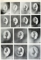
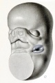
Embryonic External Ear
Images of the lateral view of the human embryonic head from week 5 (stage 14) through to week 8 (stage 23) showing development of the auricular hillocks that will form the external ear. The adult ear is also shown indicating the part of the ear that each hillock contributes.
- develops from six aural hillocks: 3 on first pharyngeal arch and 3 on the second pharyngeal arch.
- originally on neck, moves cranially during mandible development
Embryo 6mm
Embryo 12 mm
Embryo 14mm
Embryo 18mm


Movement of the external ear in human embryo (week 6 to 8)[1]
Pharyngeal Contributions

| Pharyngeal Arch | Hillock | Auricle Component |
| Arch 1 | 1 | tragus |
| 2 | helix | |
| 3 | cymba concha | |
| Arch 2 | 4 | concha |
| 5 | antihelix | |
| 6 | antitragus |
- Outer- external auditory meatus
- derived from first pharyngeal cleft
- ectodermal diverticulum
- week 5 - extends inwards to pharynx
- until week 18 has ectodermal plug - plug forms stratified squamous epithelia of canal and outer eardrum
Human Timeline
| Time | EAM Appearance |
| Embryonic period | Ectodermal cells proliferate and fill the entire lumen forming a meatal plug |
| 10 weeks | Meatal plug extends in a disc-like fashion. In the horizontal plane the meatus is boot-shaped with a narrow neck and the sole of the meatal plug spreading widely to form the future tympanic membrane medially. Proximal portion of the neck starts to be resorbed. |
| 13 weeks | Disc-like plug innermost surface in contact with the primordial malleus, contributes to the formation of the tympanic membrane. |
| 16.5 week | Meatus is fully patent throughout its length, lumen is still narrow and curved. |
| 18 week | Meatus is already fully expanded to its complete form. |
Based on data from[3]
Auricular Cartilage

|
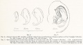
Image shows the embryonic and fetal growth of the auricular cartilage within the pinna.[4]
Fig. 6. Lateral views of left auricular cartilage, taken from reconstructions of human embryos of the Carnegie Collection: No. 460 (21 mm.), No. 417 (32 mm.), No. 886 (43 mm.). X14.
|
Human Auricle Development

Stage 12 - otic placode
Stage 13 - first and second pharyngeal arches, otic vesicle
Ventrolateral view head of human embryos
Region first cleft
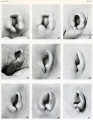
Disappearance of hillocks
Month 3 - Fetus
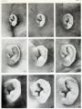
Month 4 - Fetus
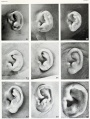
Month 5 - Fetus
Embryo ear cartilage 21 - 50 mmm CRL


External Auditory Meatus
| At Birth | Adult |
|---|---|

|

|
Development of the human external auditory meatus (EAM) begins in the late embryo and continues through the fetal second trimester. The period the "metal plug" is present has been variously described. The best EAM developmental time course is described in two studies.[5][3]
| Period | Week | Description |
| Embryo | week 8 | Funnel-shaped tube continues medially into mesenchymal tissue, forms a curved path. |
| Fetus (first trimester) | week 9 | Ectodermal cells proliferate, fill the meatus lumen and form the "meatal plug". |
| Fetus (first trimester) | week 10 | Meatal plug bottom extends in a disc-like fashion, so that in the horizontal plane the meatus is boot-shaped with a narrow neck and the sole of the meatal plug spreading widely to form the future tympanic membrane medially. At the same time, the plug in the proximal portion of the neck starts to be resorbed. |
| Fetus (second trimester) | week 13 | Meatal plug disc-like, innermost surface in contact with the primordial malleus, contributes to formation of tympanic membrane. |
| Fetus (second trimester) | week 15 | Meatal plug innermost portion splits, leaving a thin ectodermal cell layer of immature tympanic membrane. The neck of the boot forms the border between the primary and secondary meatus, and is the last part to split. |
| Fetus (second trimester) | week 16.5 | The meatus is fully patent throughout entire length. Lumen is still narrow and curved. Epithelium cornification commences. |
| Fetus (second trimester) | week 18 | The meatus is now fully expanded to its complete form. |
| Links: outer ear | hearing | timeline Reference[3] | ||
Fetal epithelium[5]
- originates as a tube derived from the epithelium of the fundus of the primary external canal
- composed of a thin, flat epithelium on the medial side
- continuous with a thicker epithelium on the lateral side
- then merges with the external epithelium of the primary external canal
Epithelium cornification begins in the second trimester, at week 16 (GA week 18), and is followed by clearing of keratinous debris to the exterior.
The adult stratified squamous epithelium lines the external auditory meatus and covers tympanic membrane.
Innervation
The auriculotemporal nerve supplies a large part of the pinna, some innervation may also arise from the trigeminus.
Postnatal Growth
Postnatally, human ears continue to grow throughout the entire lifetime and have a sexually dimorphic pattern, described in a large study.[6] Three anatomical features of the ear were found to not grow at all after birth; Concha auriculae width, Incisura intertragica width, and the helical brim diameter of the auricle.
- birth - external ear bigger than the large head in proportion to the body
- childhood - large yearly increases decrease by 8 or 10 years of age.
- adult - male increases in all parameters were greater than for female ears.
| Age | Female | Male |
| Birth | 52 (4.3) | 52 (4.1) |
| 20 yrs | 61 (3.9) | 65 (4.0) |
| Older than 70 yrs | 72 (4.6) | 78 (4.8) |
Ear Features
- Darwin's tubercle - (Woolnerian tip) is a tubercle is seen along the upper, posterior portion of the helix (upper and middle thirds).
- "railroad track" - associated with fatal alcohol syndrome, the curve at top part of outer ear is underdeveloped and folded over parallel to curve beneath.
Lobe Attachment
In the normal population, free earlobes have been described as dominant.[7] With some researchers suggesting that "attached" would be better described as "lobeless". There have been several historic studies identifying attached ear lobes in some population groups (Indian[8], Malaysian). There are a number of syndromes and genetic disorders associated with variation in lobe attachment.
- Links: OMIM 128900 | PMID 14277139
Molecular
Outer Ear Genes
- controlled by genes that regulate arch 1 and 2 development
- related to hindbrain segmentation (rhombomere 4)
- Mouse - Hoxa1/Hoxb1, goosecoid, Endothelin1, dHAND
Abnormalities

There are a range of external ear abnormalities relate to final structure, size and position. In some cases these abnormalities relate directly to pharyngeal arch development or may be part of a wider spectrum of abnormalities associated with a genetic or environmental (fetal alcohol syndrome) disorders. Some known abnormalities include: anotia, microtia, prominent ear, lop ear, cup ear, cryptotia and Stahl's ear. Other associated external ear abnormalities include the formation of the external auditory meatus (canal) and pre-auricular fistulae (pits) and appendages. Finally, a range of abnormalities can be found associated with the overlying skin of both the external ear and the ear canal.{#pmid:18261212|PMID18261212}}
Minor structural anomalies have been shown to be corrected by appropriate splinting in the early neonatal period.{#pmid:18490209|PMID18490209}}
Anotia

Upper Auricular Detachment


Microtia

Microtia (autosomal-recessive) - A mutation has been identified in HoxA2 (https://www.omim.org/entry/604685 HOXA2] 7p15.2){#pmid:18394579|PMID18394579}}
- Links: Hox
Cleft Lobule
Oculo-auricular syndrome - A mutation in the NKX5-3 (HMX3 10q26.13) human homeobox gene.{#pmid:18423520|PMID18423520}}
Stahl's Ear
A rare ear abnormality, where the rim of the ear is flattened and the upper portions deformed. More common in Oriental background and can occur from mild to severe. The skin and cartilage are both folded to different degrees that can result in a pointed upper edge. This pointed ear has been said to resemble the Star Trek television character "Vulcan" ear shape.
External Auditory Meatus
The external auditory meatus (canal) can also fail to canalise leading to a range of malformation including membranous and/or bony atresia and stenosis.
External Auditory Meatus Stenosis[9]
- Type A - a marked narrowing of the canal with an intact skin layer.
- Type B - a partial development of the canal with an atresia plate at the medial part.
- Type C - a complete bony canal atresia.
Pre-auricular Fistulae and Appendages


There are also a range of pre-auricular fistulae (pits) and appendages that generally occur in a specific region beside the tragus and crus helicis.

Auricular Pit

Posterior helix pit associated with Beckwith-Wiedemann syndrome.
Additional Images
Historical Images
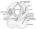
Fig. 35. Diagrammatic Section through the Cephalic region of an embryo, showing the origin of the Auditory System.
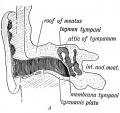
Fig. 36 A. Section of the External Auditory Meatus of the Adult.
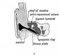
Fig. 36 B. A Section of the External Auditory Meatus at Birth. (After Symington.)
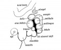
Fig. 37. Showing the Tubercles which arise round the First Visceral Cleft to form the External Ear.
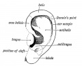
Fig. 38. Showing the part of the Adult Ear formed by each Tubercle.
(1922) Embryo 18 mm reconstruction model, Carnegie Collection No. 1390


References
- ↑ 1.0 1.1 Kagurasho M, Yamada S, Uwabe C, Kose K & Takakuwa T. (2012). Movement of the external ear in human embryo. Head Face Med , 8, 2. PMID: 22296782 DOI.
- ↑ Sforza C, Grandi G, Binelli M, Tommasi DG, Rosati R & Ferrario VF. (2009). Age- and sex-related changes in the normal human ear. Forensic Sci. Int. , 187, 110.e1-7. PMID: 19356871 DOI.
- ↑ 3.0 3.1 3.2 Nishimura Y & Kumoi T. (1992). The embryologic development of the human external auditory meatus. Preliminary report. Acta Otolaryngol. , 112, 496-503. PMID: 1441991
- ↑ Streeter GL. Development of the auricle in the human embryo. (1922) Carnegie Instn. Wash. Publ. 277, Contrib. Embryol., 14: 111-138.
- ↑ 5.0 5.1 Michaels L & Soucek S. (1989). Development of the stratified squamous epithelium of the human tympanic membrane and external canal: the origin of auditory epithelial migration. Am. J. Anat. , 184, 334-44. PMID: 2756906 DOI.
- ↑ 6.0 6.1 Niemitz C, Nibbrig M & Zacher V. (2007). Human ears grow throughout the entire lifetime according to complicated and sexually dimorphic patterns--conclusions from a cross-sectional analysis. Anthropol Anz , 65, 391-413. PMID: 18196763
- ↑ DUTTA P & GANGULY P. (1965). FURTHER OBSERVATIONS ON EAR LOBE ATTACHMENT. Acta Genet Stat Med , 15, 77-86. PMID: 14277139
- ↑ Sharma A, Sidhu NK, Sharma MK, Kapoor K & Singh B. (2007). Morphometric study of ear lobule in northwest Indian male subjects. Anat Sci Int , 82, 98-104. PMID: 17585565 DOI.
- ↑ Kösling S, Omenzetter M & Bartel-Friedrich S. (2009). Congenital malformations of the external and middle ear. Eur J Radiol , 69, 269-79. PMID: 18054456 DOI.
Reviews
Anthwal N & Thompson H. (2016). The development of the mammalian outer and middle ear. J. Anat. , 228, 217-32. PMID: 26227955 DOI.
Alasti F & Van Camp G. (2009). Genetics of microtia and associated syndromes. J. Med. Genet. , 46, 361-9. PMID: 19293168 DOI.
Torban E & Goodyer P. (2009). The kidney and ear: emerging parallel functions. Annu. Rev. Med. , 60, 339-53. PMID: 18976115 DOI.
Wood-Jones F & I-Chuan W. (1934). The Development of the External Ear. J. Anat. , 68, 525-33. PMID: 17104502
Articles
Michaels L & Soucek S. (1989). Development of the stratified squamous epithelium of the human tympanic membrane and external canal: the origin of auditory epithelial migration. Am. J. Anat. , 184, 334-44. PMID: 2756906 DOI.
Sforza C, Grandi G, Binelli M, Tommasi DG, Rosati R & Ferrario VF. (2009). Age- and sex-related changes in the normal human ear. Forensic Sci. Int. , 187, 110.e1-7. PMID: 19356871 DOI.
Nishimura Y & Kumoi T. (1992). The embryologic development of the human external auditory meatus. Preliminary report. Acta Otolaryngol. , 112, 496-503. PMID: 1441991
Search PubMed
May 2010 "Outer Ear Development" All (1478) Review (120) Free Full Text (215)
Search Pubmed: Outer Ear Development | Pinna Development
Hearing Terms
| Hearing Terms | ||
|---|---|---|
Hearing and Balance Development
|
External Links
External Links Notice - The dynamic nature of the internet may mean that some of these listed links may no longer function. If the link no longer works search the web with the link text or name. Links to any external commercial sites are provided for information purposes only and should never be considered an endorsement. UNSW Embryology is provided as an educational resource with no clinical information or commercial affiliation.
- Neuroscience Neuroscience - The External Ear
Glossary Links
- Glossary: A | B | C | D | E | F | G | H | I | J | K | L | M | N | O | P | Q | R | S | T | U | V | W | X | Y | Z | Numbers | Symbols | Term Link
Cite this page: Hill, M.A. (2024, June 26) Embryology Hearing - Outer Ear Development. Retrieved from https://embryology.med.unsw.edu.au/embryology/index.php/Hearing_-_Outer_Ear_Development
- © Dr Mark Hill 2024, UNSW Embryology ISBN: 978 0 7334 2609 4 - UNSW CRICOS Provider Code No. 00098G