Lecture - 2014 Course Introduction: Difference between revisions
mNo edit summary |
mNo edit summary |
||
| Line 58: | Line 58: | ||
! [[Australian Statistics]] | ! [[Australian Statistics]] | ||
|- | |- | ||
| | | colwidth=250px|[[File:Australia_mothers_and_babies_2011.jpg|link=Australia’s_mothers_and_babies_2011|200px]] | ||
| | | colwidth=250px|[[File:Assisted reproductive technology in Australia and New Zealand 2010.jpg|200px]] | ||
|- | |- | ||
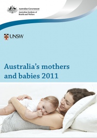
| Australia’s mothers and babies (2011) | | Australia’s mothers and babies (2011) | ||
| Line 68: | Line 68: | ||
|} | |} | ||
{| class="wikitable mw-collapsible mw-collapsed" | |||
! Victoria - 10 most reported birth anomalies | |||
|- | |||
| Based upon statistics from the Victorian Perinatal Data Collection Unit in Victoria between 2003-2004. | |||
|- | |||
| | |||
{| | |||
|- | |||
| width=120px| [[File:Hypospadia_classifications.jpg|100px|Hypospadia]] | |||
| '''Hypospadias''' (More? [[Development Animation - Genital Male External]] | [[Genital_System_-_Abnormalities#Hypospadia|Genital Abnormalities - Hypospadia]]) | |||
|- | |||
| [[File:Hydronephrosis.jpg|100px|Obstructive Defect of the Renal Pelvis]] | |||
| '''Obstructive Defects of the Renal Pelvis''' (obstructive defects of the renal pelvis, uteropelvic junction obstruction, pelvo-uterero junction obstruction) Term describing a developmental renal abnormality due to partial or complete blockage of the drainage of the kidney pelvis requiring surgical correction. The blockage can also have several causes including: unusual [[U#ureter|ureter]] twisting or bending, [[U#ureter|ureter]] compression by a blood vessel, malformations of the muscular wall. The blockage leads to an accumulation of urine in the affected region, with several potential effects: [[N#nephron|nephron]] damage from compression (hydronephrosis); decreased urine output leading to lack of amniotic fluid ([[O#oligohydramnios|oligohydramnios]]); respiratory development effects due to the lack of [[A#amniotic fluid|amniotic fluid]]. | |||
* The most common type of obstruction is at the uteropelvic junction (UPJ), between the junction of the ureter and the kidney. | |||
* Blockage lower as the ureter enters the bladder, the ureterovesicular junction (UVJ), usually involves only one kidney and the back flow enlarges the affected ureter ([[M#megaureter|megaureter]]). | |||
(More? [[Renal System - Abnormalities]] | [[Renal System Development]]) | |||
|- | |||
| [[File:Ventricular_Septal_Defect.jpg|100px|Ventricular Septal Defect]] | |||
| '''Ventricular Septal Defect''' (More? [[Cardiovascular_System_-_Abnormalities#Ventricular_Septal_Defect|Cardiovascular Abnormalities - Ventricular Septal Defect]]) | |||
[[File:Basic_Heart_Development_Timeline.jpg|600px]] | |||
Heart Development Timeline (see [[Basic Cardiac Embryology]]) | |||
|- | |||
| [[File:Congenital_dislocation_hip.jpg|100px|Congenital dislocation hip]] | |||
| '''Congenital Dislocated Hip''' (More? [[Musculoskeletal_System_-_Abnormalities#Developmental_Dysplasia_of_the_Hip|Musculoskelal Abnormalities - Congenital Dislocation of the Hip (CDH)]]) | |||
(DHH, [[C#congenital dislocated hip|congenital dislocated hip]], congenital hip dislocation, congenital hip dysplasia) Term describes a spectrum of musculoskeletal disorders of hip instability due either to the femoral head being able to move outside the acetabulum (luxation or dislocation), or abnormally within the acetabulum (subluxation or partial dislocation). This includes presentation following a normal examination of the hips in the newborn period ([[O#Ortolani test|Ortolani]] and [[B#Barlow test|Barlow]] tests). When detected can be managed with splinting (Denis-Browne splint) allows the hip joint to develop normally and does not require surgery. If undetected and left untreated, the hip joint develops abnormally and surgical reduction is required. (More? [[Musculoskeletal System Development]]) | |||
|- | |||
| [[File:Chromosome-_trisomy.jpg|100px|Trisomy 21 male]] | |||
| '''Trisomy 21 or Down syndrome''' - (More? [[Trisomy 21]]) | |||
|- | |||
| [[File:Hydrocephalus.jpg|100px|Hydrocephalus]] | |||
| '''Hydrocephalus''' (More? [[Neural_System_-_Abnormalities#Hydrocephalus|Neural Abnormalities - Hydrocephalus]] | [http://www.ninds.nih.gov/disorders/hydrocephalus/detail_hydrocephalus.htm NINDS - Hydrocephalus Fact Sheet] | [http://www.hydrocephalus.org.au Hydrocephalus Support Association] | [http://nhfonline.org/treatment.php USA National Hydrocephalus Foundation]) | |||
|- | |||
| [[File:cleft_palate.jpg|100px|Cleft palate]] | |||
| '''Cleft Palate''' (More? [[Development Animation - Palate 1]] | [[Development Animation - Palate 2]] | [[Head_Development_-_Abnormalities#Cleft_Palate|Cleft Palate]]) | |||
|- | |||
| [[File:Chromosome-_trisomy 18.jpg|100px|Trisomy 18 male]] | |||
| '''Trisomy 18 or Edward Syndrome''' - multiple abnormalities of the heart, diaphragm, lungs, kidneys, ureters and palate 86% discontinued (More? [[Trisomy 18]]) | |||
|- | |||
| | |||
| '''Renal Agenesis/Dysgenesis '''- reduction in neonatal death and stillbirth since 1993 may be due to the more severe cases being identified in utero and being represented amongst the increased proportion of terminations (approximately 31%). (More? [[Renal_System_-_Abnormalities#Renal_Agenesis.2FDysgenesis|Renal Abnormalities - Renal Agenesis]]) | |||
|- | |||
| [[File:Bilateral_cleft_palate.jpg|100px|Bilateral cleft palate]] | |||
| '''Cleft Lip and Palate''' - occur with another defect in 33.7% of cases. (More? [[Head_Development_-_Abnormalities#Cleft_Lip|Cleft Lip]]) | |||
|} | |||
==Human Development== | ==Human Development== | ||
Revision as of 16:41, 1 August 2014
Course Introduction

Course coordinator |
This first lecture will be a general introduction to the course and the subject of Embryology.
|
Objectives
| <html5media height="384" width="352">File:Human development 001.mp4</html5media> |
|
ANAT2341 Course Outline

I will spend the first half going through the current course design, online support and assessment criteria. This is an opportunity to ask the coordinator questions about the course.
Course Links: Homepage | Overview | Timetable | Moodle
Email me for any additional information or to make an appointment.
Textbooks
- Either of the textbooks listed below are recommended for this course and page references to both are given in each lecture.
- Both textbooks available at campus bookshop and online to UNSW students.
- There are additional embryology textbooks that can also be used, consult course organizer.
| The Developing Human: Clinically Oriented Embryology (10th edn) |
|---|
 UNSW Students have online access to the current 10th edn. through the UNSW Library subscription (with student Zpass log-in).
|
|

History
Australian Data

| Australian Statistics | |
|---|---|
|

|
| Australia’s mothers and babies (2011) | Assisted reproductive technology in Australia and New Zealand (2010) |
| Average maternal age in 2011 was 30.0 years, the same as 2009 but still more than the earlier years (2000, 29.0 years; 2002, 29.4 years). | Assisted Reproductive Technology (ART) was used by 3.8% (2009, 3.6%) of women who gave birth. |
| Victoria - 10 most reported birth anomalies | ||||||||||||||||||||
|---|---|---|---|---|---|---|---|---|---|---|---|---|---|---|---|---|---|---|---|---|
| Based upon statistics from the Victorian Perinatal Data Collection Unit in Victoria between 2003-2004. | ||||||||||||||||||||
Human Development
Glossary Links
Cite this page: Hill, M.A. (2026, February 27) Embryology Lecture - 2014 Course Introduction. Retrieved from https://embryology.med.unsw.edu.au/embryology/index.php/Lecture_-_2014_Course_Introduction
|


