Palate Development: Difference between revisions
mNo edit summary |
mNo edit summary |
||
| Line 2: | Line 2: | ||
== Introduction == | == Introduction == | ||
[[File:Stage18 em11.jpg|thumb|300px|Human Embryo Face ([[Week 7]], [[Carnegie stage 18]], 44 - 48 days, CRL 13 - 17 mm)]] | [[File:Stage18 em11.jpg|thumb|300px|Human Embryo Face ([[Week 7]], [[Carnegie stage 18]], 44 - 48 days, CRL 13 - 17 mm)]] | ||
The palate anatomically separates the nasal cavity from the oral cavity and structurally has a bony (hard) anterior component and a muscular (soft) posterior component. The oral side of the palate is covered with a squamous stratified (pluristratified) epithelium. The surface of the hard palate of most mammalian species is further thrown into a series of transversal palatal ridges or ''rugae palatinae''. Both the palatal ridge number and arrangement are also species specific. | The palate anatomically separates the nasal cavity from the oral cavity and structurally has a bony (hard) anterior component and a muscular (soft) posterior component ending with the uvula. The oral side of the palate is covered with a squamous stratified (pluristratified) epithelium. The surface of the hard palate of most mammalian species is further thrown into a series of transversal palatal ridges or ''rugae palatinae''. Both the palatal ridge number and arrangement are also species specific. | ||
Neural crest has a major contribution to the palate development with two main and separate times of development, during embryonic (primary) and an early fetal (secondary). There are a number of molecular, mechanical and morphological steps in involving the fusion of contributing structures including a key epithelial to mesenchymal transition. | Neural crest has a major contribution to the palate development with two main and separate times of development, during embryonic (primary) and an early fetal (secondary). There are a number of molecular, mechanical and morphological steps in involving the fusion of contributing structures including a key epithelial to mesenchymal transition. | ||
Revision as of 12:03, 16 May 2014
| Embryology - 16 Jun 2024 |
|---|
| Google Translate - select your language from the list shown below (this will open a new external page) |
|
العربية | català | 中文 | 中國傳統的 | français | Deutsche | עִברִית | हिंदी | bahasa Indonesia | italiano | 日本語 | 한국어 | မြန်မာ | Pilipino | Polskie | português | ਪੰਜਾਬੀ ਦੇ | Română | русский | Español | Swahili | Svensk | ไทย | Türkçe | اردو | ייִדיש | Tiếng Việt These external translations are automated and may not be accurate. (More? About Translations) |
Introduction

The palate anatomically separates the nasal cavity from the oral cavity and structurally has a bony (hard) anterior component and a muscular (soft) posterior component ending with the uvula. The oral side of the palate is covered with a squamous stratified (pluristratified) epithelium. The surface of the hard palate of most mammalian species is further thrown into a series of transversal palatal ridges or rugae palatinae. Both the palatal ridge number and arrangement are also species specific.
Neural crest has a major contribution to the palate development with two main and separate times of development, during embryonic (primary) and an early fetal (secondary). There are a number of molecular, mechanical and morphological steps in involving the fusion of contributing structures including a key epithelial to mesenchymal transition.
The primary palate is formed by two parts:
- maxillary components of the first pharyngeal arch (lateral)
- frontonasal prominence (midline)
The secondary palate can also be divided in two anatomical parts:
- anterior hard palate - ossified (contributions from the maxilla and palatine bones)
- posterior soft palate - muscular.
This separation of events into embryonic and fetal period corresponds closely to the classification of abnormalities associated with the palate.
Some Recent Findings

|
| More recent papers |
|---|
 This table allows an automated computer search of the external PubMed database using the listed "Search term" text link.
More? References | Discussion Page | Journal Searches | 2019 References | 2020 References Search term: Cleft Palate <pubmed limit=5>Cleft Palate</pubmed> Search term: Palate Embryology <pubmed limit=5>Palate Embryology</pubmed> |
Textbooks

- The Developing Human: Clinically Oriented Embryology (8th Edition) by Keith L. Moore and T.V.N Persaud - Moore & Persaud Chapter Chapter 10 The Pharyngeal Apparatus pp201 - 240.
- Larsen’s Human Embryology by GC. Schoenwolf, SB. Bleyl, PR. Brauer and PH. Francis-West - Chapter 12 Development of the Head, the Neck, the Eyes, and the Ears pp349 - 418.
Movies
|
|
|
|
|
|
|

- Links: Movies | Ultrasound
Development Overview
- week 4 - pharyngeal arch formation, first pharngeal arch contributes mandible and maxilla.
- week 6 - 7 - primary palate formation maxillary processes and frontonasal prominence.
- week 9 - secondary palate shelves fuse, separating oral and nasal cavities.
Embryonic Period
- (week 4) - pharyngeal arch formation in rostrocaudal sequence (1, 2, 3, 4 and 6)
- First pharyngeal arch - upper maxillary (pair) and lower mandibular prominences
- Late embryonic period - maxillary prominences fuse with frontonasal prominence forming upper jaw (maxilla and upper lip)


Fetal Period
|
|
Pharyngeal Arch Development
Major features to identify for each: arch, pouch, groove and membrane. Contribute to the formation of head and neck and in the human appear at the 4th week. The first arch contributes the majority of upper and lower jaw structures.
- Pharynx - begins at the buccopharyngeal membrane (oral membrane), apposition of ectoderm with endoderm (no mesoderm between).


- branchial arch (Gk. branchia= gill)
- arch consists of all 3 trilaminar embryo layers
- ectoderm- outside
- mesoderm- core of mesenchyme
- endoderm- inside
Neural Crest
- Mesenchyme invaded by neural crest generating connective tissue components
- cartilage, bone, ligaments
- arises from midbrain and hindbrain region
Face Development

Begins week 4 centered around stomodeum, external depression at oral membrane
5 initial primordia from neural crest mesenchyme
- single frontonasal prominence (FNP) - forms forehead, nose dorsum and apex
- nasal placodes develop later bilateral, pushed medially
- paired maxillary prominences - form upper cheek and upper lip
- paired mandibular prominences - lower cheek, chin and lower lip
Frontonasal Process
The frontonasal process (FNP) forms the majority of the superior part of the early face primordia. It later fuses with the maxillary component of the first pharyngeal arch to form the upper jaw. Failure of this fusion event during the embryonic period leads to cleft lip. Under the surface ectoderm the process mesenchyme consists of two cell populations; neural crest cells, forming the connective tissues; and the mesoderm forming the endothelium of the vascular network.
A chicken developmental model study has identified a specific surface region, the Frontonasal Ectodermal Zone (FEZ), initially induced by bone morphogenetic proteins that appears to regulate the future growth and patterning of the frontonasal process. The specific frontonasal ectodermal zone was located in the frontonasal process ectoderm flanking a boundary between Sonic hedgehog (Shh) and Fibroblast growth factor 8 (Fgf8) expression domains.[6]
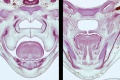
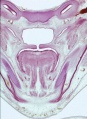
Embryonic Palate
|
Human primary palate
|

|
- EM Links: Image - stage 16 | Image - stage 17 | Image - stage 18 | Image - stage 19 | Palate Development
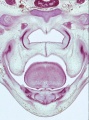
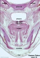
Fetal Palate
Secondary palate, fusion in the human embryo in week 9. This requires the early palatal shelves growth, elevation and fusion during the early embryonic period. The fusion event is to both each other and the primary palate. palatal shelf elevation | secondary palate
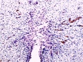
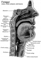
Hard and soft palate
hard palate
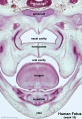
hard palate labeled
soft palate
soft palate labeled
Detail - hard and soft palate junction
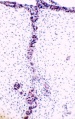
Detail - hard palate seam


Ventral aspect of hard palate of human embryo of 80 mm
Head Growth
- continues postnatally - fontanelle allow head distortion on birth and early growth
- bone plates remain unfused to allow growth, puberty growth of face
Animal Palate
Mouse Palate
- E11 - protrude from bilateral maxillary processes
- E12.5 - secondary palatal development begins
- E12.5-E14 - grow vertically along the developing tongue
- E14.5 - they elevate, meet, and fuse at the midline, to form an intact palate shelf, reflex opening and closing movements of the mouth
- E15.5 - palatal fusion is complete, mesenchymal condensation followed by osteogenic differentiation occurs.

Mouse (E13.5) Palatal Shelf Wnt5a, Osr2 and Pax9 Expression.[8]
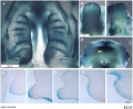
Image - Mouse E13.5 Bmp7 palate
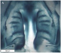
Image - palate
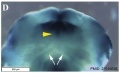
Image - palate detail


|

|
| Mouse ruga pattern (E16) | Mouse - Spry1 cleft palate |
- Links: Mouse Development | Bone Morphogenetic Protein | Wnt | Pax
Dog Palate

Newborn dog with cleft palate
Molecular
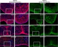
MMP25 PMID 20809987
Image - Mouse E13.5 Bmp7 palate PMID 23516636
Image - palate Bmp7 palate PMID 23516636
Image - palate detail Bmp7 palate PMID 23516636

- Links: Bone Morphogenetic Protein
Abnormalities

The way in which the upper jaw forms from fusion of the smaller upper prominence of the first pharyngeal arch leads to a common congenital defect in this region called "clefting", which may involve either the upper lip, the palate or both structures.
| Australian Palate Abnormalities (2002-2003) |
|---|
| Cleft lip with or without cleft palate (9.2 per 10,000 births) ICD-10 Q36.0, Q36.1, Q36.9, Q37.0–Q37.5, Q37.8, Q37.9 |
A congenital anomaly characterised by a partial or complete clefting of the upper lip, with or without clefting of the alveolar ridge or the hard palate. Excludes a midline cleft of the upper or lower lip and an oblique facial fissure (going towards the eye).
|
| Cleft palate without cleft lip (8.1 per 10,000 births) ICD-10 Q35.0–Q35.9 |
A congenital anomaly characterised by a closure defect of the hard and/or soft palate behind the foramen incisivum without a cleft lip. This anomaly includes sub-mucous cleft palate, but excludes cleft palate with a cleft lip, a functional short palate and high narrow palate.
|
|
International Classification of Diseases - Cleft Palate
Cleft lip and cleft palate (Q35-Q37)
Use additional code (Q30.2), if desired, to identify associated malformations of the nose. Excludes Robin's syndrome ( Q87.0 )
| Q37 | Cleft palate with cleft lip |
| Q37.0 | Cleft hard palate with bilateral cleft lip |
| Q37.1 | Cleft hard palate with unilateral cleft lip |
| Cleft hard palate with cleft lip NOS | |
| Q37.2 | Cleft soft palate with bilateral cleft lip |
| Q37.3 | Cleft soft palate with unilateral cleft lip |
| Cleft soft palate with cleft lip NOS | |
| Q37.4 | Cleft hard and soft palate with bilateral cleft lip |
| Q37.5 | Cleft hard and soft palate with unilateral cleft lip |
| Cleft hard and soft palate with cleft lip NOS | |
| Q37.8 | Unspecified cleft palate with bilateral cleft lip |
| Q37.9 | Unspecified cleft palate with unilateral cleft lip |
| Cleft palate with cleft lip NOS |
Embryonic Human Cleft Palate

|
| Stage16 (ventral view) |
Cleft Lip

|
|
| Cleft Lip Genes[10] | ||||||||||||||||||||||||||||||||||||||
|---|---|---|---|---|---|---|---|---|---|---|---|---|---|---|---|---|---|---|---|---|---|---|---|---|---|---|---|---|---|---|---|---|---|---|---|---|---|---|
Midline Cleft Lip Genes
| ||||||||||||||||||||||||||||||||||||||
|
Cleft Lip (+/− cleft palate) Genes
|
Cleft Palate


|
(Data: Congenital Malformations Australia 1981-1992 P. Lancaster and E. Pedisich ISSN 1321-8352)
Search Pubmed Now: cleft lip | cleft palate |
Cleft Risk Variants
Two genes were identified from a recent genome-wide study.[4]
- MAFB is expressed in the mouse palatal shelf.
- ABCA4 is a member of a superfamily of transmembrane proteins, and mutations in ABCA4 play a major role in the etiology of Stargardt disease and related retinopathies. Gene produces an ATP-binding cassette (ABC) superfamily trans-membrane protein
- Links: OMIM - MAFB | OMIM - ABCA4
Ten most frequently reported Birth Anomalies
- Hypospadias (More? Male movie | Genital Abnormalities - Hypospadia)
- Obstructive Defects of the Renal Pelvis (More? Renal System - Abnormalities)
- Ventricular Septal Defect (More? Cardiovascular Abnormalities - Ventricular Septal Defect)
- Congenital Dislocated Hip (More? Musculoskelal Abnormalities - Congenital Dislocation of the Hip (CDH))
- Trisomy 21 or Down syndrome - (More? Trisomy 21)
- Hydrocephalus (More? Hydrocephalus)
- Cleft Palate (More? Palate_Development)
- Trisomy 18 or Edward Syndrome - multiple abnormalities of the heart, diaphragm, lungs, kidneys, ureters and palate 86% discontinued (More? (More? Trisomy 18)
- Renal Agenesis/Dysgenesis - reduction in neonatal death and stillbirth since 1993 may be due to the more severe cases being identified in utero and being represented amongst the increased proportion of terminations (approximately 31%). (More? Renal System - Abnormalities)
- Cleft Lip and Palate - occur with another defect in 33.7% of cases.(More? Palate Development | Head Development)
(From the Victorian Perinatal Data Collection Unit in the Australian state of Victoria between 2003-2004)
Folate
A recent study of periconceptional folate supplementation using the Cochrane Pregnancy and Childbirth Group's Trials Register (July 2010) identified no statistically significant evidence of any effects on prevention of cleft palate and cleft lip at birth.[12]
References
- ↑ <pubmed>23613893</pubmed>| PLoS One.
- ↑ <pubmed>22022457</pubmed>| PLoS One.
- ↑ <pubmed>21246652</pubmed>
- ↑ 4.0 4.1 <pubmed>20436469</pubmed>
- ↑ <pubmed>17693063</pubmed>
- ↑ <pubmed>18028903</pubmed>
- ↑ <pubmed>8227288</pubmed>
- ↑ <pubmed>24433583</pubmed>| BMC Dev Biol.
- ↑ P. Lancaster and E. Pedisich, Congenital Malformations Australia 1981-1992, ISSN 1321-835.
- ↑ 10.0 10.1 <pubmed>21331089</pubmed>
- ↑ <pubmed>20537182</pubmed>| Orphanet J Rare Dis.
- ↑ <pubmed>20927767</pubmed>
Journals
- The Cleft Palate-Craniofacial Journal Homepage | Available issues
Reviews
Indian J Plast Surg. 2009 October; 42(Suppl):Cleft Lip and Palate Issue <pubmed>22186724</pubmed> <pubmed>19131313</pubmed> <pubmed>16962647</pubmed> <pubmed>3074914</pubmed> <pubmed>8714286</pubmed>
Articles
<pubmed></pubmed> <pubmed></pubmed> <pubmed></pubmed> <pubmed>20149609</pubmed> <pubmed>19341725</pubmed>
Search PubMed
Search Pubmed: palate development | cleft palate development |
Additional Images
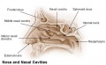
Nasal cavities and palate
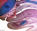
Palate, tongue and Meckel's cartilage
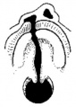
Historic Pharynx cartoon
Unilateral cleft lip and palate
Cleft palate feeder


Historic
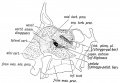
Fig. 6. Showing the structures formed in the Lateral Nasal Processes.
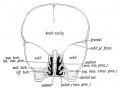
Fig. 7. Coronal section of the skull of a 7th month human foetus to show the cartilages of the Lateral and Mesial Nasal Processes and the bones formed round them.
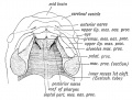
Fig. 8. Showing the ingrowth of the palatal plates of the two maxillary processes early in the 2nd month. (After Kollmann.) .
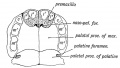
Fig. 9. Showing the Hard Palate at birth. The premaxillary part is formed from the Mesial Nasal Processes ; the remainder by the Palatal Plates of the Maxillary Processes.
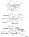
Fig. 10, a, b, c. Showing what become of the skeletons of the Mandibular Arch (Meckel's Cartilage) and Maxillary Process (Palato-quadrate Cartilage).
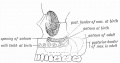
Fig. 11. Showing the manner in which the development of the Maxillary Antrum affects the size of the palate and position of the molar teeth.


Terms
- cleft - An anatomical gap or space occuring in abnormal development in or between structures. Most commonly associated with cleft lip and cleft palate. Term is also used to describe the external groove that forms between each pharyngeal arch during their formation.
- cleft lip - An abnormality of face development leading to an opening in the upper lip. Clefting of the lip and or palate occurs with 300+ different abnormalities. Depending on many factors, this cleft may extend further into the oral cavity leading to a cleft palate. In most cases clefting of the lip and palate can be repaired by surgery.
- cleft palate - An abnormality of face development leading to an opening in the palate, the roof of the oral cavity between the mouth and the nose. Clefting of the lip and or palate occurs with 300+ different abnormalities. In most cases clefting of the lip and palate can be repaired by surgery. Palate formation in the embryo occurs at two distinct times and developmental processes called primary and secondary palate formation. This leads to different forms (classifications) and degrees of clefting.
- epithelial mesenchymal transition - (EMT, epitheliomesenchymal transformation) conversion of an epithelium into a mesenchymal (connective tissue) cellular organization.
- epitheliomesenchymal transformation - (epithelial mesenchymal transition) conversion of an epithelium into a mesenchymal (connective tissue) cellular organization.
- medial edge epithelial - (MEE) opposing palatal shelves adhere to each other to form this epithelial seam.
- palate - The roof of the mouth (oral cavity) a structure which separates the oral from the nasal cavity. Develops as two lateral palatal shelves which grow and fuse in the midline. Initally a primary palate forms with fusion of the maxillary processes with the nasal processes in early face formation. Later the secondary palate forms the anterior hard palate which will ossify and separate the oral and nasal cavities. The posterior part of the palate is called the soft palate (velum, muscular palate) and contains no bone. Abnormalities of palatal shelf fusion can lead to cleft palate.
- palatogenesis - The process of palate formation, divided into primary and secondary palate development.
- pharyngeal arch - (branchial arch, Greek, branchial = gill) These are a series of externally visible anterior tissue bands lying under the early brain that give rise to the structures of the head and neck. In humans, five arches form (1,2,3,4 and 6) but only four are externally visible on the embryo. Each arch has initially identical structures: an internal endodermal pouch, a mesenchymal (mesoderm and neural crest) core, a membrane (endoderm and ectoderm) and external cleft (ectoderm). Each arch mesenchymal core also contains similar components: blood vessel, nerve, muscular, cartilage. Each arch though initially formed from similar components will differentiate to form different head and neck structures.
- philtrum - (infranasal depression, Greek, philtron = "to love" or "to kiss") Anatomically the surface midline vertical groove in the upper lip. Embryonically formed by the fusion of the frontonasal prominence (FNP) with the two maxillary processes of the first pharyngeal arch. Cleft palate (primary palate) occurs if these three regions fail to fuse during development. Fetal alcohol syndrome is also indicated by flatness and extension of this upper lip region.
- T-box 22 - (TBX22) a transcription factor that cause X-linked cleft palate and ankyloglossia in humans. Tbx22 is induced by fibroblast growth factor 8 (FGF8) in the early face while bone morphogenic protein 4 (BMP4) represses and therefore restricts its expression. (More? OMIM - TBX22)
- Transforming Growth Factor-beta - (TGFβ) factors induces both epithelial mesenchymal transition and/or apoptosis during palatal medial edge seam disintegration.
External Links
External Links Notice - The dynamic nature of the internet may mean that some of these listed links may no longer function. If the link no longer works search the web with the link text or name. Links to any external commercial sites are provided for information purposes only and should never be considered an endorsement. UNSW Embryology is provided as an educational resource with no clinical information or commercial affiliation.
- Prof Virginia Diewert - Professor of Orthodontics, University of British Columbia, who recently visited the Lab and helped with content, organisation and development of the Palate Development section.
- Medline Plus - Cleft Lip and Palate
- Better Health Channel - Cleft palate and cleft lip
- March of Dimes Birth Defects Foundation - Cleft Palate
Glossary Links
- Glossary: A | B | C | D | E | F | G | H | I | J | K | L | M | N | O | P | Q | R | S | T | U | V | W | X | Y | Z | Numbers | Symbols | Term Link
Cite this page: Hill, M.A. (2024, June 16) Embryology Palate Development. Retrieved from https://embryology.med.unsw.edu.au/embryology/index.php/Palate_Development
- © Dr Mark Hill 2024, UNSW Embryology ISBN: 978 0 7334 2609 4 - UNSW CRICOS Provider Code No. 00098G