Computed Tomography: Difference between revisions
mNo edit summary |
mNo edit summary |
||
| Line 7: | Line 7: | ||
Other potential developmental research imaging techniques include: positron emission tomography (PET), single photon emission computed tomography, magnetic resonance imaging, computed tomography, optical bioluminescence, fluorescence and high frequency ultrasound. | Other potential developmental research imaging techniques include: positron emission tomography (PET), single photon emission computed tomography, magnetic resonance imaging, computed tomography, optical bioluminescence, fluorescence and high frequency ultrasound. | ||
:'''Links:''' [[Movies#Computed Tomography|Computed Tomography]] | [[Abnormal Development - Radiation]] | [[:Category:Computed Tomography|Category:Computed Tomography]] | |||
:'''Links:''' [[Movies#Computed Tomography|Movies - Computed Tomography]] | [[Abnormal Development - Radiation]] | [[:Category:Computed Tomography|Category:Computed Tomography]] | |||
| Line 22: | Line 24: | ||
* '''Virtual histology of transgenic mouse embryos for high-throughput phenotyping.'''<ref name=PMID16683035><pubmed>16683035</pubmed>| [http://www.plosgenetics.org/article/info:doi/10.1371/journal.pgen.0020061 PLoS Genetics]</ref> "We present a novel, rapid, and inexpensive method for obtaining high-resolution virtual histology for phenotypic assessment of mouse embryos. Using osmium tetroxide to differentially stain tissues followed by volumetric X-ray computed tomography to image whole embryos, isometric resolutions of 27 mum or 8 mum were achieved with scan times of 2 h or 12 h, respectively, using mid-gestation E9.5-E12.5 embryos." | * '''Virtual histology of transgenic mouse embryos for high-throughput phenotyping.'''<ref name=PMID16683035><pubmed>16683035</pubmed>| [http://www.plosgenetics.org/article/info:doi/10.1371/journal.pgen.0020061 PLoS Genetics]</ref> "We present a novel, rapid, and inexpensive method for obtaining high-resolution virtual histology for phenotypic assessment of mouse embryos. Using osmium tetroxide to differentially stain tissues followed by volumetric X-ray computed tomography to image whole embryos, isometric resolutions of 27 mum or 8 mum were achieved with scan times of 2 h or 12 h, respectively, using mid-gestation E9.5-E12.5 embryos." | ||
|} | |} | ||
{| class="wikitable collapsible collapsed" | |||
! More recent papers | |||
|- | |||
| [[File:Mark_Hill.jpg|90px|left]] {{Most_Recent_Refs}} | |||
Search term: [http://www.ncbi.nlm.nih.gov/pubmed/?term=Embryo+Computed+Tomography ''Embryo Computed Tomography''] | |||
<pubmed limit=5>Embryo Computed Tomography</pubmed> | |||
|} | |||
==Early Mouse Development MicroCT== | ==Early Mouse Development MicroCT== | ||
Revision as of 19:04, 25 October 2013
Introduction

In embryology, the technique of micro-CT (μCT) has recently begun to become relevant in animal models and used for analysis of normal development features and those seen in genetically modified animal models, such as the mouse (More? Mouse Development).
Computed Tomography or computed axial tomography (CAT or CT scan) began in 1970's using x-ray and a computer to produce images either as individual slices or reconstructed to give three dimensional (3D) views of specific anatomical regions or structures.
Other potential developmental research imaging techniques include: positron emission tomography (PET), single photon emission computed tomography, magnetic resonance imaging, computed tomography, optical bioluminescence, fluorescence and high frequency ultrasound.
- Links: Movies - Computed Tomography | Abnormal Development - Radiation | Category:Computed Tomography
Some Recent Findings
|
| More recent papers |
|---|
 This table allows an automated computer search of the external PubMed database using the listed "Search term" text link.
More? References | Discussion Page | Journal Searches | 2019 References | 2020 References Search term: Embryo Computed Tomography <pubmed limit=5>Embryo Computed Tomography</pubmed> |
Early Mouse Development MicroCT
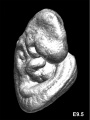
Mouse E9.5
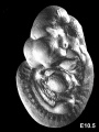
Mouse E10.5
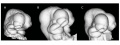
Mouse E10.5 head (fixation variations)
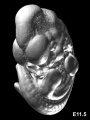
Mouse E11.5
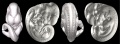
Mouse E11.5
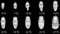
Mouse CT E9.5-E12 head
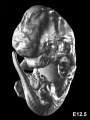
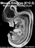
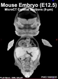
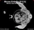
Mouse E12.5
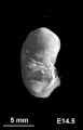
Mouse E14.5


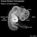
Human Embryo (week 6)
|
| Stage 17 Embryo |
| Page | Play |
Human Placenta

Right image shows computed tomography angiography (CTA) of the term human placenta viewed from the fetal side.[4]
Legend
- CA - chorionic artery
- PSA - primary stem artery
- SSA - secondary stem artery
- TSA - tertiary stem artery
Movies
|
|
|
|
| |||||||||||||||
|
|
|
|
- Links: Movies - Computed Tomography
Radiation Exposure

Computed tomography is a source of medical X-ray radiation exposure to the general population. The risk to an individual patient on the basis of dose levels of developing a malignant tumour due to CT is low and acceptable compared to the substantial benefit. There is though a large range/variation in radiation exposure between individual institutions and equipment settings.
Total radiology collective effective dose
- UK (2005-2006) approximately 60% from CT.
- Germany (2000-2005) for cancer patients from all X-ray procedures was approximately 82%.
- USA about 67%
References
- ↑ <pubmed>20163731</pubmed>| BMC Developmental Biology
- ↑ <pubmed>20190279</pubmed>
- ↑ <pubmed>16683035</pubmed>| PLoS Genetics
- ↑ <pubmed>20226038</pubmed>| BMC Physiol.
- ↑ <pubmed>21044293</pubmed>| BMC Med Imaging.
Books
Molecular Imaging and Contrast Agent Database (MICAD) NBK5330 | PMID:20641179
Search Pubmed
Search Pubmed: Embryo Computed Tomography | Computed Tomography | Micro-computed tomography apparatus
External Links
External Links Notice - The dynamic nature of the internet may mean that some of these listed links may no longer function. If the link no longer works search the web with the link text or name. Links to any external commercial sites are provided for information purposes only and should never be considered an endorsement. UNSW Embryology is provided as an educational resource with no clinical information or commercial affiliation.
- University of Calgary 3D Morphometrics Lab
- Duke Center for In Vivo Microscopy A 4D Atlas and Morphologic Database
- Normal C57BL/6 mouse embryos from embryonic day E10.5 to E19.5
- Normal C57BL/6 mouse neonates from post-natal day 0 to 32
- Mutant mouse embryos with cardiac septation defects (conditional ablation of the Smoothened receptor gene, Mef2C-AHF-Cre;Smoflox/- mutants)
- Drishti and Drishti-2 stands for vision or insight in Sanskrit, an Indian language. Ajay Limaye
Glossary Links
- Glossary: A | B | C | D | E | F | G | H | I | J | K | L | M | N | O | P | Q | R | S | T | U | V | W | X | Y | Z | Numbers | Symbols | Term Link
Cite this page: Hill, M.A. (2024, June 15) Embryology Computed Tomography. Retrieved from https://embryology.med.unsw.edu.au/embryology/index.php/Computed_Tomography
- © Dr Mark Hill 2024, UNSW Embryology ISBN: 978 0 7334 2609 4 - UNSW CRICOS Provider Code No. 00098G