Gastrointestinal Tract - Gall Bladder Development: Difference between revisions
mNo edit summary |
|||
| Line 12: | Line 12: | ||
{{Gastrointestinal Tract Links}} | [[Gastrointestinal Tract - Gall Bladder Histology|Gall Bladder Histology]] | [http://embryology.med.unsw.edu.au/Notes/git7a.htm original page] | |||
== Some Recent Findings == | == Some Recent Findings == | ||
| Line 20: | Line 20: | ||
* '''Embryology of the biliary tract'''<ref name="PMID20551648"><pubmed>20551648</pubmed></ref> "A hepatic diverticulum appears in the ventral wall of the primitive midgut early in the 4th week of intrauterine life in the development of the human embryo. This small diverticulum is the anlage for the development of the liver, extrahepatic biliary ducts, gallbladder, and ventral pancreas. By the 5th week, all elements of the biliary tree are recognizable. Marked elongation of the common duct occurs with plugging of the lumen by epithelial cells. Recanalization of the lumen of the common duct starts at the end of the 5th week and moves slowly distally. By the 6th week, the common duct and ventral pancreatic bud rotate 180 degrees clockwise around the duodenum. Early in the 7th week, the bile and pancreatic ducts end in closed cavities of the duodenum. Between the early 8th and 12th week, hepatopancreatic ducts have both superior and inferior orifices." | * '''Embryology of the biliary tract'''<ref name="PMID20551648"><pubmed>20551648</pubmed></ref> "A hepatic diverticulum appears in the ventral wall of the primitive midgut early in the 4th week of intrauterine life in the development of the human embryo. This small diverticulum is the anlage for the development of the liver, extrahepatic biliary ducts, gallbladder, and ventral pancreas. By the 5th week, all elements of the biliary tree are recognizable. Marked elongation of the common duct occurs with plugging of the lumen by epithelial cells. Recanalization of the lumen of the common duct starts at the end of the 5th week and moves slowly distally. By the 6th week, the common duct and ventral pancreatic bud rotate 180 degrees clockwise around the duodenum. Early in the 7th week, the bile and pancreatic ducts end in closed cavities of the duodenum. Between the early 8th and 12th week, hepatopancreatic ducts have both superior and inferior orifices." | ||
* '''Muscularis mucosae versus muscularis propria in gallbladder, cystic duct, and common bile duct: smoothelin and desmin immunohistochemical study'''<ref name="PMID21074688"><pubmed>21074688</pubmed></ref> "The muscle layer in the cystic duct and common bile duct is not well defined, and it is unresolved whether it represents muscularis mucosae or muscularis propria. ... Based on our findings, we conclude that, in the gallbladder wall, the muscle layer is muscularis propria and there is no muscularis mucosae present. In the cystic duct and common bile duct, only an attenuated and incomplete muscle layer of muscularis mucosae is present; because there is no muscularis propria, there probably is limited contractile function." | * '''Muscularis mucosae versus muscularis propria in gallbladder, cystic duct, and common bile duct: smoothelin and desmin immunohistochemical study'''<ref name="PMID21074688"><pubmed>21074688</pubmed></ref> "The muscle layer in the cystic duct and common bile duct is not well defined, and it is unresolved whether it represents muscularis mucosae or muscularis propria. ... Based on our findings, we conclude that, in the gallbladder wall, the muscle layer is muscularis propria and there is no muscularis mucosae present. In the cystic duct and common bile duct, only an attenuated and incomplete muscle layer of muscularis mucosae is present; because there is no muscularis propria, there probably is limited contractile function." | ||
|} | |||
{| class="wikitable collapsible collapsed" | |||
! More recent papers | |||
|- | |||
| [[File:Mark_Hill.jpg|90px|left]] {{Most_Recent_Refs}} | |||
Search term: ''Gall Bladder Embryology'' | |||
<pubmed limit=5>Gall Bladder Embryology</pubmed> | |||
|} | |} | ||
Revision as of 23:18, 5 Mayıs 2013
Introduction

This section of notes gives an overview of Gall Bladder and hillary tree development, histology and abnormalities associated with the biliary system. In the adult, the gall bladder is a site of bile salt storage and concentration, to then be released into the duodenum where they act to solubilize dietary lipids by their detergent effect. Bile salts are a cholesterol derivative (breakdown product).
The transverse septum differentiates to form the hepatic diverticulum and the hepatic primordium, these two structures together will go on to form different components of the mature liver and gall bladder.
The hepatic diverticulum divides into two parts: pars hepatica (larger cranial part, primordium of the liver) and pars cystica (smaller ventral invagination, primordium of gall bladder).
The pars cystica vacuolates and expands, the stalk becoming the cystic duct. This structure is initially hollow, then solid (by proliferation of epithelial lining), and then recanalized occurs by vacuolation of this expanded epithelium. There are several opinions as to whether the duct has a solid phase or remains patent throughout development.[1][2]
See also Gall Bladder Histology.
| Gall Bladder Histology | original page
Some Recent Findings
|
| More recent papers |
|---|
  This table allows an automated computer search of the external PubMed database using the listed "Search term" text link.
More? References | Discussion Page | Journal Searches | 2019 References | 2020 References Search term: Gall Bladder Embryology <pubmed limit=5>Gall Bladder Embryology</pubmed> |
Embryonic Development

Early embryonic gall bladder (Carnegie stage 13, Week 4)

Late embryonic gall bladder (Carnegie stage 22, Week 8)
Abnormalities
Infections
These mainly relate to postnatal infections. Recent studies in the mouse have identified that gastrointestinal tract listeria infections can relocate to the gall bladder and reside there, leading to later reinfection of the gastrointestinal tract.
- Links: Bacterial Infection
Additional Images
See also Gall Bladder Histology
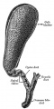
Historic drawing gall bladder anatomy
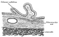
Historic drawing gall bladder transverse section
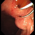
Abnormality - ectopic opening of the common bile duct (CBD) into the duodenal bulb.


References
Reviews
<pubmed>21074731</pubmed> <pubmed>20152372</pubmed> <pubmed>18484608</pubmed> <pubmed>15853977</pubmed> <pubmed>15382016</pubmed>
Articles
<pubmed>21078254</pubmed> <pubmed>20191134</pubmed> <pubmed>16273658</pubmed>
Search Pubmed
July 2010
Search Bookshelf Gall Bladder Development
Search Pubmed Now: Gall Bladder Development | Cholangiocyte Development |
Glossary Links
- Glossary: A | B | C | D | E | F | G | H | I | J | K | L | M | N | O | P | Q | R | S | T | U | V | W | X | Y | Z | Numbers | Symbols | Term Link
Cite this page: Hill, M.A. (2026, September 14) Embryology Gastrointestinal Tract - Gall Bladder Development. Retrieved from https://embryology.med.unsw.edu.au/embryology/index.php/Gastrointestinal_Tract_-_Gall_Bladder_Development
- © Dr Mark Hill 2026, UNSW Embryology ISBN: 978 0 7334 2609 4 - UNSW CRICOS Provider Code No. 00098G
