Urinary Bladder Development
| Embryology - 16 Jul 2026 |
|---|
| Google Translate - select your language from the list shown below (this will open a new external page) |
|
العربية | català | 中文 | 中國傳統的 | français | Deutsche | עִברִית | हिंदी | bahasa Indonesia | italiano | 日本語 | 한국어 | မြန်မာ | Pilipino | Polskie | português | ਪੰਜਾਬੀ ਦੇ | Română | русский | Español | Swahili | Svensk | ไทย | Türkçe | اردو | ייִדיש | Tiếng Việt These external translations are automated and may not be accurate. (More? About Translations) |
Introduction


The paired adult kidneys filter blood, reabsorb water, have endocrine functions and excrete waste. The waste in the form of urine for excretion, collects initially in the renal pelvis and flows through the ureters to the urinary bladder. The wall of the urinary bladder is composed of layers of smooth muscle and in the male has close anatomical relationship with the prostate gland. (More? prostate)
Abnormalities in renal development can lead to ureter obstruction and interfere with flow of urine to the bladder during the fetal period.
- Historic Bladder: 1912 Cloaca, Bladder, Urethra, and Urogenital Sinus | 1921 Urinary Bladder
Some Recent Findings

|
| More recent papers |
|---|
 This table allows an automated computer search of the external PubMed database using the listed "Search term" text link.
More? References | Discussion Page | Journal Searches | 2019 References | 2020 References Search term: Bladder Embryology | Bladder Development | Ureter Development | Urethra Development | Trigone Development | Detrusor Development |
| Older papers |
|---|
| These papers originally appeared in the Some Recent Findings table, but as that list grew in length have now been shuffled down to this collapsible table.
See also the Discussion Page for other references listed by year and References on this current page.
|
Textbook References
- The Developing Human: Clinically Oriented Embryology (8th Edition) by Keith L. Moore and T.V.N Persaud - Moore & Persaud Chapter 13 p303-346
- Larsen’s Human Embryology by GC. Schoenwolf, SB. Bleyl, PR. Brauer and PH. Francis-West - Chapter 10 p261-306
Movies
|
|
|
|
|
|
|
Renal System Development | All Renal Cartoons
Cloaca

|
Animation - Endoderm forming the cloaca and the primitive urinary bladder continuous with the allantois. |
- hindgut region ending at the cloacal membrane
- divided (ventro-dorsally) by the urogenital septum
- ventral - common urogenital sinus
- dorsal - rectum
Common Urogenital Sinus
- superior end continuous with allantois
- common urogenital sinus and mesonephric duct fuse (connect)
- differentiates to form the bladder
- inferior end forms urethra
- this will be different in male and female development
![]()
Urogenital Septum
| Urogenital Septum |
| Page | Play |
- Links: cloaca
Urinary Bladder Development

- early origins of the bladder at the superior end of the common urogenital sinus
- open inferiorly to the cloaca and superiorly to the allantois
- Septation of the claoca - divides the anterior region to the primordial bladder component from the posterior rectal component.
- associated ureters and urethra
Dorsal view of developing bladder
| Trigone |
| Page | Play |
- Ultrasound measurement of the bladder size can be used as a diagnostic tool for developmental abnormalities.
Bladder Epithelium
The embryonic epithelium remains unaltered for a long time varying from columnar to low cubical. The mulitlayered epithelium first begins in the early fetus (week 10, 50 mm head-foot length) in the caudal portion. The transitional epithelium appears in the fetus of 60 mm head-foot-length.
- one-layered epithelium - at the apex.
- two-layered epithelium - dorsal and ventral surface (thicker on dorsal).
- many-layered epithelium - at the fundus.
Detrusor Muscle
- The adult detrusor muscle consists of three layers of smooth (involuntary) muscle fibres.
- external layer - fibres arranged longitudinally
- middle layer - fibres arranged circularly
- internal layer - fibres arranged longitudinally
- Links: smooth muscle
Trigone Development
| Trigone |
| Page | Play |
Week 8
The Carnegie stage 22 human male embryo is 27mm (CRL) in size and approximately equal to day 54 - 56 of development. These images have been selected to show some key features of late embryo development.

|

|

|

|

|
| G5 urogenital | G6 urogenital | G7 urogenital | unlabeled | labeled |

Fetal Urinary Bladder
Fetal Development - 10 Weeks - Early female fetal bladder development. Anatomically lying behind the pubic symphysis and in front of the developing uterus. Surrounded by the developing detrusor muscle and the superior end extending towards the ventral body wall herniation.

|

|
| midline section | medial section |
- Links: Fetal Development - 10 Weeks

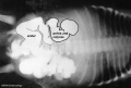
|
MRI appearance of normal fetal kidney.[6] Sagittal T2- SSFSE of a fetal abdomen at GA 25 week. Adequate volume of the amniotic fluid and the developing lungs indicate good renal function.
Note that the urinary bladder can occupy a considerable portion of the abdomen as a normal finding.
|
Ureter Development

- The adult ureter is a thick-walled muscular tube, 25 - 30 cm in length, running from the kidney to the urinary bladder.
- Anatomically can be described in two parts the abdominal part (pars abdominalis) and pelvic part (pars pelvina).
- The ureter is composed of three layers: outer fibrous layer (tunica adventitia), muscular layer (tunica muscularis) and mucous layer (tunica mucosa).
- The muscular layer can also be subdivided into 3 fibre layers: an external longitudinal, a middle circular, and an internal longitudinal.

Robert Meyer (1864 – 1947) |
The "Weigert-Meyer law" is a rule concerning the anatomical relationship of the two ureters, named after the pathologist Carl Weigert in conjunction with the embryologist Robert Meyer |
- PubMed: Ureter Development
Urethra Development
- Contrasting mechanisms of penile urethral formation in mouse and human[7] "This paper addresses the developmental mechanisms of formation of the mouse and human penile urethra and the possibility that two disparate mechanisms are at play. It has been suggested that the entire penile urethra of the mouse forms via direct canalization of the endodermal urethral plate. While this mechanism surely accounts for development of the proximal portion of the mouse penile urethra, we suggest that the distal portion of the mouse penile urethra forms via a series of epithelial fusion events. Through review of the recent literature in combination with new data, it is unlikely that the entire mouse urethra is formed from the endodermal urethral plate due in part to the fact that from E14 onward the urethral plate is not present in the distal aspect of the genital tubercle. Formation of the distal portion of the mouse urethra receives substantial contribution from the preputial swellings that form the preputial-urethral groove and subsequently the preputial-urethral canal, the later of which is subdivided by a fusion event to form the distal portion of the mouse penile urethra. Examination of human penile development also reveals comparable dual morphogenetic mechanisms. However, in the case of human, direct canalization of the urethral plate occurs in the glans, while fusion events are involved in formation of the urethra within the penile shaft, a pattern exactly opposite to that of the mouse. The highest incidence of hypospadias in humans occurs at the junction of these two different developmental mechanisms. The relevance of the mouse as a model of human hypospadias is discussed."
- Historic Urethra: 1912 Cloaca, Bladder, Urethra, and Urogenital Sinus
- Search PubMed: Urethra Development
Newborn Urinary Bladder

|

|
| The Newborn Male Bladder | The Newborn Female Bladder |
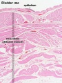
Bladder Histology

Can be described anatomically by its 4 layers from outside inward:
- Serous - the superior or abdominal surfaces and the lateral" surfaces of the bladder are covered by visceral peritoneum, the serous membrane (serosa) of the abdominal cavity, consisting of mesthelium and elastic fibrous connective tissue.
- Muscular - the detrusor muscle is the muscle of the urinary bladder wall.
- Submucosa - connects the muscular layer with the mucous layer.
- Mucosa - (mucus layer) a transitional epithelium layer formed into folds (rugae).

Animal Models
 Mouse bladder development E12.5-E16.5[4]
Abnormalities

Duplicated Bladder

|
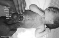
Urinary bladder duplication is an extremely rare abnormality.[8]
This MRI of a male newborn infant shows the duplicated bladder and also duplicated external genitalia (phallus).
|
Urorectal Septum Malformation
- thought to be a deficiency in caudal mesoderm which in turn leads to the malformation of the urorectal septum and other structures in the pelvic region.
- Recent research has also identified the potential presence of a persistent urachus prior to septation of the cloaca (common urogenital sinus).
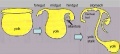
Bladder Exstrophy

{{ICD-11} LB31.3 Exstrophy of urinary bladder - Bladder exstrophy (or classic bladder exstrophy) is a congenital genitourinary malformation belonging to the spectrum of the exstrophy-epispadias complex and is characterized by an evaginated bladder plate, epispadias and an anterior defect of the pelvis, pelvic floor and abdominal wall.
- developmental abnormality associated with bladder development.
- origins appear to occur not just by abnormal bladder development, but by a congenital malformation of the ventral wall of abdomen (between umbilicus and pubic symphysis).
- There may also be other anomalies associated with failure of closure of abdominal wall and bladder (epispadias, pubic bone anomalies).
| Male | Female | = | alt=male bladder exstrophy|300px | alt=female bladder exstrophy|300px |
|---|
Absent Bladder
Absent or small bladder can be associated with renal agenesis.
Ureter and Urethra
- Ureter - Duplex Ureter
- Urethra- Urethral Obstruction and Hypospadias
Prune Belly Syndrome
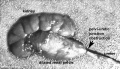
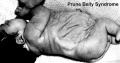
Prune_belly


- lower urinary tract obstruction
- mainly male
- fetal urinary system ruptures leading to collapse and "prune belly" appearance.
Horseshoe Kidney

- fusion of the lower poles of the kidney.
- During migration from the sacral region the two metanephric blastemas can come into contact, mainly at the lower pole.
- The ureters pass in front of the zone of fusion of the kidneys.
- The kidneys and ureters usually function adequately but there is an increased incidence of upper urinary tract obstruction or infection.
- Some horseshoe variations have been described as having associated ureter abnormalities including duplications.
References
- ↑ Liu X, Xie X, Jin ZW, Wang H, Song Y, Zhao P & Li L. (2021). The Allantois and Urachus: Histological Study Using Human Embryo and Fetuses. Fetal Pediatr Pathol , , 1-10. PMID: 34854363 DOI.
- ↑ Gredler ML, Patterson SE, Seifert AW & Cohn MJ. (2020). Foxa1 and Foxa2 orchestrate development of the urethral tube and division of the embryonic cloaca through an autoregulatory loop with Shh. Dev. Biol. , 465, 23-30. PMID: 32645357 DOI.
- ↑ Favorito LA, Pazos HM, Costa SF, Costa WS & Sampaio FJ. (2014). Morphology of the fetal bladder during the second trimester: comparing genders. J Pediatr Urol , 10, 1014-9. PMID: 25434295 DOI.
- ↑ 4.0 4.1 Islam SS, Mokhtari RB, Kumar S, Maalouf J, Arab S, Yeger H & Farhat WA. (2013). Spatio-temporal distribution of Smads and role of Smads/TGF-β/BMP-4 in the regulation of mouse bladder organogenesis. PLoS ONE , 8, e61340. PMID: 23620745 DOI.
- ↑ Tanaka ST, Ishii K, Demarco RT, Pope JC, Brock JW & Hayward SW. (2010). Endodermal origin of bladder trigone inferred from mesenchymal-epithelial interaction. J. Urol. , 183, 386-91. PMID: 19914648 DOI.
- ↑ Saleem SN. (2014). Fetal MRI: An approach to practice: A review. J Adv Res , 5, 507-23. PMID: 25685519 DOI.
- ↑ Liu G, Liu X, Shen J, Sinclair A, Baskin L & Cunha GR. (2018). Contrasting mechanisms of penile urethral formation in mouse and human. Differentiation , 101, 46-64. PMID: 29859371 DOI.
- ↑ Gajbhiye V, Nath S, Ghosh P, Chatterjee A, Haldar D & Das SK. (2015). Complete duplication of the urinary bladder: An extremely rare congenital anomaly. Urol Ann , 7, 91-3. PMID: 25657554 DOI.
- ↑ Ebert AK, Reutter H, Ludwig M & Rösch WH. (2009). The exstrophy-epispadias complex. Orphanet J Rare Dis , 4, 23. PMID: 19878548 DOI.
Reviews
Shen J, Overland M, Sinclair A, Cao M, Yue X, Cunha G & Baskin L. (2016). Complex epithelial remodeling underlie the fusion event in early fetal development of the human penile urethra. Differentiation , 92, 169-182. PMID: 27397682 DOI.
Shapiro E. (2009). Clinical implications of genitourinary embryology. Curr Opin Urol , 19, 427-33. PMID: 19461520 DOI.
Brenner-Anantharam A, Cebrian C, Guillaume R, Hurtado R, Sun TT & Herzlinger D. (2007). Tailbud-derived mesenchyme promotes urinary tract segmentation via BMP4 signaling. Development , 134, 1967-75. PMID: 17442697 DOI.
Costantini F. (2006). Renal branching morphogenesis: concepts, questions, and recent advances. Differentiation , 74, 402-21. PMID: 16916378 DOI.
Search Bookshelf bladder development | trigone development ureter development
Articles
Viana R, Batourina E, Huang H, Dressler GR, Kobayashi A, Behringer RR, Shapiro E, Hensle T, Lambert S & Mendelsohn C. (2007). The development of the bladder trigone, the center of the anti-reflux mechanism. Development , 134, 3763-9. PMID: 17881488 DOI.
Search PubMed
Search Pubmed: urinary bladder development | bladder development
Images
Endoderm cartoon
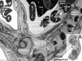
Fetal urogenital region most lateral right
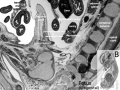
Fetal urogenital region lateral right
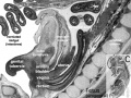
Fetal urogenital region medial
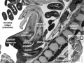
Fetal urogenital region midline
Fetal kidney (10 weeks)
Bladder histology

Prune belly
Renal outflow obstruction
Bladder Exstrophy


Historic Images
| Historic Disclaimer - information about historic embryology pages |
|---|
|
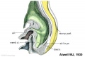
Stage 11 historic Atwell (1930)
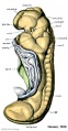
Stage 11 historic Heuser (1930)
retroperitoneal
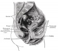
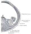
Fig. 1139 Adult Female Bladder


Bailey FR. and Miller AM. Text-Book of Embryology (1921) New York: William Wood and Co.
Text-Book of Embryology. Bailey, F.R. and Miller, A.M. (1921). New York: William Wood and Co. The Urinary Bladder
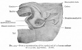
Cloaca and surrounding structures human embryo of 6.5 mm
Cloacal region human embryo
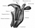
Caudal end human embryo of 11.5 mm
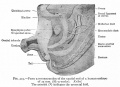
Caudal end human embryo of 25 mm


Keith A. Human Embryology and Morphology. (1902) London: Edward Arnold. The Uro-genital System
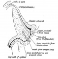
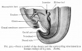
Fig. 99. A section of the male bladder and urethra at birth

Terms
- bladder exstrophy - A congenital malformation with bladder open to ventral wall of abdomen (between umbilicus and pubic symphysis) and may have other anomolies associated with failure of closure of abdominal wall and bladder (epispadias, pubic bone anomolies).
- hydronephrosis - (congenital hydronephrosis, Greek, hydro = water) A kidney abnormality due to partial or complete obstruction at the pelvi-ureteric junction. This leads to a grossly dilated renal pelvis causing extensive renal damage before birth.
- mesonephric duct - (= Wollfian duct) An early developing urogenital duct running the length of the embryo that will differentiate and form the male reproductive duct system. In females this duct degenerates (some remnants may remain associated in broad ligament).
- proteinuria - The abnormal presence of protein in the urine and an indicator of diesease including diabetic kidney disease (DKD, diabetic nephropathy).
- renal - (Latin, renes = kidney) Term used in relation to the kidney and associated structures (renal pelvis, renal artery)
- ureter - The two ureters are hollow tubes that link and carries urine from kidney to the bladder. The tubes have a muscular wall lined with transitional epithelium.
- urethra - The single muscular tube that links and carries urine from the bladder to the exterior. In humans, the urethral length differs between the sexes (male longer, female shorter).
- urinary - Term used to describe all components of the kidney system including the bladder, ureters and urethra.
- urine - Term used to describe the liquid waste produced by the kidney, stored in the bladder and excreted from teh body through the urethra.
- urorectal septum - (URS) The structure which develops to separate the cloaca (common urogenital sinus) into an anterior urinary part and a posterior rectal part.
- Wolffian duct - (= mesonephric duct, preferred terminology), runs from the mesonephros to cloaca, differentiates to form the male vas deferens and in the female regresses. Named after Caspar Friedrich Wolff (1733-1794), a German scientist and early embryology researcher and is said to have established the doctrine of germ layers. (More? Caspar Friedrich Wolff)
Glossary Links
- Glossary: A | B | C | D | E | F | G | H | I | J | K | L | M | N | O | P | Q | R | S | T | U | V | W | X | Y | Z | Numbers | Symbols | Term Link
Cite this page: Hill, M.A. (2026, July 16) Embryology Urinary Bladder Development. Retrieved from https://embryology.med.unsw.edu.au/embryology/index.php/Urinary_Bladder_Development
- © Dr Mark Hill 2026, UNSW Embryology ISBN: 978 0 7334 2609 4 - UNSW CRICOS Provider Code No. 00098G