Salivary Gland Development
| Embryology - 27 Apr 2024 |
|---|
| Google Translate - select your language from the list shown below (this will open a new external page) |
|
العربية | català | 中文 | 中國傳統的 | français | Deutsche | עִברִית | हिंदी | bahasa Indonesia | italiano | 日本語 | 한국어 | မြန်မာ | Pilipino | Polskie | português | ਪੰਜਾਬੀ ਦੇ | Română | русский | Español | Swahili | Svensk | ไทย | Türkçe | اردو | ייִדיש | Tiếng Việt These external translations are automated and may not be accurate. (More? About Translations) |
Introduction
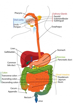
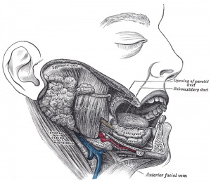
The salivary glands arise as epithelial buds in the oral cavity between week 6 to 7 (GA week 8 to 9) and extend into the underlying mesenchyme. The three paired groups of salivary glands are named by their anatomical location: parotid, submandibular and sublingual.
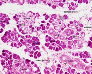
The adult glands are mucoserous tubuloacinar glands, with secretory acini and the initial part of the duct system also participates in the secretory process.Secretions from glands close to the oral cavity are mainly mucous, while glands located further away from the oral cavity (parotid) are mainly serous. Structurally, each salivary gland is divided by connective tissue septa into lobes, which are in turn subdivided into lobules.
| Additional Links | ||||||||||
|---|---|---|---|---|---|---|---|---|---|---|
|
Some Recent Findings
|
| More recent papers |
|---|
|
This table allows an automated computer search of the external PubMed database using the listed "Search term" text link.
More? References | Discussion Page | Journal Searches | 2019 References | 2020 References Search term: Salivary Gland Development | Salivary Gland Embryology | Submandibular Gland Development | Parotid Gland Development | Sublingual Gland Development |
| Older papers |
|---|
| These papers originally appeared in the Some Recent Findings table, but as that list grew in length have now been shuffled down to this collapsible table.
See also the Discussion Page for other references listed by year and References on this current page.
|
Salivary Ducts
Gland ducts
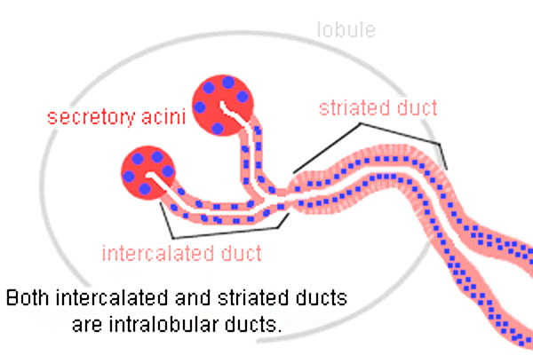
intercalated -> striated -> excretory -> main excretory ducts
- Intercalated duct - region close to acinar neck are thought to contain salivary gland stem cells.
Submandibular Gland
The submandibular gland is also called the submaxillary gland are located along the side of the mandible.
Timeline
Embryonic development of submandibular gland during week 7 and 8 based upon Streeter.[5]
- Carnegie stage 19 - Short, club-like duct entering mesenchymal primordium of gland.
- Carnegie stage 20 - Longer, knobby duct well in gland. Duct beginning to form knob-like branches.
- Carnegie stage 21 - Simple, stubby primary branching of duct.
- Carnegie stage 22 - Secondary branching of duct. Practically solid duct; suggestion of lumen in distal part. Definite lumen in oral part of duct.
- Carnegie stage 23 - Long duct, much branched. Lumen deep in gland. Lumina in many terminal branches of ducts. Beginning orientation of epithelial tree. Angiogenesis beginning around epithelium. Mesoblast begins to form layer around gland.
Anatomy
- locate inferior space of the mylohyoid.
- submandibular duct (Wharton’s duct)
- runs forward along the lingual nerve in the sublingual space
- opens in the sublingual caruncle
Histology
|
|
Parotid Gland
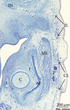
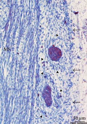
| Overview neck region (week 8) | Parotid gland (week 8) |
|---|---|
|
|
| Parotid gland primordial of capsule propia (arrowheads). |
- C - Meckel’s cartilage
- CL - Cervical lamina
- I - Inferior alveolar nerve
- L - Lingual nerve
- M - Mandible
- MS - Masseter muscle
- P - Anlage of the parotid gland
Anatomy
- largest salivary gland
- located in the triangle surrounded by:
- superiorly by the zygomatic arch
- anteriorly by the masseter
- posteriorly by the sternocleidomastoid
- inferior pole mostly confined to the angle of mandible.
- medial pole mostly confined to the temporomandibular joints.
- parotid duct (Stenon’s duct) leaves the anterior border, passes anteriorly on the masseter, penetrates the buccinator, and opens into the buccal cavity.
- named after Niels Stensen (1638 - 1686) a Danish anatomist, natural scientist, and theologist.
- facial nerve (CN VII) penetrates the gland
Sublingual Gland
Anatomy
- locate superior of the mylohyoid
- superior border of sublingual gland appears as the sublingual fold in the oral floor.
- major sublingual duct (Bartholin’s duct) opens in the sublingual caruncle.
- numerous minor sublingual ducts on the sublingual fold.
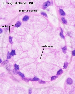
Histology
|
|
Lingual Gland
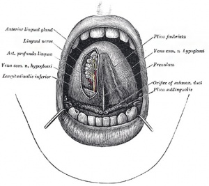
- Mucous glands are similar in structure to the labial and buccal glands. They are found especially at the back part behind the vallate papillæ, but are also present at the apex and marginal parts. In this connection the anterior lingual glands (Blandin or Nuhn) require special notice. They are situated on the under surface of the apex of the tongue, one on either side of the frenulum, where they are covered by a fasciculus of muscular fibers derived from the Styloglossus and Longitudinalis inferior. They are from 12 to 25 mm long, and about 8 mm. broad, and each opens by three or four ducts on the under surface of the apex.
- Serous glands (von Ebner's gland) occur only at the back of the tongue in the neighborhood of the taste-buds, their ducts opening for the most part into the fossæ of the vallate papillæ. These glands are racemose (clustered), the duct of each branching into several minute ducts, which end in alveoli, lined by a single layer of more or less columnar epithelium. Their secretion is of a watery nature, and probably assists in the distribution of the substance to be tasted over the taste area.
(text modified from Gray's anatomy)
Animal Models
Mouse
Submandibular salivary gland developmental timeline data below from [6]
- E11.5 - thickening of the primitive oral epithelium that grows into the first branchial (mandibular) arch mesenchyme to form the solid epithelial placode.
- E12 - placode protrudes into the mesenchyme forming a single, solid mass of cells connected to the tongue epithelium by a stalk of immature duct epithelial cells.
- E12.5 - indentations (clefts) start to form on the surface of the epithelial bud accompanied by alterations in the basement membrane. Clefts separate the primary bud into multiple buds and the epithelium proliferates. The base of the cleft becomes the primordial ductal structure, salivary branching morphogenesis is repeated multiple times over the following days.
- E14 - simple one-bud one-duct salivary gland has both grown and branched significantly, and the main duct begins to lumenize. The end buds undergo reorganization and begin to form acini – the main secretory units of the salivary gland.
- E15 – E16 - lumenization of the main secretory duct is nearly complete.
- E17 - the acini complete lumenization, so that the gland has a continuous network of lumenized ducts connecting the acini to the oral cavity. Both nerves and blood vessels populate the gland in association with the branching epithelium.
- Glands continue to mature after birth with cellular differentiation occurs in parallel with branching morphogenesis.
- Links: Mouse Development
Molecular
Recent studies have identified molecular mechanisms for salivary gland branching as similar to those used for development of the respiratory system branching using the molecules FGF10 and Sox9.[2]
FGF10
Fibroblast Growth Factor 10 (FGF10) is a secreted protein growth factor acting through a membrane receptor. Wnt and Fgf10 receptors combine with Kit signalling to promote the expansion of the gland distal tip cells.
- Links: Fibroblast Growth Factor
SOX9
Sox9 is a transcription factor that establishes the identity of the developing gland distal compartment before the initiation of branching morphogenesis.[2]
Histology
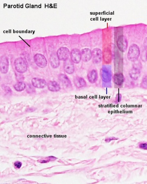
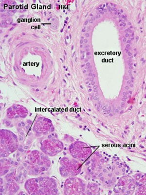
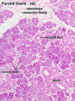
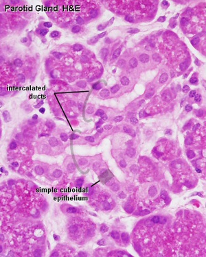
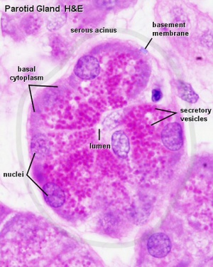
Parotid gland
|
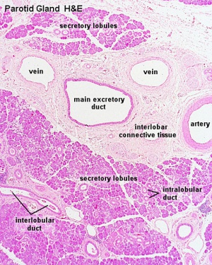
|
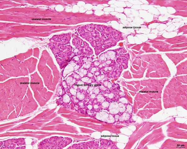
Lingual salivary gland
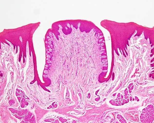
Circumvallate Papilla
Human Tongue ( lingual salivary gland, white adipose tissue, skeletal muscle)
Abnormalities
LB10 Structural developmental anomalies of salivary glands or ducts
| ICD-11 LB10 Structural developmental anomalies of salivary glands or ducts - Any condition caused by failure of the salivary glands and ducts to correctly develop during the antenatal period. |
Cystic Fibrosis
| ICD-11 CA25 Cystic fibrosis - [https://icd.who.int/browse11/l-m/en#/http://id.who.int/icd/entity/290257878
CA25.0 Classical cystic fibrosis] | CA25.1 Atypical cystic fibrosis | 1323966141 CA25.2 Subclinical cystic fibrosis
|
The absence of the transmembrane conductance regulator (CFTR) affects not only respiratory tract exocrine secretion, but also secretion in salivary glands.[7]
In the mouse, CFTR expression begins at E15 and within ducts but not in acini.[8]
Primary Sjögren's syndrome
ICD-11 4A43.2 Sjögren syndrome - [https://icd.who.int/browse11/l-m/en#/http://id.who.int/icd/entity/899463360 4A43.20 Primary Sjögren syndrome
|
Primary Sjögren's syndrome (pSS) results in the destruction of the salivary and the lacrimal glands through a mononuclear cell infiltration.[9]
Links: PubMed - salivary gland abnormality
References
- ↑ Ono Minagi H, Sarper SE, Kurosaka H, Kuremoto KI, Taniuchi I, Sakai T & Yamashiro T. (2017). Runx1 mediates the development of the granular convoluted tubules in the submandibular glands. PLoS ONE , 12, e0184395. PMID: 28877240 DOI.
- ↑ 2.0 2.1 2.2 Chatzeli L, Gaete M & Tucker AS. (2017). Fgf10 and Sox9 are essential for the establishment of distal progenitor cells during mouse salivary gland development. Development , 144, 2294-2305. PMID: 28506998 DOI.
- ↑ Kwon HR, Nelson DA, DeSantis KA, Morrissey JM & Larsen M. (2017). Endothelial cell regulation of salivary gland epithelial patterning. Development , 144, 211-220. PMID: 28096213 DOI.
- ↑ Amano O, Mizobe K, Bando Y & Sakiyama K. (2012). Anatomy and histology of rodent and human major salivary glands: -overview of the Japan salivary gland society-sponsored workshop-. Acta Histochem Cytochem , 45, 241-50. PMID: 23209333 DOI.
- ↑ Streeter GL. Developmental Horizons In Human Embryos Description Or Age Groups XIX, XX, XXI, XXII, And XXIII, Being The Fifth Issue Of A Survey Of The Carnegie Collection. (1957) Carnegie Instn. Wash. Publ. 611, Contrib. Embryol., 36: 167-196.
- ↑ Larsen M, Yamada KM & Musselmann K. (2010). Systems analysis of salivary gland development and disease. Wiley Interdiscip Rev Syst Biol Med , 2, 670-82. PMID: 20890964 DOI.
- ↑ da Silva Modesto KB, de Godói Simões JB, de Souza AF, Damaceno N, Duarte DA, Leite MF & de Almeida ER. (2015). Salivary flow rate and biochemical composition analysis in stimulated whole saliva of children with cystic fibrosis. Arch. Oral Biol. , 60, 1650-4. PMID: 26351748 DOI.
- ↑ Shin YH, Lee SW, Kim M, Choi SY, Cong X, Yu GY & Park K. (2016). Epigenetic regulation of CFTR in salivary gland. Biochem. Biophys. Res. Commun. , 481, 31-37. PMID: 27833020 DOI.
- ↑ Aqrawi LA, Jensen JL, Øijordsbakken G, Ruus AK, Nygård S, Holden M, Jonsson R, Galtung HK & Skarstein K. (2018). Signalling pathways identified in salivary glands from primary Sjögren's syndrome patients reveal enhanced adipose tissue development. Autoimmunity , , 1-12. PMID: 29504848 DOI.
- 1897 Glands draft only.
Reviews
Hauser BR & Hoffman MP. (2015). Regulatory Mechanisms Driving Salivary Gland Organogenesis. Curr. Top. Dev. Biol. , 115, 111-30. PMID: 26589923 DOI.
Patel VN & Hoffman MP. (2014). Salivary gland development: a template for regeneration. Semin. Cell Dev. Biol. , 25-26, 52-60. PMID: 24333774 DOI.
Articles
Chen Z, Huang J, Liu Y, Dattilo LK, Huh SH, Ornitz D & Beebe DC. (2014). FGF signaling activates a Sox9-Sox10 pathway for the formation and branching morphogenesis of mouse ocular glands. Development , 141, 2691-701. PMID: 24924191 DOI.
Patel VN, Likar KM, Zisman-Rozen S, Cowherd SN, Lassiter KS, Sher I, Yates EA, Turnbull JE, Ron D & Hoffman MP. (2008). Specific heparan sulfate structures modulate FGF10-mediated submandibular gland epithelial morphogenesis and differentiation. J. Biol. Chem. , 283, 9308-17. PMID: 18230614 DOI.
Search PubMed
Search term: Head Development | Parotid Development | Submandibular Gland Development | Sublingual Gland Development
Terms
Abbreviations: ( ) plural form in brackets, A. Arabic, abb. abbreviation, c. circa =about, F. French adj. adjective, G. Greek, Ge. German, cf. compare, L. Latin, dim. diminutive, NA. Nomina anatomica, q.v. which see, OF. Old French
- acinus (-i) - L. = a juicy berry, a grape; applied to small, rounded terminal secretory units of compound exocrine glands that have a small lumen (adj. acinar).
- Bartholin’s duct - see sublingual duct.
- buccal - L. bucca = cheek; related to cheek or mouth.
- circumvallate papillae - (vallate papillae) tongue largest and least numerous papillae (human 8 to 12) occur in depressions of the surface of the tongue and are surrounded with a trench formed by the infolding of the epithelium. Taste buds are numerous on the lateral surfaces of these papillae and the excretory ducts of serous glands (glands of von Ebner) open into the trenches surrounding the papillae.
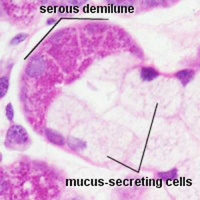
- demilune - L. dimidius = half + luna = moon; crescent-shaped cap of serous cells over mucous alveolus in some salivary glands.
- ductus (-us) - L. = passage from L. ducere = to lead; tube lined by epithelium for exocrine glandular secretions to reach surface.
- filiform papillae - tongue smallest and most numerous papillae, provide a rough surface to aid in the manipulation and processing of foods.
- fungiform - L. fungus = mushroom + forma = a shape; of lingual papillae.
- fungiform papillae - tongue single evenly spaced between the filiform papillae. Epithelium is slightly thinner than on the remaining surface of the tongue and connective tissue core is richly vascularised.
- gland - L. glandula , dim of L. glans = an acorn, a pellet; term used to describe mesenteric lymph nodes (Herophilus, c. 300 BC).
- intercalated - L. inter = between + calare = to proclaim, calatus = inserted; of a duct inserted between the end of the gland (acinus, or alveolus) and a larger duct. Partially covered by myoepithelial cells
- intercalated duct - cuboidal epithelium, modify saliva, add bicarbonate ions (buffering) and absorb chloride.
- intramural - L. intra = within + murus = wall; within the wall of an organ.
- labial - adj. L. labialis = of the lips, L. labium = lip, rim of a vessel.
- lacteal - L. lac = milk ( lacteus = of milk, lactare = to suckle); intestinal lymphatic, containing chyle after a fatty meal.
- lining epithelium - non-keratinised stratified squamous epithelium, covers the remaining (non-masticatory epithelium) surfaces of the oral cavity.
- lingual - adj. L. lingua = tongue.
- lips - outside (lined by skin) and inside (oral mucosa), vermilion border (prolabium) transition from the skin to the oral mucosa, forms only small part of the anatomical lips.
- lumen - L. = light; space enclosed by tubular or vesicular structure; hence luminal.
- masticatory epithelium - covers the tongue, gingivae and hard palate. Keratinised epithelium to different degrees depending on the extent of physical forces exerted on the epithelium.
- microvilli - epithelial cell apical surface specialisation seen in the small intestinal. These microfilament filled structures increase the surface area for absorption and secretion.
- mucin - L. mucus from G. muxa = snot, slime; protein constituent of all mucus; occurs as granules in secretory cells.
- mucosa - (-ae) L. = mucous membrane.
- mucus - L. = slime (adj. mucous).
- oesophagus - G. oiso = future of phero = I carry + pahgein = to eat; G. oisophagos = gullet, or tube carrying food from pharynx to stomach.
- oesophageal glands - located in the submucosa produce a mucous secretion, which lubricates the epithelium and aids the passage of food. Oesophagus closest to the stomach may also be mucosal mucus-producing glands, similar to the glands in the adjacent mucosa of the stomach.
- oral - adj. L. os, oris = mouth.
- palate - L. palatum = roof of mouth.
- palisade - L. palus = stake; like a fence of stakes.
- papilla - (-ae) L. = a teat, a nipple; a nipple-like projection, e.g., on the tonge (Malpighi, c. 1670; cf. circumvallate, filiform, foliate, fungiform, vallate); duodenal papilla (containing duodenal ampulla).
- parotid - G. para = beside + otos = of the ear; a salivary gland.
- parotid duct - (Stenson's duct) the major duct of the parotid gland that allows salivary gland secretions to empty into the oral cavity.
- ruga - (-ae) L. = a fold or wrinkle, e.g., in stomach.
- sac - L. saccus = sack, bag, from G. sakkos .
- salivary - L. saliva = spittle.
- secretion - L. secretus = separated; production of materials by glandular activity.
- serous - adj. L. = having nature of serum, watery fluid.
- Stenson's duct - see parotid duct. Named after Niels Stensen (1638 - 1686) a Danish anatomist, natural scientist, and theologist.
- striated ducts - modifies saliva (secretion of potassium and the absorption of sodium), columnar cells with the nucleus of located approximately midways between the apical and basal cell surfaces. Striations are found in the basal part of the cytoplasm of the cells, where numerous mitochondria are found between infoldings of the basal cell membrane. These cells can also take up a secretable form of antibodies and release them into the saliva.
- sublingual caruncle - location on either side of the frenulum linguae on the sublingual surface of the tongue.
- sublingual duct - (major sublingual duct, Bartholin’s duct) from the sublingual gland that opens in the sublingual caruncle. There are also numerous minor sublingual ducts opening on the sublingual fold.
- submandibular - adj. L. " + mandibula = jaw.
- submandibular duct - (Wharton’s duct) from the submandibular gland, runs forward along the lingual nerve in the sublingual space and opens in the sublingual caruncle.
- tongue muscles - skeletal muscle organized into strands oriented more or less perpendicular to each other.
- tongue nerves - movement (XII, hypoglossal nerve - motor) and sensory information. (V, trigeminal nerve - sensory - anterior two-thirds; VII, facial nerve - taste; IX, glossopharyngeal nerve - sensory/taste - posterior one-third).
- tonsil - L. tonsilla (origin obscure); mass of lymphocytes close to an epithelium, e.g., lingual tonsil, palatine tonsil (the "tonsil"), pharyngeal tonsil (adenoid, tonsil of Luschka, q.v.), tubal tonsil (of auditory tube).
- tubuloacinar gland - secretory acini with the first part of the duct system also participating in the secretory process.
- vallate papillae - see circumvallate papillae.
- von Ebner's glands - serous glands associated with circumvallate papillae, their ducts open into the trenches surrounding the papillae ("rinsing glands").
- Wharton’s duct - see submandibular duct. Named after Thomas Wharton (1614–1673) an English anatomist also known for umbilical cord Wharton’s jelly.
| GIT Links: Introduction | Medicine Lecture | Science Lecture | endoderm | mouth | oesophagus | stomach | liver | gallbladder | Pancreas | intestine | mesentery | tongue | taste | enteric nervous system | Stage 13 | Stage 22 | gastrointestinal abnormalities | Movies | Postnatal | milk | tooth | salivary gland | BGD Lecture | BGD Practical | GIT Terms | Category:Gastrointestinal Tract | ||||
|
Glossary Links
- Glossary: A | B | C | D | E | F | G | H | I | J | K | L | M | N | O | P | Q | R | S | T | U | V | W | X | Y | Z | Numbers | Symbols | Term Link
Cite this page: Hill, M.A. (2024, April 27) Embryology Salivary Gland Development. Retrieved from https://embryology.med.unsw.edu.au/embryology/index.php/Salivary_Gland_Development
- © Dr Mark Hill 2024, UNSW Embryology ISBN: 978 0 7334 2609 4 - UNSW CRICOS Provider Code No. 00098G
