Placenta - Stage 13
| Embryology - 23 Apr 2024 |
|---|
| Google Translate - select your language from the list shown below (this will open a new external page) |
|
العربية | català | 中文 | 中國傳統的 | français | Deutsche | עִברִית | हिंदी | bahasa Indonesia | italiano | 日本語 | 한국어 | မြန်မာ | Pilipino | Polskie | português | ਪੰਜਾਬੀ ਦੇ | Română | русский | Español | Swahili | Svensk | ไทย | Türkçe | اردو | ייִדיש | Tiếng Việt These external translations are automated and may not be accurate. (More? About Translations) |
Embryo cardiovascular system movie
Serial Labeled Images
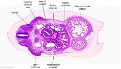
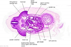
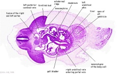
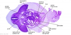
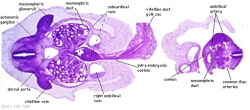
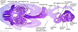
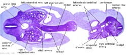
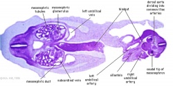
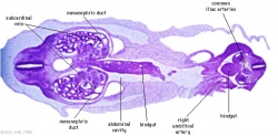
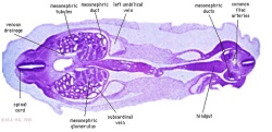
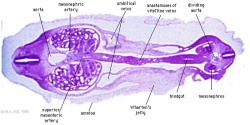
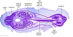
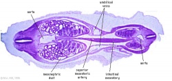
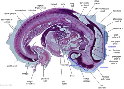
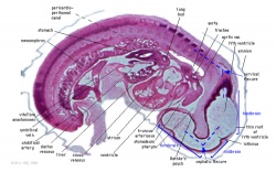
Carnegie Stage 13 Embryo the outer dark line in the sections is the amniotic membrane and the fluid-filled space between the membrane and the embryo is the amniotic sac filled with amniotic fluid.
Clicking the section image or label will open a full page view (use the browser back button to return here).
Table Legend
Features: identifies, reading in a left to right sequence, major anatomical structures visible in the specific embryo section.
- Bullet points relate to placental structures.
- R = right, L = left.
|
Features: dorsal body wall, spinal cord, dorsal aorta, posterior cardinal veins, stomach, liver, heart ventricles, and ventral body wall.
|
|
Features: dorsal body wall, spinal cord, upper limb buds, dorsal aorta, posterior cardinal veins, stomach, liver, heart ventricle, and ventral body wall.
|
|
Features: dorsal body wall, spinal cord, upper limb buds, dorsal aorta fusing, posterior cardinal veins, duodenum, liver, heart ventricle, and ventral body wall.
|
|
Features: dorsal body wall, spinal cord, upper limb buds, dorsal aorta, posterior cardinal veins, mesonephros, duodenum, liver, transverse septum, and ventral body wall.
|
|
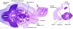
Features: dorsal body wall, spinal cord, upper limb buds, dorsal aorta, posterior cardinal veins, mesonephros, intestine, umbilicus, ventral body wall and lower end of embryo with cloaca.
|
|
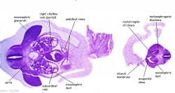
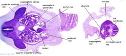
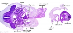
Features: dorsal body wall, spinal cord, upper limb buds, dorsal aorta, mesonephros, intestine, umbilicus, ventral body wall and lower end of embryo with cloaca.
|
|
|
|
|
|
|
| |
|
|
|
|
|
|
|
|
F5L |
|
|
|
|
|
|
|
|
All Sections
About Stage 13 Embryo Sections - This image is from a serial section of a 6mm CRL pig embryo with some features of the Stage 14 embryo. This embryo is approximately equal to the day 42 human embryo. Use these serial images to identify internal features and relationships that exist within the embryo at this stage. Then compare these images with the later features of the Carnegie stage 22 human embryo.
| Stage 13 Serial unlabeled images | Embryo Stage 13 Serial labeled images |
Glossary Links
- Glossary: A | B | C | D | E | F | G | H | I | J | K | L | M | N | O | P | Q | R | S | T | U | V | W | X | Y | Z | Numbers | Symbols | Term Link
Cite this page: Hill, M.A. (2024, April 23) Embryology Placenta - Stage 13. Retrieved from https://embryology.med.unsw.edu.au/embryology/index.php/Placenta_-_Stage_13
- © Dr Mark Hill 2024, UNSW Embryology ISBN: 978 0 7334 2609 4 - UNSW CRICOS Provider Code No. 00098G