Musculoskeletal System - Bone Development Timeline
| Embryology - 26 Jul 2026 |
|---|
| Google Translate - select your language from the list shown below (this will open a new external page) |
|
العربية | català | 中文 | 中國傳統的 | français | Deutsche | עִברִית | हिंदी | bahasa Indonesia | italiano | 日本語 | 한국어 | မြန်မာ | Pilipino | Polskie | português | ਪੰਜਾਬੀ ਦੇ | Română | русский | Español | Swahili | Svensk | ไทย | Türkçe | اردو | ייִדיש | Tiếng Việt These external translations are automated and may not be accurate. (More? About Translations) |
Introduction
The adult human skeleton has about 206 different bones, each develop with their own specific bone timeline. Many prenatal bones fuse postnatal developing neonate and child (about 275).
The two main forms of ossification occur in different bones, intramembranous (eg skull) and endochondral (eg vertebra) ossification. Ossification in general continues postnatally, through puberty until mid 20s.
- Without postnatal growth in the long bones and axial skeleton, we would be as high as when we are born!

|

Appendicular skeleton (126 bones in limbs, shoulders, pelvis) |
Endochondral ossification within the limb begins at Carnegie stage 18 and also occurs throughout embryo skeleton. This process is the replacement of a cartilage "template" with bone (week 5-12) that continues through postnatal development, with a second surge of growth at puberty.
These notes summarise the timecourse of development of some of these bones in humans. Historically, the ossification timeline in human embryos was published by Mall in 1906[1] and was the basis of his later publication in his 1910 embryology textbook Manual of Human Embryology.
Some Recent Findings

|
| More recent papers |
|---|
 This table allows an automated computer search of the external PubMed database using the listed "Search term" text link.
More? References | Discussion Page | Journal Searches | 2019 References | 2020 References Search term: Bone Development Timeline |
Ossification Stages

The process of ossification as determined postnatally clinically has been divided into a series of stages.[5]
- Stage 1 - non-ossified epiphysis
- Stage 2 - discernible ossification centre
- Stage 3 - partial fusion
- Stage 4 - total fusion
- Stage 5 - an additional stage recently added is the disappearance of the epiphyseal scar after total fusion.
Clavicle Ossification
The following identifies the process of ossification of the medial clavicular epiphyseal cartilage.[5]
- Stage 3 - 16 years
- Stage 4 - 20 years (women), 21 years (men)
- Stage 5 - 26 years
| Age | No. of instances in girls | No. of instances in boys | No. of centres in girls | No. of centres in boys |
|---|---|---|---|---|
| 11 | 12 | 2 | 1 | 0 |
| 12 | ll | 9 | 0 | 1 |
| 13 | 3 | 15 | 0 | 0 |
| 14 | 8 | 10 | 2 | 2 |
| 15 | 10 | 1l | 2 | 1 |
| 16 | 16 | 14 | 4 | 1 |
| 17 | 12 | 12 | 5 | 5 |
| 18 | 10 | 15 | 2 | 3 |
| 19 | 25 | ll | ll | 7 |
| 20 | 28 | 19 | 12 | 7 |
| 21 | 14 | 20 | 7 | 1l |
| 22 | 18 | 11 | 5 | 4 |
| 23 | 17 | 16 | 4 | 4 |
| 24 | 18 | 15 | 4 | 1 |
| 25 | 19 | 18 | 2 | 2 |
| 26 | 10 | 8 | 1 | 0 |
| Table Data: Flecker (1932)[6] | ||||
Medial Clavicular Epiphysis Ossification
The following table compares male and female timing in years of ossification based upon CT analysis.[2]
| Event | Male | Female |
|---|---|---|
| Transition from unfused to fusing | 20.60 | 19.19 |
| Transition from fusing to complete fusion | 21.92 | 21.47 |
Long Bone Ossification

Humerus
Appearance and fusion of bone secondary ossification centres, proximal is closer to body and distal is further away from the body.
Appearance
- Proximal epiphysis gestation week 36 - 4 years
- Distal epiphysis 6 months - 10 years
Fusion
- Proximal epiphysis 12 - 20 years
- Distal epiphysis 11 - 19 years
Femur
Appearance
- Proximal epiphysis 1 - 12 years
- Distal epiphysis Gestation week 36 - 40
Fusion
- Proximal epiphysis 11 - 19 years
- Distal epiphysis 14 - 19 years
Data from reference Table 1.[7]
Historic Limb Data
Manual of Human Embryology by Franz Keibel and Franklin P. Mall (1910)
Upper Limb
| Bone | Centres | Time of appearance of centre | Union of primary and secondary centres; remarks. |
|---|---|---|---|
| Clavicle | Diaphysis | 6th week | There are two centres in the shaft, a medial and a lateral. These blend on the 45th day (Mall). Shaft and epiphysis unite between the 20th and 25th years. |
| Sternal epiphysis | 18th to 20th year | ||
| Scapula | Primary centres: | The chief centre appears near the lateral angle. The subcoracoid centre appears at the base of the coracoid process and also gives rise to a part of the superior margin of the glenoid fossa. The coracoid process joins the body about the age of puberty. The acromial epiphysis centres (two or three in number) fuse with one another soon after their appearance and with the spine between the 22nd and 25th years (Quain); 20th year (Wilms). The subcoracoid and the epiphysis of the coracoid process, the glenoid fossa, the inferior angle, and the vertebral margin join between the 18th and 24th years in the order mentioned (Sappey). | |
| 1. That of the body, the spine, and the base of the glenoid cavity. | 8th week (Mall) 1 | ||
| 2. Goraooid process | 1st year | ||
| 3. Subcoracoid | 10th to 12th year | ||
| Epiphyses: | |||
| Acromial epiphyses | 15th to 18th year | ||
| Epiphysis of the inferior angle. | 16 to 18th year | ||
| Epiphyses of the vertebral border. | 18th to 20th year | ||
| Epiphyses of upper surface of coracoid. | 16th to 18th year. | ||
| Epiphysis of surface of glenoid fossa. | 16th to 18th year. | ||
| Humerus | Diaphysis | 6th to 7th week (Mall) | The epiphyses of the head, the tuberculum majus and the tuberculum minus (the last is inconstant) unite with one another in 4th-6th year and with the shaft in 20th-25th year. The epiphyses of the capitulum, lateral epicondyle, and trochlea unite with one another and then in the 16th-17th year join the shaft. The epiphysis of the medial epicondyle joins the shaft in the 18th year. |
| Epiphyses: | |||
| Head | 1st to 2d year | ||
| Tuberculum majus | 2d to 3d year | ||
| Tuberculum minus | 3d to 5th year | ||
| Capitulum | 2d to 3d year | ||
| Epioondylus med | 5th to 8th year | ||
| Lateral margin of trochlea | 11th to 12th year | ||
| Epicondylus lat | 12th to 14th year | ||
| Radius | Diaphysis | 7th week (Mall) | The superior epiphysis and shaft unite between the 17th and 20th years. The inferior epiphysis and shaft about the 21st year (Pryor); M 21st year, F 21st-25th year (Sappey). Sometimes an epiphysis is found m the tuberosity (R. and K.) and in the styloid process (Sappey). |
| Epiphyses: | |||
| Carpal end | F 8th month - M 15th month (Pryor) | ||
| Humeral end | 6th-7th year | ||
| Ulna | Diaphysis | 7th week | The centre for the shaft of the ulna arises a few days later than that for the radius. The proximal epiphysis is united to the shaft about the 17th year; the inferior epiphysis between the 18th and 20th years; F 20th - 21st years, M 21st - 24th years (Sappey). There is sometimes an epiphysis in the styloid process (Sohwegel) and in the tip of the olecranon process (Sappey). |
| Epiphyses: | |||
| Carpal end | F 6th-7th year - M 7th-8th year (Pryor) | ||
| Humeral end | 10th year | ||
| Carpus | Os capitatum | F 3d-6th month M 4th-10th month | The navicular sometimes has two centres of ossification (Serres. Rambaud and Renault). Serres and Pryor have described two centres of ossification in the lunatum. Debierre has described two centres in the pisiform, one in a girl of eleven, the other in a boy of twelve. The OS hamatum may have a special centre for the hamular process. Pryor has found two centres in the triquetrum. Pryor (1908), describes the centres of ossification of the carpal bones as assuming shapes characteristic of each bone at an early period. |
| Os hamatum | F 5th-10th month M 6th-12th month | ||
| Os triquetrum | F 2d-3d year M about 3 years | ||
| Os lunatum | F 3rd-4th year M about 4 years | ||
| Os naviculare | F at 4 years, or early in 5th year M about 5 years | ||
| Os mult. maj. | F 4th-5th year M 5th-6th year | ||
| Osmult. min. | F 4th-5th year M 6th-6th year | ||
| Os pisiforme | F 9th-10th year M 12th-3th year | ||
| Metacarpals | Diaphyses | 9th week (Mall) | The centres for the shafts of the second and third metacarpals are the first to appear. There may be a distal epiphysis for the first metacarpal and a proximal epiphysis for the second. Pryor (1906). found the distal epiphysis of the first metacarpal in about 6 per cent, of cases. It is a family characteristic. It arises before the 4th year and unites later. Pryor found the proximal epiphysis of the second metacarpal in six out of two hundred families. It unites with the shaft between the 4th and 6th-7th year; sometimes, however, not until the 14th year. In the seal and some other animals all the metacarpals have proximal and distal epiphyses (Quain). The epiphyses join the shafts between the 15th and 20th years. There may bean independent epiphysis for the styloid process of the 5th metacarpal. The epiphysis of the metacarpal of the index finger appears first. This is followed by those of the 3d, 4th, 5th, and 1st digits. |
| Proximal epiphysis of the first metacarpal | 3d year | ||
| Distal epiphyses of the metacarpals | 2d year | ||
| Phalanges | Diaphyses | 9th week (Mall) | |
| First row | Proximal epiphyses | 1st-3rd year (Pryor) | The shafts of the phalanges of the second and third fingers are the first to show centres of ossification. The phalanges of the little finger are the last, the epiphysis in the middle finger is the first to appear. This is followed by those of the 4th, 2d, 5th, and 1st digits. |
| Middle row | Diaphyses | 11th-12th week (Mall) | The centres in the shafts of this row are the last to appear. The epiphysis of the phalanx of the middle finger is the first to appear. This is followed by those of the ring, index, and little finger (Pryor). |
| Proximal epiphyses | 2nd-3rd year | ||
| Terminal row | Diaphyses | 7th-8th week | The terminal phalanx of the thumb is the first to show a centre of ossification in the shaft. This is the first centre of ossification in the hand. It is developed in connective tissue while the centres of the other phalanges are developed in cartilage (Mall). The epiphysis of the ungual phalanx of the thumb is followed by those of the middle, ring, index, and little fingers. The fusion of the epiphyses of the phalanges with the diaphyses takes place in the 18th-20th year. |
| Proximal epiphyses | 2nd-3rd year | ||
| Sesamoid bones | Ossification begins generally in the 13th - 14th years, and may not take place until after middle life (Thilenius). For table of relative frequency in the embryo and adult see p. 385. | ||
| Days and weeks refer to the prenatal, years to the postnatal period. M = male F = female. According to Poirier, Traite d'Anatomie, p. 138, two centres appear in the eighth week, and unite in the third month to form a centre of ossification for the body of the scapula. | |||
| Links: limb | bone | upper limb ossification timeline | lower limb ossification timeline | Historic - Chapter 11 Development of the Skeleton | timeline | Category:Timeline Table Data Reference[8] | |||
Lower Limb
| Bone | Centres | Time of appearance of centre | Time of fusion: general remarks |
|---|---|---|---|
| Os coxae | Os ilium | 56th day (Mall) | The rami of the ischium and the pubis are united by bone in the 7th or 8th year (Quain) ( 12-14 year Sappey). In the acetabulum the three hip bones are separated by a Y-shaped cartilage until after puberty. In this cartilage between the ilium and pubis the "os acetabuli" appears between the ninth and twelfth years. This bone, variable in size, forms a greater or less part of the pubic portion of the articular cavity. Leche (1884). Krause (1885), and many others consider it primarily an independent bone. About puberty between the ilium and ischium and over the acetabular surfaces of these bones small irregular epiphyseal centres appear. The os acetabuli becomes imited to the pubic bone about puberty and soon afterwards the acetabular portions of the ilium and ischium and the ischium and pubis begin to become united by bone. The acetabular portions of the pubis and ilium are unite a little later. Osseous union takes place earlier on the pelvic than on the articular surface of the acetabulum. The union of the several primary centres and the epiphyses is usually completed about the twentieth year. |
| Os ischii | 105th day (Mall) | ||
| Os pubis | 4th to 5th fetal month | ||
| Os acetabuli. | 9th to 12th year | ||
| Epiphyses:
Those of the acetabulum |
Soon after puberty | ||
| Crest of ilium | Soon after puberty | Fuses with main bone 20th to 25th year | |
| Tuberosity of ischium | Soon after puberty | Fusion begins in the 17th year and is completed between the 20th and 24th years (Sappey) | |
| Ischial spine | Soon after puberty | 18th to 20th year (Poirier). | |
| Ant. inf. spine of ilium | Soon after puberty | 18th to 20th year (Poirier) | |
| Symphysis end of os pubis (1 or 2 centres) | 18th to 20th year (Sappey) | After the 20th year | |
| Femur | Diaphysis | 43d day (Mall) | |
| Epiphyses:
Distal end |
Shortly before birth1 | 20th to 24th year | |
| Head | 1st year | 18th to 19th year | |
| Great trochanter | 3d to 4th year (Osseous granules soon after birth, (Poirier) | 18th year | |
| Small trochanter | 13th to 14th year
8th year (Sappey) |
17th year (Quain)
Proximal epiphysis 18th to 22d year (Poirier) | |
| Patella | 3d to 5th year | The osseous patella reaches its definitive form soon before puberty | |
| Tibia | Diaphysis | 44th day (Mall) | |
| Epiphyses:
Proximal end |
About birth | 19th to 24th year (Sappey) | |
| Distal end | 2d year | 16th to 19th year | |
| Tubercle (occas.) | 13th year | Fuses with epiphysis of the proximal end and then with this to the diaphysis | |
| Fibula | Diaphysis | 55th day (Mall). | |
| Epiphyses:
Distal end |
2d year | 20th to 22d year | |
| Proximal end | 3d to 5th year | 22d to 24th year | |
| Calcaneus | Chief centre | 6th fetal month | The chief nucleus is endochondral. A periosteal nucleus appears frequently in the 4-5 fetal month (Hasselwander) |
| Epiphysis (distal end) | 10th year (Quain)
7th-8th year ( Sappey) |
15th-16th year (Quain)
16th-18th year (Poirier) M 17-21, average 20 years F 13-17, average 16 years (Hasselwander) | |
| Talus | 6th fetal month (Hasselwander) | In the 7th-8th year the posterior part of the talus, the os trigonum, is frequently ossified from a special centre (v. Bardeleben). It fuses about the 18th year. | |
| Cuboid | About birth | ||
| Cuneiform III | 1st year | ||
| Cuneiform I | 2d-3d year | ||
| Cuneiform II | 3d-4th year | ||
| Navicular | 4th-5th year | ||
| Metatarsals | Diaphyses | 8th-10th week | According to v. Bardeleben a second centre of ossincation appears much later than the primary in the navicular, and finally about the time of puberty a medial epiphyseal centre arises. |
| Epiphyses | 3d-8th year | The centre for the 2d metatarsal usually appears first, then come the 3rd, 4th, 1st and 5th. The epiphysis of the 1st metatarsal appears at the proximal end of the bone: the other epiphyses arise at the distal ends of the metatarsals. There may be a distal epiphysis in the first metatarsal also.2 In some instances a proximal epiphysis is formed on the tuberosity of the fifth metatarsal (Gruber). The epiphyses unite with the shafts in the 17-21 year in males and in the 14-19 year in females. (Hasselwander). | |
| Phalanges: | |||
| Terminal row | Diaphyses | 58th day (Mall) | |
| Epiphyses (distal) | 4th year | M 13-23, average 16-21 year.
F 13-17, average 14-17 year (Hasselwander). | |
| Middle row | Diaphyses | 4th-10th fetal month | |
| Epiphyses | 3d year | M 15-19 year
F 13-16 year (Hasselwander) | |
| Proximal row | Diaphyses | 3d fetal month | |
| Epiphyses | 3d year | M 15-17 year.
F 14-15 year (Hasselwander) The centres for the shafts of the phalanges often appear double, one for the dorsal and one for the plantar surface. The centres for the medial phalanges in each row usually appear before the more laterally placed centres. The centre for the 5th terminal phalanx appears much later than the other centres in this row (Mall). According to Rambaud and Renault the epiphyses arise each from two centres which fuse together. In the terminal phalanx of the great toe the ossification centre of the epiphysis often appears as early as the second or even the first year. (Hasselwander) | |
| Sesamoid bones of the great toe | M 14th year
F 12th-13th year |
Ossification may begin in the 8th year in females, in the 11th in males (Hasselwander). | |
| |||
| Days and weeks refer to the prenatal, years to the postnatal period. M = male F = female. According to Poirier, Traite d'Anatomie, p. 138, two centres appear in the eighth week, and unite in the third month to form a centre of ossification for the body of the scapula. | |||
| Links: limb | bone | upper limb ossification timeline | lower limb ossification timeline | Historic - Chapter 11 Development of the Skeleton | timeline | Category:Timeline Table Data Reference[8] | |||
Hip

- Triradiate cartilage 14 - 16 years.
- Y-shaped growth plate region within the developing hip seen in childhood x-rays.
Mandible Ossification

Prenatal
Week 6 - Intramembranous ossification center develops lateral to Meckel's cartilage.
Week 7 - Coronoid process begins differentiating.
Week 8 - Coronoid process fuses with main mandibular mass.
- Stage 21[9]
- bone-formation has taken place and formed a plate on the lateral side of Meckel's cartilage
- plate is in the substance of, and is surrounded by, that mesodermal condensation that outlines the mandible and precedes the formation of bone.
- plate of bone is confined to the region of what approximately corresponds to the future body of the mandible
- mesodermal condensation can be traced further backwards always on the lateral side of Meckel's cartilage and the associated branches of the mandibular nerve.
- beyond the limit of bone-formation the lateral pterygoid muscle can be seen running into and outlining the terminal part of the mesodermal condensation.
- rudimentary condylar area - Meckel's cartilage with the inferior dental and lingual nerves on its upper surface lies medial to this area and inferior to the lateral pterygoid muscle.
Week 10 (approx) - Both condylar and coronoid processes are recognizable and anterior portion of Meckel's cartilage begins to ossify.
Weeks 12-14 - Secondary cartilages for the condyle, coronoid, and symphysis appear.
Weeks 14-16 - Deciduous tooth germs start to form.
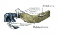
Embryo CRL 24 mm (outer aspect, about Carnegie stage 22)
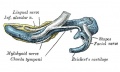
Embryo CRL 24 mm (inner aspect, about Carnegie stage 22)
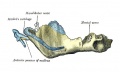
Embryo CRL 95 mm (outer aspect, about Fetal week 12, GA week 14)
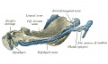
Embryo CRL 95 mm (inner aspectt, about Fetal week 12, GA week 14)


Birth

At birth mandible still has separate right and left halves.
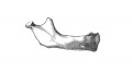
Birth
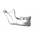
Childhood
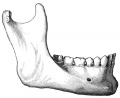
Adult
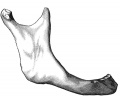
Old Age


Postnatal
Year 1 - Fusion of right and left halves of mandible at the symphysis.
Infancy and childhood - Increase in both size and shape of the mandible; eruption and replacement of teeth.
Year 12-14 - All permanent teeth emerged except third molars.
Data source- Table 4[7]
Carpal Bones
Data from a wrist study of 57 human embryonic (stages 17-23) and fetal (9-14 weeks).[10]
- stage 17 - undifferentiated mesenchyme
- stage 18 and stage 19 - condensated mesenchyme
- stage19 and stage 20 - pre-chondrogenic
- stages 21 and over - chondrogenic. Chondrification begins with capitate and hamate (stage 19) and ends with pisiform (stage 22).
- week 14 - vascular bud penetrates into the lunate cartilage.
Related nerve, muscle and joint development from the same study.
- stage 18 - hand plate median, ulnar and radial nerves and thenar eminence.
- stages 21 - indications of the interosseous muscles.
- stages 22 - flexor digitorum superficialis, flexor digitorum profundus and lumbrical muscles, transverse carpal ligament and collateral ligaments emerge.
- stages 23 - articular disc, radiocarpal and ulnocarpal ligaments and deep palmar arterial arch.
- week 9 - radiate carpal and interosseous ligaments appear.
- week 10 - dorsal radiocarpal ligament and articular capsule.
- week 13 - synovial membrane present.
Animal Models
Mouse
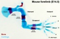
Forelimb E14.5
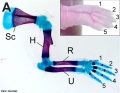
Forelimb E18.5
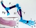
Hindlimb E18.5
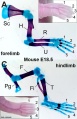
Limb E18.5


References
- ↑ Mall FP. On ossification centers in human embryos less than one hundred days old. (1906) Amer. J Anat. 5:433-458.Mall, F. P. On Ossification Centers in Human Embryos. The American Journ. of Anat. Vol. 5. 433-458 1906.
- ↑ 2.0 2.1 Franklin D & Flavel A. (2015). CT evaluation of timing for ossification of the medial clavicular epiphysis in a contemporary Western Australian population. Int. J. Legal Med. , 129, 583-94. PMID: 25398635 DOI.
- ↑ Skórzewska A, Grzymisławska M, Bruska M, Lupicka J & Woźniak W. (2013). Ossification of the vertebral column in human foetuses: histological and computed tomography studies. Folia Morphol. (Warsz) , 72, 230-8. PMID: 24068685
- ↑ Pafundi D, Lee C, Watchman C, Bourke V, Aris J, Shagina N, Harrison J, Fell T & Bolch W. (2009). An image-based skeletal tissue model for the ICRP reference newborn. Phys Med Biol , 54, 4497-531. PMID: 19556686 DOI.
- ↑ 5.0 5.1 Schmeling A, Schulz R, Reisinger W, Mühler M, Wernecke KD & Geserick G. (2004). Studies on the time frame for ossification of the medial clavicular epiphyseal cartilage in conventional radiography. Int. J. Legal Med. , 118, 5-8. PMID: 14534796 DOI.
- ↑ Flecker H. (1932). Roentgenographic observations of the times of appearance of epiphyses and their fusion with the diaphyses. (1932) J Anat. 67: 118-164.3 PMID 17104405
- ↑ 7.0 7.1 Zoetis T, Tassinari MS, Bagi C, Walthall K & Hurtt ME. (2003). Species comparison of postnatal bone growth and development. Birth Defects Res. B Dev. Reprod. Toxicol. , 68, 86-110. PMID: 12866701 DOI.
- ↑ 8.0 8.1 Keibel F. and Mall FP. Manual of Human Embryology I. (1910) J. B. Lippincott Company, Philadelphia.
- ↑ SYMONS NB. (1952). The development of the human mandibular joint. J. Anat. , 86, 326-32. PMID: 12980883
- ↑ Hita-Contreras F, Martínez-Amat A, Ortiz R, Caba O, Alvarez P, Prados JC, Lomas-Vega R, Aránega A, Sánchez-Montesinos I & Mérida-Velasco JA. (2012). Development and morphogenesis of human wrist joint during embryonic and early fetal period. J. Anat. , 220, 580-90. PMID: 22428933 DOI.
Online Textbooks
- Developmental Biology by Gilbert, Scott F. Sunderland (MA): Sinauer Associates, Inc.; c2000 Paraxial and intermediate mesoderm | Osteogenesis: The Development of Bones
- Molecular Biology of the Cell Alberts, Bruce; Johnson, Alexander; Lewis, Julian; Raff, Martin; Roberts, Keith; Walter, Peter New York and London: Garland Science; c2002 Search Molecular Biology of the CellBone Is Continually Remodeled by the Cells Within ItImage: Figure 22-52. Deposition of bone matrix by osteoblasts.Image: Figure 22-56. The development of a long bone.
Reviews
Laor T & Jaramillo D. (2009). MR imaging insights into skeletal maturation: what is normal?. Radiology , 250, 28-38. PMID: 19092089 DOI.
Land C & Schoenau E. (2008). Fetal and postnatal bone development: reviewing the role of mechanical stimuli and nutrition. Best Pract. Res. Clin. Endocrinol. Metab. , 22, 107-18. PMID: 18279783 DOI.
Articles
PARK EA. (1964). THE IMPRINTING OF NUTRITIONAL DISTURBANCES ON THE GROWING BONE. Pediatrics , 33, SUPPL:815-62. PMID: 14152896
Search PubMed
- Bone Development
Search Pubmed: Human Bone Development Timeline | Human Bone Development
External Links
External Links Notice - The dynamic nature of the internet may mean that some of these listed links may no longer function. If the link no longer works search the web with the link text or name. Links to any external commercial sites are provided for information purposes only and should never be considered an endorsement. UNSW Embryology is provided as an educational resource with no clinical information or commercial affiliation.
Terms
- growth recovery lines (growth arrest lines, Harris lines, Parks lines) - lines visible on x-ray images of increased bone density that represent the position of the growth plate at the time of insult to the organism and formed on long bones due to growth arrest. Insults can include juvenile malnutrition, disease or trauma.
Additional Images
Historic Images
| Historic Disclaimer - information about historic embryology pages |
|---|
|
- Mall

Fig 1 Embryo No. 333
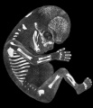
Fig 2 Embryo No. 284
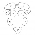
Fig 3 Occipital Ossification
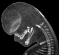
Fig 4 Embryo No. 284 Head
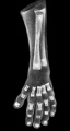
Fig 5 Embryo No. 300 Hand
Fig 6 Embryo No. 300 Leg


Glossary Links
- Glossary: A | B | C | D | E | F | G | H | I | J | K | L | M | N | O | P | Q | R | S | T | U | V | W | X | Y | Z | Numbers | Symbols | Term Link
Cite this page: Hill, M.A. (2026, July 26) Embryology Musculoskeletal System - Bone Development Timeline. Retrieved from https://embryology.med.unsw.edu.au/embryology/index.php/Musculoskeletal_System_-_Bone_Development_Timeline
- © Dr Mark Hill 2026, UNSW Embryology ISBN: 978 0 7334 2609 4 - UNSW CRICOS Provider Code No. 00098G