Lecture - Week 3 Development
| Embryology - 2 Jul 2026 |
|---|
| Google Translate - select your language from the list shown below (this will open a new external page) |
|
العربية | català | 中文 | 中國傳統的 | français | Deutsche | עִברִית | हिंदी | bahasa Indonesia | italiano | 日本語 | 한국어 | မြန်မာ | Pilipino | Polskie | português | ਪੰਜਾਬੀ ਦੇ | Română | русский | Español | Swahili | Svensk | ไทย | Türkçe | اردو | ייִדיש | Tiếng Việt These external translations are automated and may not be accurate. (More? About Translations) |
Week 3 | Placentation | Gastrulation | Notochord
Introduction
In this lecture I will talk about week 3 of embryonic development (GA week 5). I will cover concepts including early placentation, gastrulation and embryonic folding.
Objectives

- Understand the process early placentation, villi formation
- Understand broadly the events of week 3 of human development
- Understand the process of gastrulation
- Understand the process of axis formation and embryo folding
- Brief understanding of early cardiogenesis.
Lecture Resources
| Movies | |||||||||||||||||||
|---|---|---|---|---|---|---|---|---|---|---|---|---|---|---|---|---|---|---|---|
|
|
|
|
| |||||||||||||||
|
|
|
|
|
| References | |
|---|---|
 |
|
 |
The following chapter links only work with a UNSW connection.
|
 |
The following chapter links only work with a UNSW connection. |
| 2016 Lecture Video Recording |
|---|
| This 2016 lecture video recording is similar in content to the current 2018 lecture.
<html5media height="600" width="800">File:2016Lecture-Week-3.mp4</html5media> Click to play new window - 2016 Lecture Video (43.52 MB) |
| Recent Research |
|---|
|
Gao LR, Wang G, Zhang J, Li S, Chuai M, Bao Y, Hocher B & Yang X. (2018). High salt-induced excess reactive oxygen species production resulted in heart tube malformation during gastrulation. J. Cell. Physiol. , 233, 7120-7133. PMID: 29574800 DOI. Williams ML & Solnica-Krezel L. (2017). Regulation of gastrulation movements by emergent cell and tissue interactions. Curr. Opin. Cell Biol. , 48, 33-39. PMID: 28586710 DOI. Buers I, Pennekamp P, Nitschke Y, Lowe C, Skryabin BV & Rutsch F. (2016). Lmbrd1 expression is essential for the initiation of gastrulation. J. Cell. Mol. Med. , 20, 1523-33. PMID: 27061115 DOI. Scialdone A, Tanaka Y, Jawaid W, Moignard V, Wilson NK, Macaulay IC, Marioni JC & Göttgens B. (2016). Resolving early mesoderm diversification through single-cell expression profiling. Nature , 535, 289-293. PMID: 27383781 DOI. |
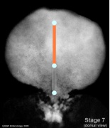
- Embryo Stage 7 (dorsal)
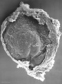
Embryonic disc (SEM)
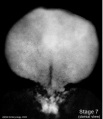
Embryonic disc
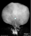
Primitive node and streak
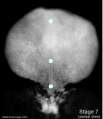
Oral and cloacal membranes
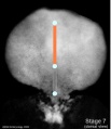
Axial process


Early Placentation

The trophoblast layer has now differentiated into two morphologically distinct cellular layers.
- Syncytiotrophoblasts - form a multinucleated cytoplasmic mass by cytotrophoblast cell fusion and both invade the decidua and secrete hCG
- Cytotrophoblasts - form a cellular layer around the blastocyst, proliferates and extends behind syncitiotrophoblasts
Early Utero-Placental exchange
- transfer of nutrition from maternal lacunae filled with secretions from uterine glands and maternal blood from blood vessels.
- development of trophoblast villi extending into the uterine decidua.
- ongoing process with placental development (covered in future lecture)

There are three stages of villi development:
- Primary Villi - cytotrophoblast
- Secondary Villi - cytotrophoblast + extraembryonic mesoderm
- Tertiary Villi - cytotrophoblast + extraembryonic mesoderm + blood vessels
There are two main types of early villi:
- Anchoring villi - attached to decidua
- Floating villi - not attached to decidua, floating in maternal lacunae.
Gastrulation
Embryonic Disc
- Gastrulation, (Greek, Gastrula = belly) means the formation of gut.
- used in a more looser sense to to describe the formation of the trilaminar embryo.
- Epiblast layer - consists of totipotential cells forming all 3 embryo layers (germ layers) that form the entire embryo.
- Historically, gastrulation was one of the earliest observable morphological event occurring in the frog embryo.
- Currently, the molecular and physical mechanisms that regulate patterning and migration during this key event are being investigated in several different animal models.
- In humans, it is proposed that similar mechanisms regulate gastrulation to those found in other vertebrates.

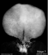
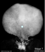
Embryonic Disc showing primitive streak (Carnegie stage 7) |

|
|

|
|
Primitive StreakThe primitive streak is the visible feature which represents the site of cell migration to form the additional layers.
|

|
Primitive Node
|
|

|
- Links: Gastrulation
Epithelial to Mesenchymal Transition

|

|
- Epithelial cells (organised cellular layer) which loose their organisation and migrate/proliferate as a mesenchymal cells (disorganised cellular layers) are said to have undergone an Epithelial Mesenchymal Transition (EMT).
- Mesenchymal cells have an embryonic connective tissue-like cellular arrangement
- cells that have undergone this process may at a later time and under specific signaling conditions undergo the opposite process, mesenchyme to epithelia.
- In development, this process can be repeated several times during tissue differentiation.
This process occurs at the primitive streak where epiblast cells undergo an epithelial to mesenchymal transition in order to delaminate and migrate.
MH - there are a number of common cellular changes that occur during embryonic development at different times and in different tissues, which we can classify into "developmental mechanisms".
Notochord
- The notochord is a structure which has an early mechanical role in embryonic disc folding and a major signaling role in patterning surrounding embryonic tissue development.
- A developmental feature not present in the adult anatomy.
- This signaling role patterns many different tissues (neural plate, neural tube, somites, endodermal organs).
- Has own sequence of development from a primitive axial process
- axial process an initial epiblast hollow epithelial tube which extends in the midline from the primitive pit, cranially in the embryonic disc (toward the oral membrane).
- neuroenteric canal is a transient communication between the amnionic cavity and the yolk sac cavity formed by the axial process.
- notochordal plate forms from the axial process merging with the endoderm layer.
- notochord forms from the notochordal plate which then separates back into the mesoderm layer as a solid column of cells lying in the midline of the embryonic disc and running rostro-caudally (head to tail).
- An alternate name for the notochord is "axial mesoderm".
MH - Much of our knowledge of this structure comes from the study of animal models of development.
- Embryo Stage 7 (dorsal)
Embryonic disc (SEM)
Embryonic disc
Primitive node and streak
Oral and cloacal membranes
Axial process
|
|
- Links: Notochord Movie | Mesoderm
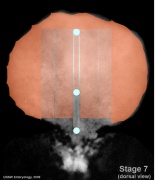
Embryo Folding
|

Embryonic Disc Folding - all edges of the embryonic disc will fold ventrally, forming a rostro-caudal "C" shaped tube. |
|
Mesoderm
- Embryo Stage 7 (dorsal)
Dorsal view
Primitive streak and node
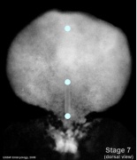
Oral and cloacal membranes
Axial mesoderm
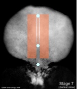
Paraxial mesoderm
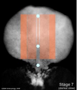
Intermediate mesoderm
Lateral plate


Cardiogenesis
- Heart - heart in week 3 begins as paired heart tubes.
- formed from prechordal splanchnic mesoderm.

Timeline

|
|
Next: Lab 1 - Preimplantation and Early Implantation |