File:Crowder1957 fig01.jpg
Crowder1957_fig01.jpg (515 × 463 pixels, file size: 91 KB, MIME type: image/jpeg)
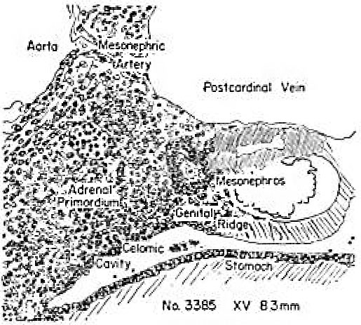
Fig. 1
Carnegie Embryo no. 3385 Carnegie stage 15
Figures 1 to 5, drawn to the same scale, X too, illustrate the relative size of the adrenal in horizons xiv to xvii, compared with surrounding structures. Figures 6 and 7 represent the disruption of the cortex by the neural elements in horizon xxiii.
Earliest evidence of adrenal primordium and change in character of cells in celomic epithelium over adrenal and gonadal areas.
| Week: | 1 | 2 | 3 | 4 | 5 | 6 | 7 | 8 |
| Carnegie stage: | 1 2 3 4 | 5 6 | 7 8 9 | 10 11 12 13 | 14 15 | 16 17 | 18 19 | 20 21 22 23 |
- Links: fig 1 | fig 2 | fig 3 | fig 4 | fig 5 | fig 6 | fig 7 | fig 1-7 | plate 1 | plate 2 | plate 3 | 1957 Crowder | Adrenal Development
References
Crowder RE. The development of the adrenal gland in man, with special reference to origin and ultimate location of cell types and evidence in favor of the "cell migration" theory. (1957) Contrib. Embryol., Carnegie Inst. Wash. 36, 193-210.
Cite this page: Hill, M.A. (2024, April 26) Embryology Crowder1957 fig01.jpg. Retrieved from https://embryology.med.unsw.edu.au/embryology/index.php/File:Crowder1957_fig01.jpg
- © Dr Mark Hill 2024, UNSW Embryology ISBN: 978 0 7334 2609 4 - UNSW CRICOS Provider Code No. 00098G
File history
Click on a date/time to view the file as it appeared at that time.
| Date/Time | Thumbnail | Dimensions | User | Comment | |
|---|---|---|---|---|---|
| current | 22:26, 21 June 2017 | | 515 × 463 (91 KB) | Z8600021 (talk | contribs) |
You cannot overwrite this file.
File usage
The following 2 pages use this file: