Endocrine - Placenta Development
| Embryology - 27 Apr 2024 |
|---|
| Google Translate - select your language from the list shown below (this will open a new external page) |
|
العربية | català | 中文 | 中國傳統的 | français | Deutsche | עִברִית | हिंदी | bahasa Indonesia | italiano | 日本語 | 한국어 | မြန်မာ | Pilipino | Polskie | português | ਪੰਜਾਬੀ ਦੇ | Română | русский | Español | Swahili | Svensk | ไทย | Türkçe | اردو | ייִדיש | Tiếng Việt These external translations are automated and may not be accurate. (More? About Translations) |
Introduction
For complete notes on placenta development and function see Placenta Development.
Lecture - Placenta Development
| Placenta Links: placenta | Lecture - Placenta | Lecture Movie | Practical - Placenta | implantation | placental villi | trophoblast | maternal decidua | uterus | endocrine placenta | placental cord | placental membranes | placenta abnormalities | ectopic pregnancy | Stage 13 | Stage 22 | placenta histology | placenta vascular | blood vessel | cord stem cells | 2013 Meeting Presentation | Placenta Terms | Category:Placenta | ||
|
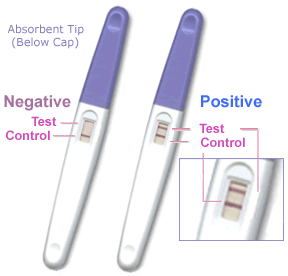
- Human chorionic gonadotrophin (hCG) - like leutenizing hormone, supports corpus luteum in ovary, pregnant state rather than menstrual, maternal urine in some pregnancy testing
- Human chorionic somatommotropin (hCS) - or placental lactogen stimulate (maternal) mammary development
- Human chorionic thyrotropin (hCT)
- Human chorionic corticotrophin (hCACTH)
- progesterone and estrogens - support maternal endometrium
- Progesterone-Associated Endometrial Protein (PAEP, pregnancy protein-14, PP14) - glycoprotein hormone from decidualized endometrium with possible immunoregulatory activities.
- IGF binding protein-1 (IGFBP-1) peptide hormone from decidualized endometrium increases during the first trimester, peaks during the second trimester, falls briefly, and peaks a second time before term.[1]
- Relaxin - corpus luteum then placenta in the first trimester, then declines in the second trimester. Binds to relaxin receptors on both the decidua and cytotrophoblasts.
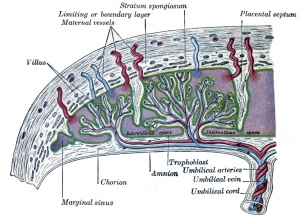
- Placenta - Maternal (decidua) and Fetal (trophoblastic cells, extra-embryonic mesoderm) components
- Endocrine function - maternal and fetal precursors, synthesis and secretion
- Protein Hormones - chorionic gonadotropin (hCG), chorionic somatomammotropin (hCS) or placental lactogen (hPL), chorionic thyrotropin (hCT), chorionic corticotropin (hCACTH)
- hCG - up to 20 weeks, fetal adrenal cortex growth and maintenance
- hCS – rise through pregnancy, stimulates maternal metabolic processes, breast growth
- Steroid Hormones - progesterone (maintains pregnancy), estrogens (fetal adrenal/placenta)
- Protein Hormones - chorionic gonadotropin (hCG), chorionic somatomammotropin (hCS) or placental lactogen (hPL), chorionic thyrotropin (hCT), chorionic corticotropin (hCACTH)
Some Recent Findings
|
| More recent papers |
|---|
|
This table allows an automated computer search of the external PubMed database using the listed "Search term" text link.
More? References | Discussion Page | Journal Searches | 2019 References | 2020 References Search term: Endocrine Placenta
|
| Older papers |
|---|
| These papers originally appeared in the Some Recent Findings table, but as that list grew in length have now been shuffled down to this collapsible table.
See also the Discussion Page for other references listed by year and References on this current page.
|
Human Chorionic Gonadotrophin
Human chorionic gonadotrophin (hCG) like leutenizing hormone, supports corpus luteum in ovary, pregnant state rather than menstrual. Presence in the maternal urine is the basis of some pregnancy testing.
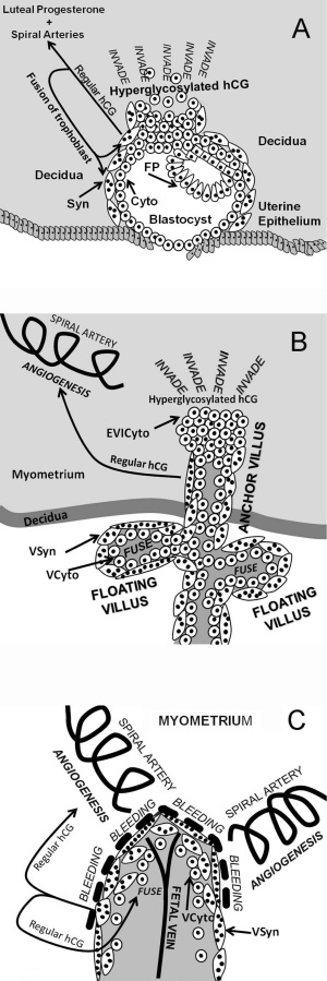
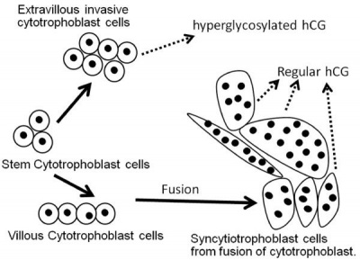
The level of the hyperglycosylated form (HCG-H) is high during the implantation phase and then decreases to the end of the first trimester.[8] In addition to the implantation role it has suggested immune cell modulation and endothelial functions.
Trophoblast cell hCG
- hCG Links: hCG | Trophoblast hCG function | Trophoblast cell hCG | implantation | placenta | NIH - The History of the Pregnancy Test
Placental Estrogen
Maternal late pregnancy shows a large increase in estriol levels.[9]
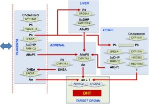
- fetal adrenal - gland production of dehydroepiandrosterone sulphate (DHEAS)
- fetal liver - conversion to 16α-OH-DHEAS
- placenta - sulphate removal to allow aromatization to estriol.
Placental estrogen, mainly estriol, suppresses gonadotropin secretion from the maternal pituitary gland. Maternal estrogen levels are often a useful indicator of fetal well being.[10][11]
- Uterus - stimulates growth of the myometrium, antagonizes the myometrial-suppressing activity of progesterone.
- Mammary Gland - stimulates mammary gland ductal and alveolar growth.
- Fetal Ovary - stimulates development of female fetal ovary.[12]
A second role for fetal adrenal DHEA-S is possible regulation of the effects of glucocorticoids on the developing brain.[5]
- Links: adrenal
Placental Progesterone
The fetal placenta production of progesterone throughout the second and third trimesters is a key to maintaining the uterus in the non-menstral pregnant state.[13] A second identified immunological role is in modulation of cytokine production.[14]
Human Chorionic Somatommotropin
Human chorionic somatommotropin hormone (hCS, CSH) is also called placental lactogen, due to its role in stimulating maternal mammary development, there may be little growth (somatogen) effects of this hormone. The hormone is structurally similar to both growth hormone (GH) and prolactin (PRL} and binds strongly to PRL receptors but weakly to GH receptors.
- Links: mammary gland
Cortiticotropin Releasing Hormone
The placenta synthesises urocortins (Ucn 1, Ucn 2, Ucn 3), cortiticotropin releasing hormone (CRH) analogues.[15] Appears to be produced by chorio-decidual cells.
Relaxin
The placenta and corpus luteum produce relaxin, a 6 kDa peptide hormone structurally similar to insulin. The hormone stimulates in early pregnancy both uterine growth and vascularization associated with implantation. It is also postulated to have other roles in the menstrual cycle[16]
- Links: implantation
Other Factors
- placental tachykinins
- neurokinin B
- endokinin
References
- ↑ Rutanen EM. (2000). Insulin-like growth factors in obstetrics. Curr. Opin. Obstet. Gynecol. , 12, 163-8. PMID: 10873115
- ↑ 2.0 2.1 O'Shaughnessy PJ, Antignac JP, Le Bizec B, Morvan ML, Svechnikov K, Söder O, Savchuk I, Monteiro A, Soffientini U, Johnston ZC, Bellingham M, Hough D, Walker N, Filis P & Fowler PA. (2019). Alternative (backdoor) androgen production and masculinization in the human fetus. PLoS Biol. , 17, e3000002. PMID: 30763313 DOI.
- ↑ Hong SH, Kim SC, Park MN, Jeong JS, Yang SY, Lee YJ, Bae ON, Yang HS, Seo S, Lee KS & An BS. (2019). Expression of steroidogenic enzymes in human placenta according to the gestational age. Mol Med Rep , 19, 3903-3911. PMID: 30896833 DOI.
- ↑ Costa MA. (2016). The endocrine function of human placenta: an overview. Reprod. Biomed. Online , 32, 14-43. PMID: 26615903 DOI.
- ↑ 5.0 5.1 Quinn TA, Ratnayake U, Dickinson H, Castillo-Melendez M & Walker DW. (2016). The feto-placental unit, and potential roles of dehydroepiandrosterone (DHEA) in prenatal and postnatal brain development: A re-examination using the spiny mouse. J. Steroid Biochem. Mol. Biol. , 160, 204-13. PMID: 26485665 DOI.
- ↑ Yamaguchi M, Ohba T, Tashiro H, Yamada G & Katabuchi H. (2013). Human chorionic gonadotropin induces human macrophages to form intracytoplasmic vacuoles mimicking Hofbauer cells in human chorionic villi. Cells Tissues Organs (Print) , 197, 127-35. PMID: 23128164 DOI.
- ↑ Kim JH, Shin MS, Yi G, Jee BC, Lee JR, Suh CS & Kim SH. (2012). Serum biomarkers for predicting pregnancy outcome in women undergoing IVF: human chorionic gonadotropin, progesterone, and inhibin A level at 11 days post-ET. Clin Exp Reprod Med , 39, 28-32. PMID: 22563548 DOI.
- ↑ Evans J. (2016). Hyperglycosylated hCG: a Unique Human Implantation and Invasion Factor. Am. J. Reprod. Immunol. , 75, 333-40. PMID: 26676718 DOI.
- ↑ Raeside JI. (2017). A Brief Account of the Discovery of the Fetal/Placental Unit for Estrogen Production in Equine and Human Pregnancies: Relation to Human Medicine. Yale J Biol Med , 90, 449-461. PMID: 28955183
- ↑ Rainey WE, Rehman KS & Carr BR. (2004). The human fetal adrenal: making adrenal androgens for placental estrogens. Semin. Reprod. Med. , 22, 327-36. PMID: 15635500 DOI.
- ↑ Parker CR. (1999). Dehydroepiandrosterone and dehydroepiandrosterone sulfate production in the human adrenal during development and aging. Steroids , 64, 640-7. PMID: 10503722
- ↑ Albrecht ED & Pepe GJ. (2010). Estrogen regulation of placental angiogenesis and fetal ovarian development during primate pregnancy. Int. J. Dev. Biol. , 54, 397-408. PMID: 19876841 DOI.
- ↑ Pavlicev M & Norwitz ER. (2018). Human Parturition: Nothing More Than a Delayed Menstruation. Reprod Sci , 25, 166-173. PMID: 28826363 DOI.
- ↑ Raghupathy R & Szekeres-Bartho J. (2016). Dydrogesterone and the immunology of pregnancy. Horm Mol Biol Clin Investig , 27, 63-71. PMID: 26812877 DOI.
- ↑ Fekete EM & Zorrilla EP. (2007). Physiology, pharmacology, and therapeutic relevance of urocortins in mammals: ancient CRF paralogs. Front Neuroendocrinol , 28, 1-27. PMID: 17083971 DOI.
- ↑ Marshall SA, Senadheera SN, Parry LJ & Girling JE. (2017). The Role of Relaxin in Normal and Abnormal Uterine Function During the Menstrual Cycle and Early Pregnancy. Reprod Sci , 24, 342-354. PMID: 27365367 DOI.
Reviews
Costa MA. (2016). The endocrine function of human placenta: an overview. Reprod. Biomed. Online , 32, 14-43. PMID: 26615903 DOI.
De Groot LJ, Chrousos G, Dungan K, Feingold KR, Grossman A, Hershman JM, Koch C, Korbonits M, McLachlan R, New M, Purnell J, Rebar R, Singer F, Vinik A, Tal R, Taylor HS, Burney RO, Mooney SB & Giudice LC. (2000). Endocrinology of Pregnancy. , , . PMID: 25905197
John RM. (2013). Epigenetic regulation of placental endocrine lineages and complications of pregnancy. Biochem. Soc. Trans. , 41, 701-9. PMID: 23697929 DOI.
Evain-Brion D & Malassine A. (2003). Human placenta as an endocrine organ. Growth Horm. IGF Res. , 13 Suppl A, S34-7. PMID: 12914725
Articles
Search PubMed
Search Pubmed: endocrine placenta development
Additional Images
External Links
External Links Notice - The dynamic nature of the internet may mean that some of these listed links may no longer function. If the link no longer works search the web with the link text or name. Links to any external commercial sites are provided for information purposes only and should never be considered an endorsement. UNSW Embryology is provided as an educational resource with no clinical information or commercial affiliation.
- National Institutes of Health (USA) Office of History - The History of the Pregnancy Test | of Pregnancy Testing
Terms
| Endocrine Terms | ||
|---|---|---|
| ||
|
Glossary Links
- Glossary: A | B | C | D | E | F | G | H | I | J | K | L | M | N | O | P | Q | R | S | T | U | V | W | X | Y | Z | Numbers | Symbols | Term Link
Cite this page: Hill, M.A. (2024, April 27) Embryology Endocrine - Placenta Development. Retrieved from https://embryology.med.unsw.edu.au/embryology/index.php/Endocrine_-_Placenta_Development
- © Dr Mark Hill 2024, UNSW Embryology ISBN: 978 0 7334 2609 4 - UNSW CRICOS Provider Code No. 00098G
