Embryology History - Hans Spemann
| Embryology - 30 Jul 2026 |
|---|
| Google Translate - select your language from the list shown below (this will open a new external page) |
|
العربية | català | 中文 | 中國傳統的 | français | Deutsche | עִברִית | हिंदी | bahasa Indonesia | italiano | 日本語 | 한국어 | မြန်မာ | Pilipino | Polskie | português | ਪੰਜਾਬੀ ਦੇ | Română | русский | Español | Swahili | Svensk | ไทย | Türkçe | اردو | ייִדיש | Tiếng Việt These external translations are automated and may not be accurate. (More? About Translations) |
Introduction
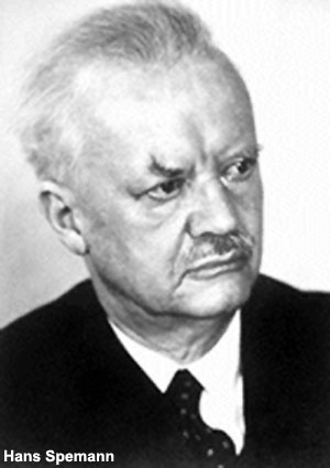
Hans Spemann (1869 - 1941) was a German embryologist who worked extensively on amphibian development and was the discoverer of the organiser region (or primitive node) the controller of gastrulation (1924). This region was also called the "Spemann's organiser". The same region in birds it is known as "Hensen's node" named for Victor Hensen (1835 – 1924) and is also known generally as the primitive node or knot.
Spemann received the 1935 Nobel Prize in Physiology or Medicine "for his discovery of the organizer effect in embryonic development". Below is the transcript from his Nobel Lecture 1935.
Viktor Hamburger was a graduate student in Spemann’s department at Freiburg during the years that the organizer graft experiments were being performed. Later in 1988 he wrote the book "The Heritage of Experimental Embryology: Hans Spemann and the Organizer".
- Links: Gastrulation | Frog Development | Week 3 | Historic - Manual of Human Embryology | Wilhelm Roux
| Embryologists: William Hunter | Wilhelm Roux | Caspar Wolff | Wilhelm His | Oscar Hertwig | Julius Kollmann | Hans Spemann | Francis Balfour | Charles Minot | Ambrosius Hubrecht | Charles Bardeen | Franz Keibel | Franklin Mall | Florence Sabin | George Streeter | George Corner | James Hill | Jan Florian | Thomas Bryce | Thomas Morgan | Ernest Frazer | Francisco Orts-Llorca | José Doménech Mateu | Frederic Lewis | Arthur Meyer | Robert Meyer | Erich Blechschmidt | Klaus Hinrichsen | Hideo Nishimura | Arthur Hertig | John Rock | Viktor Hamburger | Mary Lyon | Nicole Le Douarin | Robert Winston | Fabiola Müller | Ronan O'Rahilly | Robert Edwards | John Gurdon | Shinya Yamanaka | Embryology History | Category:People | ||
|
The Organizer-Effect in Embryonic Development

(Nobel Lecture, December 12, 1935)
The experiments which finally led to the discovery of the phenomena which are now designated as "organizer-effect" were prompted by a question which actually goes back to the beginnings of developmental mechanics, indeed to the beginnings of the history of evolution in general. How does that harmonious interlocking of separate processes come about which makes up the complete process of development? Do they go on side by side independently of each other (by "self-differentiation", Roux), but from the very beginning so in equilibrium that they form the highly complicated end product of the complete organism, or is their influence on each other one of mutual stimulation, advancement or limitation?
These questions, various answers to which constitute the theories of preformation or epigenesis, were lifted out of the realm of speculation up into that of an exact science when first Wilhelm Roux and then Hans Driesch used experimental methods in their research into development. The first experiments consisted in separating the individual parts of the embryo from each other and culturing them in isolation. This would show what each part was capable of by itself, while at the same time showing how far the developmental processes depending on them were dependent on or independent of each other.
In this way Roux was able after taking a frog's egg, pricking and destroying one of its two blastomeres, to obtain half an embryo from the other. Driesch, on the other hand, took a sea-urchin's egg, separated one segmental cell from the other and obtained a smaller but complete embryo. Further experiments showed that the differing results depended not on the material but on the method. The completely isolated segmental cell which has been reduced by half can grow into a whole in the case not only of the sea-urchin's egg, but also of amphibian's egg. This growth is inhibited if the dead cell is left attached; when this happens, the cell grows in accordance with its original determination, forming, first at least, half an embryo.
Even in those early days of research into developmental mechanics a second method of enquiry into this same question was discovered - that of "embryonic transplantation". Gustav Born observed that portions of young larval amphibians united if their freshly cut edges happened to come into contact with each other. He followed up this phenomenon and found that the individual portions were capable of self-differentiation to an astonishing degree.
It was from these premises that I began my experiments. They were all carried out on young amphibian embryos, mostly those of the common striped newt (Triton taeniatus). To make these experiments intelligible to the non-specialist it will be necessary in the first place to describe the main features in the normal development of these eggs.
Development begins immediately after fertilization, with a fairly protracted period of cell division which is called segmentation on account of the furrowing which appears on the surface. By the formation of an inner cavity or blastocoele, the blastocyst or blastula comes into being. Its lower, vegetative half (the thick floor of the blastocyst) consists of large cells rich in yolk, while the upper, animal half (the thin roof) is made up of numerous small cells poorer in yolk. Between the two is the marginal zone - a ring of medium-sized cells.
Next begins a very complicated and in many ways puzzling process: the so-called gastrulation. The end result of it is that all the material of the marginal zone and of the vegetative half of the blastula becomes invaginated and is thus covered over by animal material. Then along the line of invagination, i.e. the primitive orifice or blastopore, runs the outer layer of cells or ectoderm into the two invaginated layers, the mesoderm (originating from the marginal zone), and entoderm (corresponding to the vegetative half of the blastula, rich in yolk).
With this the primordia of the most important organs, the skin and central nervous system, vertebral column and musculature, gut and body cavity have in the main achieved their final dispositions. Their visible differentiation occupies the next phase of development.
The primordium of the central nervous system originates in the ectoderm of the dorsal surface, starting from the blastopore and coming forward as a thickened plate shaped like a shield with its anterior half broader than its posterior. This is the neural plate, and its lateral margins rise up as the neural folds. The neural folds are brought closer to each other and fused together so that the neural plate becomes a tube - the neural tube. This becomes separated from the epidermis and sinks below the surface. Its front end, which is thicker and originated in the broader anterior part of the neural plate will become brain; its thinner posterior part will become spinal cord. The neural plate lies over mesoderm. When the plate forms the neural tube, separates off and sinks below the surface, the mesoderm divides into five longitudinal strips lying side by side. The median strip is destined to be the axial skeleton or notochord. To the right and left of it is a row of mesodermal blocks or somites. These in turn are flanked on either side by the lateral plates from which arises the primordium of the coelum.
Finally, the entoderm first forms a broad open gutter, which is shaped like a trough. Its margins then bend inward towards the middle, and, along the mid-line - that is, just beneath the notochord - it completes the intestinal tube.
All these processes which, given a favourable temperature, go forward surprisingly quickly depend essentially not on the production of new material from the embryo substance but on the rearrangement of what is already there. It is therefore possible, and W. Vogt did this to perfection by means of staining, to show in the blastula or early gastrula, as it were, a topography of the rudiments of the presumptive organs.
In the face of this sort of topographical map we are again confronted with the question whether there is a real diversity in these parts which corresponds to the pattern of the presumptive rudiments in the early gastrula; whether they are more or less predestined, i.e. "determined", for their subsequent fate or whether they are still indifferent and do not have their ultimate determination impressed on them until later.
The first answer to this question was given by experiments in isolation. Thus, if the bisection is not made as early as between the two cells after the first segmentation but later, even at the blastula stage, or at that of the very young gastrula, you can still get twins. So up to this stage the cell material must still be to a large degree indifferent and capable of being used for various purposes in constructing the body. This becomes especially clear when the bisection is made in such a way that it separates the ventral half of the embryo from the dorsal half. Even then the latter half can develop into a miniature embryo of normal proportions. Here the new allocation of the material becomes perfectly clear. According to the evidence of our topographical map, the dorsal half contains almost all the material for the neural plate, i.e. much too much for a half-sized embryo; on the other hand, it lacks all of the presumptive epidermis. This latter must therefore be made good by material from the former.
Now if presumptive neural plate and presumptive epidermis are interchangeable, they must therefore also be interchangeable without prejudicing further normal development. Embryonic transplantation at this early stage must therefore produce different consequences than it would if performed in the later stages in which Gustav Born experimented.
It was on these thoughts and on the development of a way to facilitate the manipulation of these uncommonly fragile young embryos and operation upon them that the success of the new experiments rested.
The first experiment consisted in exchanging a portion of presumptive epidermis and neural plate between two embryos of the same age, each being at the beginning of gastrulation. The grafts took so smoothly and development proceeded so normally that their margins left no trace except that the grafted tissue itself was distinguishable for a while by means of its natural pigmentation, or by artificial vital staining. From this it was obvious hat, as we had expected, the portions were interchansable - that is to say, presumptive epidermis could become neural plate and presumptive neural plate could become epidermis.
From this we can infer not only the very indifferent nature of the cells at this early stage of development; the result allows the much more important conclusion that the transplanted portion must in its new environment be subjected to some kind of influence which determines its subsequent development.
It is here that the analytical superiority of this experiment is shown over the previous ones, whereby use was made of the regulation power of the embryo. For it was now possible to examine all the parts of the embryo separately for their active and reactive induction capacity, and also to vary the age and species of the implant with great latitude.
At the same time this opens important fresh possibilities: first of all in the matter of procedures. The interchangeability may be undertaken not only between embryos of the same species but also between those of different species, e.g. between embryos of Triton taeniatus which have a fair amount of pigmentation and those of Triton cristatus which have little or none. This allows us to distinguish the implant more or less clearly for a very long time even in sections and often to define its limits in terms of its cells. Let me describe a case of this kind in more detail.
A portion of presumptive neural plate was removed from an embryo Triton taeniatus at the beginning of gastrulation and exchanged with a portion of presumptive epidermis from a Triton cristatus embryo of the same age. The embryo in which the host was taeniatus later showed anteriorly and to the left in the neutral plate a smoothly grafted oblong area of white cristatus tissue which developed further into parts of the brain and eye. The other embryo with cristatus as the host showed on the right-hand side in the epidermis of the gill area a long streak of dark taeniatus tissue which developed further as epidermis and formed the covering of the outer gills. Since the portions have been exchanged, and since one portion is now settled where the other came from, we can see at once from sections that brain substance has come from presumptive epidermis, and epidermis has come from presumptive brain substance.
Because the implant in this "heteroplastic" transplantation remains distinguishable for a fairly long time it is possible to test the interchangeability of those parts of the embryo which develop inwards during gastrulation. We can, for example, establish whether the exchange is feasible not only as between one and the same layer of cells but also as between two different layers.
By and large this is in fact the case. So O. Mangold was able to show that mesodermal organs such as notochord, somites and pronephric ducts could arise from presumptive ectoderm by suitable transplantation at the beginning of gastrulation.
Now, when random samples were taken from the whole surface of the gastrula and transplanted in this way in an indifferent place it became apparent that a limited area, namely the region of the upper and lateral blastopore lip did not conform. A portion of this kind, transplanted in an indifferent place in another embryo of the same age did not develop according to its new environment but rather persisted in the course previously entered upon and constrained its environment to follow it. It invaginates altogether as if it were still in its old place, builds up part of the axial organs and completes itself out of the mesodermal environment. Above all, it induces in the overlying ectoderm a neural plate which closes to a tube, in favourable cases bulges out into optical vesicles and adds lenses and auditory vesicles.
First carried out at my instigation by Hilde Mangold, this experiment shows, therefore, that there is an area in the embryo whose parts, when transplanted into an indifferent part of another embryo, there organize the primordia for a secondary embryo. These parts were therefore given the name of "organizers" and the region of the embryos in which they are gathered together at the beginning of gastrulation was called the "centre of organization". H. Bautzmann has defined the limits of this area by systematic probing outwards and has found that it coincides more or less with the area of the presumptive notochord-mesoderm which invaginates later.
From these two facts - the development of an indifferent piece in conformity with its location and the inductive effect of an organizer - several series of experiments proceeded, connected with obvious questions. We will just touch on a few of them.
Since at first the organizer becomes invaginated, that is, completes the gastrulation it has begun, so that material in the neighbourhood can be included in the process, one might suppose that it is this process itself which causes further determination of the parts it has affected. But this is, to say the least, extremely unlikely, because the induction of neural plate takes place even though it has not itself been invaginated. This can be proved by a method which is highly significant for the whole progress of research. That is to say, those parts of the embryo which are being examined for their inductive capacity can be made to bypass the activte invagination and can be made effective by inserting them in the blastocoele through a small slit in the roof of the blastula or young gastrula which quickly heals over. The gastrulation does not suffer any essential disturbance from this and while it goes on, the blastocoele disappears and the piece we are examining comes to lie directly under the ectoderm and there shows what it is capable of. Thus a portion of the upper marginal zone of the blastula or early gastrula, or else a piece of the roof of the archenteron of the mature gastrula was planted in the blastocoele of a young gastrula and so brought beneath the ectoderm from the beginning; it was demonstrated that these portions were able to induce neural plate.
Now, these methods made it also possible to examine for their inductive capacity pieces which could not be embodied in the host embryo by any other means, either because they differed too much in age and origin or else because they were no longer living, or even not of living origin. We will have a look at these experiments next.
It had already been demonstrated in my early experiments that host and donor did not need to be exactly the same age in order to be able to work together. It was O. Mangold in particular who followed up this question and made the important discovery that the inductive reaction capacity is strictly limited in time while the inductive action capacity remains for a long time, far beyond the stage necessary for normal development.
This is true not only, as H. Bautzmann showed, for the notochord which normally induces in the earlier stages, but strangely enough also for a portion of embryo in which there would otherwise be no question if this kind of induction, viz. the neural plate. Both O. Mangold and I found simultaneously but independently, and starting from different lines of enquiry, that it can induce after transplantation. To this, O. Mangold added the important statement that the inductive capacity of this tissue persists into late stages, until there is a functioning brain in the hatched out larva.
Associated with this is the question whether and how far the inductive influence is specific in nature. Also, and this is connected with the other question, what role the action and reaction system plays in bringing about the highly complicated product of development. I had already expressed the opinion earlier that the inductive stimulus does not prescribe the specific character but releases that already inherent in the reaction system. The inductive potential already adduced of parts which have far exceeded the stage of observed normal effectiveness also points in the same direction. Still more is this true of the more recent experiments by Holtfreter which prove the extensive diffusion of factors which are able to induce a neural plate in the ectoderm of the young gastrula. So pretty well the whole animal kingdom from tapeworms to human beings was examined by the implantation method and shown to be capable of induction.
However, this does not only make obvious the largely unspecific character of the inducting agent; it also seems probable that it is chemical in nature. It was always thought to be so from the beginning. To make quite sure, experiments had to be made in which the inductor had been destroyed in various ways - by desiccation, freezing, or boiling. We got no clearly positive result from these first experiments; not until later similar ones by Holtfreter. It became apparent that this kind of treatment did not destroy the capacity of the inductors and, further, quite paradoxically, that this can in fact call forth such capacity in non-inductors.
The first experiment with a chemically treated inductor was carried out by Else Wehmeier and proved that an inductor immersed in 96% alcohol for 3f minutes did not lose its capacity.
After this, the chemical analysis was tackled in various quarters: in Germany by F. G. Fischer and E. Wehmeier, later with H. Lehmann, L. Jühling, and K. Hultzsch; in England by J. Needham, D. M. Needham, and C. H. Waddington. From the large number of separate results which still seem to be coming in I should like to draw attention to one only which is of the utmost importance in this connection. Chemically simple substances as, for example, synthetic oleic acid can nevertheless induce a complicated and in a certain sense complete structure such as a neural plate which will close over into a neural tube. Again, that would therefore indicate, as do some of the results from abnormal inductors, that most of the complication is based in the structure of the reaction system, and that the inductor has only a triggering and in some circumstances directing effect. Whether and, if so, how far and in what way such "unorganized inductors" (for it would be a contradiction in terms to speak here of "organizers") determine the direction is at the moment one of the most interesting but also most difficult questions.
But this broaches a new complex of questions which goes right back to the first induction experiments. It had already turned out in Hilde Mangold's experiments that the induced embryonic primordia were in the main arranged in the same direction as the primary ones and on a level with them. This seemed to emerge either from a general structural plan of the embryo or else from an influence of the primary embryonic primordia.
To investigate the former phenomenon, the similarity of direction of the constituents of the two embryos, two different experiments were set up. Upper blastopore lip still engaged in invagination was implanted in a different orientation in relation to the host embryo - crosswise and opposite to the orientation of the later primary primordia. With crosswise implantation it was shown that the invaginating cells of the graft were carried along by the gastrulating movements of the host and that thus the substratum was laid down along the long axis of the embryo. With opposite implantation the cells of the graft migrating against the stream get jammed but are not deflected. A controlling structure of the embryo, therefore, only works in so far as it determines the direction of the gastrulation movements both of the host embryo and the graft. It becomes even more obvious when a piece of the roof of the archenteron is planted in the blastocoele. The graft does not lie fixed in the cell formation of the host embryo so it can rather keep its original position and the induced secondary embryo primordia can be either crosswise or entirely opposite to those of the primary.
Of even greater interest, perhaps, is the result of the experiments which were to explain how the secondary primordia of the embryo were on the same level. For example, it can be seen that the auditory vesicles of both lie in nearly the same cross section of the embryo. In order to find out the cause of this regional determination or at least to establish its position the implantation was varied in two ways. To understand this we must remember one simple fact about development. In the course of gastrulation the invaginating material is rolled inwards around the upper lip of the blastopore. Thus, the material first invaginated lies farthest towards the front underneath the subsequent brain, while material invaginating later underlies the future spinal cord. Now it could be that the substratum of the head also determines the brain character of the anterior end of the neural plate ("head-organizer") and the substratum of the trunk area determines the character of the spinal cord ("trunk-organizer"). In order to test this, a portion of upper blastopore lip at the beginning of gastrulation (head-organizer) and one from an advanced and mature gastrula (trunk-organizer) were transplanted in the same place in an early gastrula, i.e. at the site where the lower blastopore lip would later develop; this was done also at different sites - in the head and trunk areas. It was shown that in fact something like a head- and trunk-organizer does exist, since the former is able to induce a brain also in the trunk region. It was shown moreover that the level in the embryo at which the induction takes place co-determines its nature, since at the head level even a trunk-organizer can induce a brain.
We have already indicated above that this last could have two different reasons. It could be that the disposition for building the head surrounds the whole embryo at head level in a broad circular band. But it could equally well be that a regional differentiating influence is exerted by the primary embryo primordia which co-determines the shape of the secondary embryo. In the region of the primary brain, respectively its primordia, there would be a "brain area" in which neural substance which had been stimulated by induction would develop into brain.
On the basis of definite facts established by experiment, Holtfreter has decided against the first and in favour of the second possibility. Moreover he has in addition discovered some more extremely interesting examples of these "embryonic areas". As we have seen, inducing tissues retain their induction capacity for a long time, and far beyond the stage of development required in the normal course. That being so, in a normal-embryo neural substance would have to be induced afresh in the epidermis which lies over the neural tube or the somites, unless that tissue had already exceeded its ephemeral period of reaction capacity. We could therefore infer, what Holtfreter discovered in a different enquiry, that a young portion still capable of reaction would in fact behave differently in this site. And it really is true that in particles of ectoderm from early gastrula implanted superficially at different levels in older gastrula a great variety of inherent potencies is activated. It depends on the region, so that in an anterior area, brain with optic and aural vesicles is induced, while further back, notochord and pronephric ducts are induced, and further back still, little tails. That shows that even the older embryo is still riddled with "embryonic areas" which do not normally come to light but can be detected at any time by indicators rich in potencies.
These inductions between parts of different ages do not complete the embryo by replacing what has been taken away; they are not "complementary" (O. Mangold) as in the case of a graft of the same age from an exactly similar site. Rather do the induced parts develop according to site only in a general sense, through "autonomic" induction; they are produced in excess and have a certain independence (O. Mangold).
A still further series of questions and experiments arose out of the first induction experiments and we will just touch on these in conclusion. As said earlier the induction effect is also possible with heteroplastic transplantation, i.e. between embryos of different species. For example; presumptive brain of a Triton taeniatus embryo can be made into epidermis in the gill area of a Triton cristatus. But the outer gills covered by it will have taeniatus properties; that is to say, they will be similar not to those of the species which has caused their development (instead of that of brain) but will resemble that of the species from which the implant originates. Potencies are not transferred to the "gill area" of the host; it is merely that those potencies relevant to its location are awakened. And in heteroplastic transplantation these diverge somewhat from those of the host. If an exchange between samples of different genus or even between systematic groups remote from each other (xeno-plastic) were possible and followed by induction effects, very valuable conclusions could be expected.
In this respect there is another question that must be dealt with which cropped up during those first experiments: whether in fact the induced organ is laid down part for part or as a whole. From the example of the outer gills we were not able to answer the question, but we could do so from two other organs - the lens and the balancers.
In the Triton, as with most amphibia, the lens of the eye arises as a sequel to the optic cup and its size depends strictly on it. Thus, if the optic cup diminishes in size so does the lens. So it follows that the smaller eye of the Triton taeniatus has a smaller lens than the larger eye of Triton cristatus at the same stage of development. E. Rotmann now interchanged presumptive lens epidermis with presumptive ventral epidermis in each of the two species at the beginning of gastrulation. The lenses which are formed at a certainmoment thereafter follow the size and degree of development of the donor. This can be seen very clearly in the constricted lens primordia with early fibre development; but even quite early stages show lens growth in the epidermis which in one case is too large for the optic cup and in the other case too small. The lens potencies therefore react in the field that activates them not only qualitatively but also quantitatively in accordance with the heredity of the species to which they belong. The lens potencies are not stimulated by the optic cup to the extent within which, with its drawn-in retina layer, it comes into contact with the epidermis. Rather is the lens more or less put in hand as a whole with the epidermis.
The balancers behave in the same way in a further completely analogous experiment of Rotmann's. In its structure and in its angle to the head it is similar to the species from which the transplanted ectoderm is derived and not to the other from which the induction has proceeded.
Added to this problem of uniformity according to species there is another in those cases of xenoplastic transplantation in which organs of different morphological significance are situated in the same region. This is so, for instance, when the ectoderm of the presumptive mouth region is exchanged between the embryos of Urodela and Anura. In the newt larva, lateral to the head and beneath the eyes are two balancers, while the tadpole has beneath the mouth near the ventral mid-line two lower suction cups. Moreover, the newt has real teeth in its mouth which both in origin and structure are comparable to our own teeth. The tadpole's mouth, on the other hand, is furnished with horny jaws and little horny processes. These are quite different in origin and structure from real teeth and indeed have nothing to do with them morphologically. It has been an old dream of mine to substitute for the presumptive mouth region of a newt the foreign ectoderm which comes from a frog early in gastrulation, since I wanted to find out what kind of "armoury" the mouth would form then. This experiment has now been successfully carried out several times since then, and also the other way round. It was first performed at my instigation and in my Institute by O. Schottt, later by Holtfreter, O. Mangold, and E. Rotmann with results we expected but hardly dared hope for. In the mouth region of a Triton larva there arose from transplanted Anura ectoderm of the early gastrula, suction cups and horny jaws; in a tadpole, balancers arose from Urodela ectoderm. When the foreign implant was so narrow that it left the place of origin of the characteristic organs wholly or partly free, these could then themselves develop alongside.
After these results we can say with all certainty of the inducing stimulus that as regards what arises, it must be of a very special nature; but as to how it arises, it must be of a very general character. We have, however, no idea at all how the "mouth area" releases potencies of the "mouth structures", even when they are of an entirely different species.
The Nobel Prize in Physiology or Medicine 1935
Hans Spemann and the Organizer
The Heritage of Experimental Embryology - By Embryology History - Viktor Hamburger|Viktor Hamburger]]. Oxford University Press, New York, 1988. xii, 196 pp., illus. $29.95. Monographs on the History and Philosophy of Biology.
As reviewed by Jan A. Witkowski. Banbury Center, Cold Spring Harbor Laboratory.
Experimental embryology was one of the most exciting fields of research in biology in the early part of this century, when the application of experimental techniques to the embryo promised a rigorous, causal understanding of the processes involved in development. In Europe the leader in this field was Hans Spemann, and of all Spemann’s research none had a greater impact than the so-called organizer experiment. Viktor Hamburger has taken this experiment and the work it inspired as the central theme of a book describing what might be called the golden age of experimental embryology.
The organizer experiment was performed by Hilde Proescholdt, who, in 1921, began a series of experiments transplanting the dorsal lip of the blastopore from the gastrulae of Triturus cristatus to gastrulae of T. taeniatus. The transplanted tissue induced the formation of a second embryonic axis and in the most famous example included neural tube, notochord, intestine, and kidney tubules. There were two remarkable features of this second embryo; it was composed of both donor and host cells and its tissues were appropriately arranged. Spemann argued that the transplanted tissue contained a center that was endowed with the power to induce and organize the formation of an embryonic axis.
Hamburger describes Spemann’s experiments in detail, showing how they were designed with great ingenuity and _performed with the simplest of tools. He remarks that Spemann’s greatest strength was his analytical acumen—his ability to interpret the data from these simple experiments and to design new experiments to explore further his new insights. One of the highlights of Hamburger’s writing is his attempt to reconstruct the process of Spemann’s thinking, reconstructions that are plausible and illuminating. Hamburger also makes a valiant effort to explicate the increasing number of concepts—“differentiation cen ter,” “organization center,” “double assurance,” “labile determination,” “assimilative induction,” and so on—that came into use.
But while Spemann and the organizer experiment are central characters in the book, Hamburger describes the work of many others who were entranced by the developing embryo. Hamburger 1s _ well placed to do this, as he was himself one of the players, spending almost 10 years with Spemann. Some scientists whose work is described, such as Schotte, Mangold, and Vogt, are well known, but one of the pleasures of this book is that Hamburger gives due credit to minor players in the drama who are usually forgotten. A large part of the book is devoted to Johannes Holtfreter, whose genius and capacity for hard work are evident from his work on the organizer. As Hamburger says, Holtfreter was “simply bolder and more inventive and willing to take risks” than those who remained more closely associated with Spemann. Such inventiveness led to Holtfreter’s in vitro experiments and the analysis of heterologous inducers and the regional specificity of induction. Hamburger takes the story of the organizer through to the 1960s and the work of Chuang, Saxen, Toivonen, Yamada, and Tiedemann, and in the last chapter he discusses his views of the organizer phenomenon as a gradient system.
It is strange that Spemann has not received more attention, for he is the only embryologist to have been awarded a Nobel Prize. Spemann wrote an autobiography that has not been translated into English, and it is a pity that Hamburger does not provide more information about Spemann’s life outside science. Hamburger’s book is strictly a “scientific” biography and does not discuss what influence, if any, Spemann’s cultural background had on his approach to science.
But this is a minor point. The story of Spemann’s scientific work is fascinating and well worth telling. It is a historically interesting episode, for what began with such high expectations entered a period of what Saxen and Toivonen have called “post-war depression.” Hamburger shows how this disillusionment arose as a consequence of the difficulties of applying a reductionist approach (and especially biochemical analysis) to the problem of the organizer. By 1939, Joseph Needham was predicting that at least 50 years would be needed to obtain “certain knowledge about the chemical nature .. . of the substance involved in embryonic induction.” Those 50 years have gone, and although there are promising beginnings (J. Gurdon, Development 99, 285-306 [1987]) that goal has not yet been met. Hamburger hopes that his book may encourage the present generation to return to these old problems with the new, powerful techniques of modern biological research. But despite Hamburger’s efforts to reformulate and clarify the major concepts of experimental embryology, I am left with the impression that the molecular-cellular-developmental biologist will feel that the time is not yet nght for an all-out assault on embryonic induction.
References
De Robertis EM. (2009). Spemann's organizer and the self-regulation of embryonic fields. Mech. Dev. , 126, 925-41. PMID: 19733655 DOI.
Harland R. (2008). Induction into the Hall of Fame: tracing the lineage of Spemann's organizer. Development , 135, 3321-3. PMID: 18820177 DOI.
Lagercrantz H. (2006). Hans Spemann (1869-1941): discoverer of the neuronal organizer. Acta Paediatr. , 95, 386-7. PMID: 16720481 DOI.
Mikhailov AT & Gorgolyuk NA. (2001). Consequences of the Spemann-Mangold organizer concept for embryological research in Russia: personal impressions. Int. J. Dev. Biol. , 45, 83-96. PMID: 11291874
Harland R & Gerhart J. (1997). Formation and function of Spemann's organizer. Annu. Rev. Cell Dev. Biol. , 13, 611-67. PMID: 9442883 DOI.
Fässler PE. (1996). Hans Spemann (1869-1941) and the Freiburg School of Embryology. Int. J. Dev. Biol. , 40, 49-57. PMID: 8735910
Steinbeisser H. (1996). The impact of Spemann's concepts on molecular embryology. Int. J. Dev. Biol. , 40, 63-8. PMID: 8735912
Allen GE. (1993). Inducers and 'organizers': Hans Spemann and experimental embryology. Hist Philos Life Sci , 15, 229-36. PMID: 8153264
Search Pubmed
Search PubMed: Spemann H Author | Spemann
External Links
External Links Notice - The dynamic nature of the internet may mean that some of these listed links may no longer function. If the link no longer works search the web with the link text or name. Links to any external commercial sites are provided for information purposes only and should never be considered an endorsement. UNSW Embryology is provided as an educational resource with no clinical information or commercial affiliation.
Glossary Links
- Glossary: A | B | C | D | E | F | G | H | I | J | K | L | M | N | O | P | Q | R | S | T | U | V | W | X | Y | Z | Numbers | Symbols | Term Link
Cite this page: Hill, M.A. (2026, July 30) Embryology Embryology History - Hans Spemann. Retrieved from https://embryology.med.unsw.edu.au/embryology/index.php/Embryology_History_-_Hans_Spemann
- © Dr Mark Hill 2026, UNSW Embryology ISBN: 978 0 7334 2609 4 - UNSW CRICOS Provider Code No. 00098G