Book - Normal Plates of the Development of Vertebrates 11
| Embryology - 26 Jul 2026 |
|---|
| Google Translate - select your language from the list shown below (this will open a new external page) |
|
العربية | català | 中文 | 中國傳統的 | français | Deutsche | עִברִית | हिंदी | bahasa Indonesia | italiano | 日本語 | 한국어 | မြန်မာ | Pilipino | Polskie | português | ਪੰਜਾਬੀ ਦੇ | Română | русский | Español | Swahili | Svensk | ไทย | Türkçe | اردو | ייִדיש | Tiếng Việt These external translations are automated and may not be accurate. (More? About Translations) |
Eycleshymer AC. and Wilson JM. Normal Plates of the Development of the Salamander Embryo (Nectürüs maculosus). Vol. 11 in series by Keibel F. Normal plates of the development of vertebrates (Normentafeln zur Entwicklungsgeschichte der Wirbelthiere) Fisher, Jena., Germany.
| Online Editor |
|---|
 This is the eleventh volume by Eycleshymer and Wilson published in 1910 in the series Normal Plates of the Development of Vertebrates edited by Franz Keibel.
|
| Historic Disclaimer - information about historic embryology pages |
|---|
|
Normal Plates of the Development of the Salamander Embryo (Nectürüs maculosus)
Normentafeln zur Entwicklungsgeschichte der Wirbeltiere by Franz Keibel

By
Albert C. Eycleshymer and James M. Wilson.
St. Louis University, St. Louis Mo., U.S.A.
With 3 Plates.
Jena,
Verlag Von Gustav Fischer.
1910.
Preface
The preparation of the normal tables and plates on Nedurus was begun several years ago by Professor C. O. Whitman but on account of unavoidable circumstances the work was delayed. It was later taken up by Professor Eycleshymer upon the suggestion of both Professor Whitman and Professor Keibel. While Professor Whitman has not directly participated in the later work, he has furnished the senior author with material and Information without which it would have been impossible to complete the work. In the completion of the work the senior author has been fortunate in having the able Cooperation of Professor James M. Wilson.
Table of Contents
|

|
Description of lllustrations
The series of eggs, embryos and larvae of Necturus, from which the following descriptions and the appended illustrations were made, were collected May I5th, 1903 and kept at a water temperature of 17o — 18o C. The illustrations are copied from the original water colored pictures which were made by Mr. Leonard H. Wilder, under the direction of the senior author. It should be emphasized that the ages, measurements and illustrations are all made from the living objects.
Note - Magnifications refer to original print versions.
Plate 1
| Fig. I. (X 10.) Side view of egg 1 day 4 hrs. after deposition. The first cleavage groove has reached the lower pole of the egg. Second grooves extend to level of the equator of the egg.
Fig. 2. (X 10.) Side view of egg 1 day 8 hrs. after deposition. The second cleavage grooves have reached the equator. The grooves of the third cleavage pass in meridional planes, but have not yet reached the equator. Fig. 3. (X 10.) Side view of egg 1 day 12 hrs. old. Five cleavage grooves have reached lower pole, dividing lower hemisphere into six segments. Fig. 4. (X 10.) Side view of egg 1 day 16 hrs, old. The greater number of cleavage grooves pass in meridional planes, many are latitudinal and some nearly radial. The upper surface of the egg shows sixteen segments, the lower nine. Fig. 5. (X 10.) Side view of egg 1 day 20 hrs. old. The upper surface of the egg shows some fifty segments, the lower nine. Fig. 6. (X 10.) Side view of egg 2 days 2 hrs. old. The upper surface of the egg shows more than one hundred segments, the lower twelve. Fig. 7. (X 10.) Side view of egg 2 days 7 hrs. old. The upper surface of egg shows about two hundred cells. The lower portion is in about same stage as described in Fig. 6.
Side view of egg 2 days I2 hrs. old. The upper surface of egg shows some five hundred cells, the lower about forty. Fig. 9. (X 10.) Top view of egg 4 days 4 hrs. old. Segmentation cavity shows through thin translucent roof. Blastopore not präsent. Fig. 10. (X 10.) Bottom view of egg 6 days 16 hrs. old. Crescentic blastopore. Line of invagination sharply separates large yolk cells from small cells of blastodisc. Fig. II. (X 10.) Dorso-lateral view of egg 10 days 10 hrs. old. Large circular blastopore; faint indication of embryonic anläge. Fig. 12. (X 10.) Side view of egg 10 days 16 hrs. old. Large circular blastopore. Anlage of mesial portion of embryo above dorsal lip of blastopore. Segmentation cavity faintly outlined. |

|
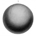
Fig. 1. (X 10.) Side view of egg 1 day 4 hrs. after deposition. The first cleavage groove has reached the lower pole of the egg. Second grooves extend to level of the equator of the egg.
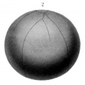
Fig. 2.
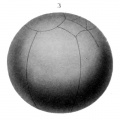
Fig. 3.
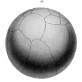
Fig. 4.
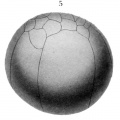
Fig. 5.
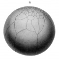
Fig. 6.
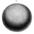
Fig. 7.
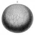
Fig. 8.
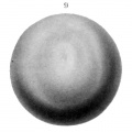
Fig. 9.
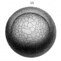
Fig. 10.
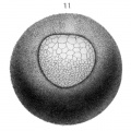
Fig. 11.
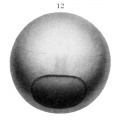
Fig. 12.


Plate 2
| Fig. 13. (X 10.)
Top view of egg 13 days 3 hrs. old. Small circular blastopore. Embryonic anläge triangulär in outline ; lateral boundaries indistinct. First appearance of neural groove. Roof of segmentation cavity thinner, making its boundaries distinct. Fig. 14. (X 10.) Top view of egg 14 days 4 hrs. old. Blastopore smaller, lateral margins of anterior portion of embryo bounded by short broad ridges which are the beginnings of the lateral portions of the neural fold. At anterior margin of embryo there is a transverse crescentic ridge which is beginning of transverse portion of neural fold. Neural groove deep but does not extend either to transverse portion of neural fold or to blastopore. Segmentation cavity crescentic.
Fig. 16. (X 10.) Top view of egg 15 days 15 hrs. old. Blastopore small, circular; yolk plug visible. Neural fold prominent, its free ends extend nearly to blastopore. Neural groove deep and narrow at anterior end, broad and shallow at posterior end, fades out just in front of blastopore. A part of the segmentation cavity is still apparent in front of the embryo. Fig. 17. (X 10.) Top view of egg 16 days 6 hrs. old. Blastopore reduced to a very minute circular aperture. Neural plate narrower than in preceding stage. Neural fold prominent, its free ends coalescing at blastopore. Neural groove extends to transverse portion of fold but does not reach blastopore. Segmentation cavity no longer visible in surface views. Fig. 18. (X 10.) Top view of embryo 17 days 2 hrs. old. Blastopore an elongated narrow aperture between ends of neural fold. Neural plate narrower than in preceding stage. The constricted portion represents in a general way the division between head and trunk. Neural fold most prominent in head region. Fig. 19. (X 10.) Top view of egg 17 days 17 hrs. old. Blastopore no longer visible. Neural plate narrowest posteriorly; broad in head region, showing boundary zone between head and trunk. Lateral portions of fold coalesced at posterior end of embryo. At anterior end of embryo a deep groove partially separates the two halves of the neural fold. Fig. 20. (X 10.) Top view of egg 18 days 13 hrs. old. Lateral portions of neural fold almost united except in head region where they are still widely separated. In the antero-lateral portions of the fold are slight evaginations which are the beginnings of the optic vesicles. Fig. 21. (X 10.) Top view of egg 18 days 15 hrs. old, 3 or 4 pairs of myotomes. Lateral portions of neural fold widely separated in head region, more closely approximated in anterior trunk region, coalesced in taii. Fig. 22. (X 10.) Dorso-lateral view of embryo 20 days 10 hrs. old, length 6 mm, 6 pairs of myotomes. Outline of body conforms to curvature of egg. Head end of embryo shows three longitudinal ridges; middle ridge lies slightly above level of lateral ridges. The middle one is common anläge of fore, mid and bind brain. The lateral ones are the common anläge of the optic vesicles and branchial arches. Anus formed. Fig. 23. (X 10.) Side view of embryo 21 days 2 hrs. old, length 7 mm, 10 — 12 pairs of myotomes. General outline of body conforms to curvature of egg. Head slightly raised above surface of yolk. Slight enlargement at end of tail. A distinct enlargement of anterior end of head shows optic vesicles; just posterior to this enlargement is the anläge of the branchial arches. Anus shows just below tip of tail. Fig. 24. (X 10.) Dorso-lateral view of embryo 22 days 17 hrs. old, length 8 mm, l6~i8 pairs of myotomes. Embryo much curved laterally. Anterior half of head free from yolk. Caudal enlargement more prominent. Optic vesicles and mandibular arch well defined. The hyoid and first branchial arches are discernible ; also the common anläge of the second and third branchial arches. |

|
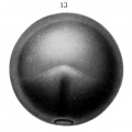
Fig. 13.
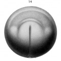
Fig. 14.
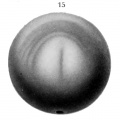
Fig. 15.
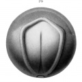
Fig. 16.
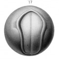
Fig. 17.
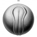
Fig. 18.
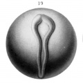
Fig. 19.
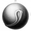
Fig. 20.
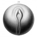
Fig. 21.
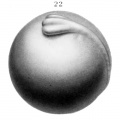
Fig. 22.
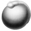
Fig. 23.
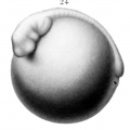
Fig. 24.


Plate 3
| Fig. 25. (X 5.)
Side view of embryo 23 days 10 hrs. old, length 9 mm, 20 — 22 pairs of myotomes. General outline of the body straighter. Head free from yolk. Caudal enlargement becoming free. Optic vesicles and forebrain much larger. Mandibular, hyoid, first branchial, and common anläge of second and third branchial arches well defined. Otic vesicle visible above hyoid arch.
Side view of embryo 24 days 22 hrs. old, length 10 mm, 23 — 24 pairs of myotomes. General outline of body of embryo straighter, less curved laterally. Head and caudal extremities free from yolk. Yolk becoming oval. Optic vesicles prominent. Ear better defined. Olfactory pits present. The mandibular, hyoid and first branchial arches are distinct. The second and third branchial arches are not yet differentiated, a slight process on the first branchial indicates the beginning of the gill bar. The anläge of the heart is visible just beneath the arches. Fig. 27. (X 5.) Side view of embryo 26 days old , length 1 1 mm , 26—27 myotomes. General outline of body straighter than in preceding stage. Head projects some 3 mm beyond margin of yolk; tail projects 1.2 mm, is thinner laterally but broader dorso-ventrally. Eye, ear, nasal pits and mouth well defined. Maxillary process discernible. Mandibular arches longer, but ventral ends widely separated. Second and third branchial arches formed. Gill bars present on three branchial arches. Anterior limb buds indicated; faint anläge of posterior limb buds. Yolk pear-shaped. Heart prominent. First surface capillaries present although not indicated in figure. Fig. 28. (X 5.) Side view of embryo 30 days 8 hrs. old, length 13 mm, 30 — 31 myotomes. The trunk of the embryo is nearly straight. At level of the posterior gill there is a pronounced neck bend and at the level of the posterior limbs a striking downward bend of the tail. The epiphysis shows in surface views. The lens is discernible. The ear is still visible. The external nasal openings are sharply defined. The boundaries of the mouth are better outlined owing to the approximation of the ventral ends of the mandibular arches. The hyoid arch is becoming obscured. The gill bars are prominent on the three branchial arches. The anterior limb buds project dorsally about .5 mm above the surface of the bod3\ The posterior limb buds are but slight elevations. The yolk is pear-shaped with its dorsal surface much flattened. The auricular and ventricular portions of the heart are apparent. The surface of the yolk is covered by a dense network of capillaries which for the most part convey blood antero-ventrally to the abdominal vein. Considerable pigment is present in the trunk region although but little has reached the outer portion of the dermis. Fig. 29. (X 5.) Side view of embryo 36 days 16 hrs. old, length 16 mm, 36-38 myotomes. In general outline the embryo shows a number of striking changes. The neck bend is not so pronounced. The tail bend is scarcely noticeable. There is a striking increase in dorso-ventral width of tail. The cerebral hemispheres are well defined. The eye is now prominent and the lens better defined. The ear is no longer visible in surface views. The mouth is well defined. The ends of the mandibular arches are closely approximated but not united. The hyoid and branchial arches are more obscure. Anlagen of gill fiiaments present on gill bars. Anterior limbs project dorsally. Posterior limbs are short ridges extending in horizontal plane. The yolk is elongated and reduced in diameter both dorso-ventrally and laterally. Surface blood vessels as in preceding stage, excepting that they are now apparent in the gill bars. The chromatophores are most numerous in the anterior and dorsal portions of the head.
Fig- 31. (X 5.) Side view of larva 49 days old, lergth 21 mm. General outline of body decidedly different. Head bend obliterated, slight upward curve in trunk. Tail broader. Eye more deeply pigmented. Gill bars very long, extending to level of end of anterior limb. From three to five lateral filaments on each gill bar. Anterior limbs project postero-ventrally ; three digits formed. Posterior limbs directed caudad ; no trace of digits. Yolk much elongated. Network of capillaries denser. Large lateral arteries, at level of upper margin of yolk, very prominent. Well defined longitudinal bands of pigment. Fig: 32. (X 5.) Side view of larva 61 days old, length 25 mm. General outline of body shows less dorsal curvature of trunk. Tail much longer in proportion to length of trunk and much broader dorso-ventrally. Gill bars longer, each posessing six to eight lateral filaments. Anterior and posterior limbs directed postero-ventrally. Anterior 3 mm long, posterior 2 mm long. Each limb shows four digits. The distribution of pigment is essentially similar to that observed in the 21 mm larva, the bands however are more sharply defined. Chromatophores in the gill bars and limbs and beginning to extend over the dorsal surface of the yolk. Fig. 33. (X 5.) Side view of larva 70 days 4 hrs. old, length 28 mm. The general outline of the body is slenderer than at any time preceding. The rapid absorption of the yolk has brought its ventral surface nearly to the level of the ventral surfaces of the head and tail. The gill bars curve dorsally and possess fi;om ten to twelve pairs of lateral filaments. The tail is somewhat constricted at the level of the posterior limbs. The limbs and digits are better developed and are now used in locomotion. Pigmentation is denser than in 25 mm larva, but same general arrangement of bands prevails. Fig. 34. (X 5.) Side view of larva 97 days old, length 34 mm. In general outline the larva begins to resemble the adult. The yolk is well absorbed. The tail is very broad and now used as a powerful caudal fin in swimming. The gill bars project dorsally and have a large number of filaments. The legs project far below the ventral surface of the body. In color the same general pattern prevails as in the 28 mm larva. There are some minor changes, the Hght band is broader and better defined, and extensions of pigment over the yolk have been so uneven that a number of irregulär oval areas are left unpigmented, causing a mottled appearance in this region. Fig. 35. (X 5.) Side view of larva 126 days old, length 39 mm. The young Nedurus now conforms in outline to the adult. In color hovvever it is decidedly different. |
|
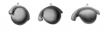
Fig. 25-27.
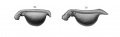
Fig. 28-29.
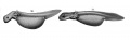
Fig. 30-31.
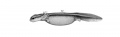
Fig. 32.
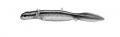
Fig. 33.
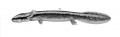
Fig. 34.
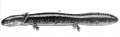
Fig. 35.
| Historic Disclaimer - information about historic embryology pages |
|---|
|
| Embryologists: William Hunter | Wilhelm Roux | Caspar Wolff | Wilhelm His | Oscar Hertwig | Julius Kollmann | Hans Spemann | Francis Balfour | Charles Minot | Ambrosius Hubrecht | Charles Bardeen | Franz Keibel | Franklin Mall | Florence Sabin | George Streeter | George Corner | James Hill | Jan Florian | Thomas Bryce | Thomas Morgan | Ernest Frazer | Francisco Orts-Llorca | José Doménech Mateu | Frederic Lewis | Arthur Meyer | Robert Meyer | Erich Blechschmidt | Klaus Hinrichsen | Hideo Nishimura | Arthur Hertig | John Rock | Viktor Hamburger | Mary Lyon | Nicole Le Douarin | Robert Winston | Fabiola Müller | Ronan O'Rahilly | Robert Edwards | John Gurdon | Shinya Yamanaka | Embryology History | Category:People | ||
|
Reference
Eycleshymer AC. and Wilson JM. Normal Plates of the Development of the Salamander Embryo (Nectürüs maculosus). Vol. 11 in series by Keibel F. Normal plates of the development of vertebrates (Normentafeln zur Entwicklungsgeschichte der Wirbelthiere) Fisher, Jena., Germany.
Glossary Links
- Glossary: A | B | C | D | E | F | G | H | I | J | K | L | M | N | O | P | Q | R | S | T | U | V | W | X | Y | Z | Numbers | Symbols | Term Link
Cite this page: Hill, M.A. (2026, July 26) Embryology Book - Normal Plates of the Development of Vertebrates 11. Retrieved from https://embryology.med.unsw.edu.au/embryology/index.php/Book_-_Normal_Plates_of_the_Development_of_Vertebrates_11
- © Dr Mark Hill 2026, UNSW Embryology ISBN: 978 0 7334 2609 4 - UNSW CRICOS Provider Code No. 00098G