ANAT2241 Respiratory System

| ANAT2241 This practical support page content is not part of the virtual science practical class and provides additional information for student self-directed learning purposes. All practical class pages are located on Moodle - ANAT2241 |
General Objective

To know the histological and cytological components of the conducting and respiratory portions of the respiratory system.
Specific Objectives
- To know the cytology of the mucosa of the nasal cavity and upper respiratory tract.
- To know the structure of the trachea and extrapulmonary bronchi.
- To know the microanatomy of the lung and histological features of intrapulmonary bronchi, bronchioles, terminal bronchioles and respiratory bronchioles.
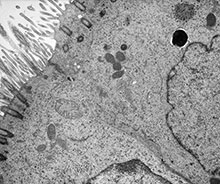
- To know alveolar ducts and alveoli, and the ultrastructure of their walls.
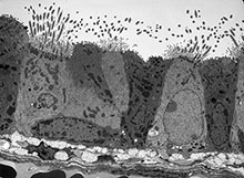
- To know the structure and ultrastructure of olfactory epithelium.
Learning activities
Examine the following virtual slides, and in your course manual identify, draw and label the following structures and note their function:
Virtual Slides: Respiratory System
Nasal Respiratory

|

|
| Nasal respiratory epithelium (inferior concha) | Nasal respiratory epithelium (detail) |

|

|
| Nasal cavity olfactory | Nasal cavity olfactory (detail) |
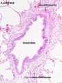
Larynx
Virtual Slide - Larynx (annotated)
Larynx Anatomy


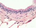
Trachea


Bronchiole


Lung

|

|
|

|


|

|

|

|
| Alveolar Duct | Alveoli | Alveoli Elastin | Lung Reticular Fibres |
Electron Micrograph
|
|
| |||||||||
|
|
Gallery
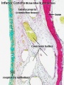
Nasal respiratory epithelium (inferior concha)
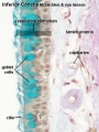
Nasal respiratory epithelium (detail)
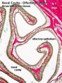
Nasal cavity olfactory
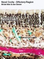
Nasal cavity olfactory (detail)
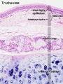
Trachea 1
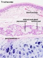
Trachea 2
Bronchiole
Respiratory bronchiole
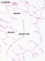
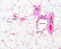
Alveolar Duct
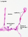
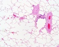
Alveoli

Alveoli Elastin
labeled lung
unlabeled lung
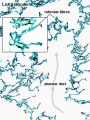
Lung reticular fibres
- Respiratory Histology: Bronchiole | Alveolar Duct | Alveoli | EM Alveoli septum | Alveoli Elastin | Trachea 1 | Trachea 2 | labeled lung | unlabeled lung | Respiratory Bronchiole | Lung Reticular Fibres | Nasal Inferior Concha | Nasal Respiratory Epithelium | Olfactory Region overview | Olfactory Region Epithelium | Histology Stains
Developmental
- Fetal Respiratory: late canalicular | unlabeled late canalicular | Hyaline cartilage | Respiratory Histology
Course Links
- Histology Glossary: A | B | C | D | E | F | G | H | I | J | K | L | M | N | O | P | Q | R | S | T | U | V | W | X | Y | Z | ANAT2241 Support | Histology | Histology Stains | Embryology Glossary
| Common Histology Stains | ||||||||||||||||||||||||||||||||||||||||||||||||||||||||||||||||||||||||||||||||||||||||||||||||||||||||||||||||||||||||||||||||||||||||||||||||
|---|---|---|---|---|---|---|---|---|---|---|---|---|---|---|---|---|---|---|---|---|---|---|---|---|---|---|---|---|---|---|---|---|---|---|---|---|---|---|---|---|---|---|---|---|---|---|---|---|---|---|---|---|---|---|---|---|---|---|---|---|---|---|---|---|---|---|---|---|---|---|---|---|---|---|---|---|---|---|---|---|---|---|---|---|---|---|---|---|---|---|---|---|---|---|---|---|---|---|---|---|---|---|---|---|---|---|---|---|---|---|---|---|---|---|---|---|---|---|---|---|---|---|---|---|---|---|---|---|---|---|---|---|---|---|---|---|---|---|---|---|---|---|---|---|
| ||||||||||||||||||||||||||||||||||||||||||||||||||||||||||||||||||||||||||||||||||||||||||||||||||||||||||||||||||||||||||||||||||||||||||||||||
| ||||||||||||||||||||||||||||||||||||||||||||||||||||||||||||||||||||||||||||||||||||||||||||||||||||||||||||||||||||||||||||||||||||||||||||||||
Practical Support
- Pages can be accessed from any internet connected computer.
ANAT2241 Support Links: The Virtual Microscope | Covering and Lining Epithelia | Glandular Epithelia | CT Components | CT Types | Bone, Bone Formation and Joints | Muscle | Nervous | Blood | Eye | Cardiovascular | Respiratory | Integumentary | Gastrointestinal | Gastrointestinal Organs | Lymphatic and Immune | Endocrine | Urinary | Female Reproductive | Male Reproductive | Histology Stains | Histology Drawings | Practicals Health and Safety 2013 | Moodle - 2019
ANAT2241 This practical support page content is not part of the science practical class and provides only background information for student self-directed learning purposes.
Cite this page: Hill, M.A. (2026, July 6) Embryology ANAT2241 Respiratory System. Retrieved from https://embryology.med.unsw.edu.au/embryology/index.php/ANAT2241_Respiratory_System
- © Dr Mark Hill 2026, UNSW Embryology ISBN: 978 0 7334 2609 4 - UNSW CRICOS Provider Code No. 00098G