Search results
From Embryology
- --[[User:Z8600021|Mark Hill]] 15:11, 22 April 2012 (EST) Embryo Number Category Members Carnegie Embryo 1380 (2 members)3 KB (212 words) - 15:11, 22 April 2012
File:Stage14 bf12.jpg ==Human Embryo Carnegie Stage 14== Carnegie Collection Embryo No.7394(416 × 600 (25 KB)) - 13:09, 22 April 2012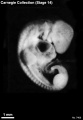
File:Stage14 bf14.jpg ==Human Embryo Carnegie Stage 14== Carnegie Collection Embryo No.7400(416 × 600 (22 KB)) - 13:04, 22 April 2012
File:Stage14 bf10.jpg ==Human Embryo Carnegie Stage 14== Carnegie Collection Embryo No.4154(416 × 600 (24 KB)) - 15:54, 21 April 2012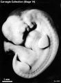
File:Stage14 bf8.jpg ==Human Embryo Carnegie Stage 14== Carnegie Collection Embryo No.6502(458 × 618 (33 KB)) - 15:56, 21 April 2012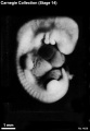
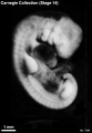
File:Stage14 bf11.jpg ==Human Embryo Carnegie Stage 14== Carnegie Collection Embryo No.{{CE4692}}(416 × 600 (24 KB)) - 14:27, 31 January 2019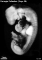
File:Stage14 bf13.jpg ==Human Embryo Carnegie Stage 14== Carnegie Collection Embryo No.6848(416 × 600 (26 KB)) - 13:09, 22 April 2012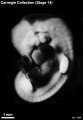
File:Stage14 bf15.jpg ==Human Embryo Carnegie Stage 14== Carnegie Collection Embryo No.7394(416 × 600 (23 KB)) - 13:08, 22 April 2012
File:Stage14 bf16.jpg ==Human Embryo Carnegie Stage 14== Carnegie Collection Embryo No.1620(416 × 600 (23 KB)) - 13:08, 22 April 2012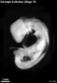
File:Stage14 bf17.jpg ==Human Embryo Carnegie Stage 14== Carnegie Collection Embryo No.7400(416 × 600 (23 KB)) - 13:05, 22 April 2012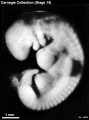
File:Stage14 bf5.jpg ==Human Embryo Carnegie Stage 14== Carnegie Collection Embryo No. {{CE6830}}(458 × 618 (32 KB)) - 10:46, 13 November 2018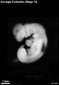
File:Stage14 bf9.jpg ==Human Embryo Carnegie Stage 14== Carnegie Collection Embryo No.5654(416 × 600 (18 KB)) - 15:54, 21 April 2012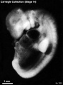
File:Stage14 bf7.jpg ==Human Embryo Carnegie Stage 14== Carnegie Collection Embryo No.7333(458 × 618 (29 KB)) - 15:53, 21 April 2012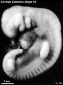
File:Stage14 bf6.jpg ==Human Embryo Carnegie Stage 14== Carnegie Collection Embryo No.1380(458 × 618 (35 KB)) - 18:00, 25 June 2015- ...negie Collection|Carnegie Embryos]] - [[Book - Contributions to Embryology Carnegie Institution No.50|Lineback (1920) - Human Colon Muscle]] ! width=200px|Carnegie No.1 KB (139 words) - 22:51, 6 February 2017
- ===All Carnegie Embryos listed=== [[Category:Carnegie Embryo 6]]5 KB (496 words) - 11:36, 29 July 2018
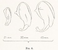
File:Streeter1922-fig06.jpg ...embryos of the Carnegie Collection: No. {{CE460}} (21 mm), No. {{CE417}} (32 mm), No. {{CE886}} (43 mm). X14. ...rnegie Embryo 460]] [[Category:Carnegie Embryo 417]] [[Category:Carnegie Embryo 886]](737 × 632 (42 KB)) - 13:48, 22 November 2017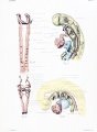
File:Congdon1922-plate01.jpg ...on of the second, are just appearing. Embryo No. {{CE2053}}, length 3 mm. (Carnegie Stage {{CS11}}) ===Figs. 31 and 32===(877 × 1,200 (145 KB)) - 21:21, 6 November 2018- ! width=200px|Carnegie No. | 32708 bytes (75 words) - 16:08, 30 January 2018
- ...images that relate to the [[Carnegie Collection]] Embryo No. {{CE6488}} [[Carnegie stage 12]] in [[Week 4]]. ! colspan=11| [[Carnegie Collection]] - [[Carnegie stage 12|Stage 12]] 2 members (0 subcategories, 2 files) - 17:40, 7 August 2017
- ...{{Embryology}} category shows [[Carnegie Collection]] embryo {{CE5154}}, [[Carnegie stage 23]] [[Week 8]]. {{Carnegie stage 23 links}}0 members (0 subcategories, 0 files) - 13:10, 3 July 2018
File:Streeter1957 plate04.jpg Fig. 32. No. {{CE4638}} [[Category:Carnegie Embryo 576]][[Category:Carnegie Embryo 8092]](1,500 × 1,986 (736 KB)) - 22:07, 30 March 2017
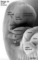
File:Stage14 sem5.jpg == Human Embryo Carnegie Stage 14== [[Carnegie stage 14|Carnegie Stage 14]] [[Week 5|day]] 32 somite 35, right dorsolateral view.(2,553 × 2,220 (313 KB)) - 09:21, 1 December 2016
File:Stage14 sem5a.jpg == Human Embryo Carnegie Stage 14== [[Carnegie stage 14|Carnegie Stage 14]] [[Week 5|day]] 32 somite 35, right dorsolateral view.(1,000 × 870 (85 KB)) - 09:21, 1 December 2016
File:Stage14 sem5b.jpg == Human Embryo Carnegie Stage 14== [[Carnegie stage 14|Carnegie Stage 14]] [[Week 5|day]] 32 somite 35, right dorsolateral view.(920 × 800 (75 KB)) - 09:21, 1 December 2016
File:Stage14 sem5c.jpg == Human Embryo Carnegie Stage 14== [[Carnegie stage 14|Carnegie Stage 14]] [[Week 5|day]] 32 somite 35, right dorsolateral view.(690 × 600 (48 KB)) - 09:21, 1 December 2016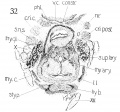
File:Lisser1911 fig32.jpg ==Fig. 32 Cross section of human Embryo 22 thyreoid cartilage, cricoid cartilage, and hyoid bone== [[:Category:Carnegie Embryo 22|'''Embryo no. 22''']] (20 mm. ) to show thyreoid cartilage, cricoid cartilage, and h(1,070 × 998 (242 KB)) - 21:04, 15 June 2016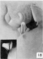
File:Spaulding-fig18.jpg ==Fig. 18. Carnegie Embryo No.1022d== 32 mm., male. X 6(445 × 607 (36 KB)) - 00:07, 15 April 2015
File:Stage14 sem2bl.jpg Human Embryo Carnegie stage 14, day 32, 35 somites(378 × 600 (43 KB)) - 10:09, 3 September 2009
File:Stage14 sem2b-limb.jpg ==Human Embryo Carnegie stage 14== Carnegie stage 14, day 32, 35 somites(378 × 600 (26 KB)) - 07:55, 9 May 2012
File:Stage14 sem2c-limb.jpg ==Human Embryo Carnegie stage 14== Carnegie stage 14, day 32, 35 somites(252 × 400 (14 KB)) - 14:27, 6 June 2011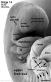
File:Stage14-sem2 limb.jpg ==Human Embryo Carnegie stage 14== Carnegie stage 14, day 32, 35 somites(630 × 1,000 (57 KB)) - 14:27, 6 June 2011
File:Stage14 sem2a-limb.jpg ==Human Embryo Carnegie stage 14== Carnegie stage 14, day 32, 35 somites(504 × 800 (40 KB)) - 14:27, 6 June 2011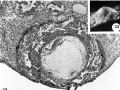
File:Hertig1956 fig32-33.jpg ==Fig. 32-33.== ...spaces and the large exocoelomic cavity. [[:Category:Carnegie Embryo 8330|Carnegie 8330]], Section 9-2-6. X 100.(1,280 × 958 (232 KB)) - 21:00, 23 February 2017
File:Stage14 sem4c.jpg == Human Embryo Carnegie Stage 14== Carnegie Stage 14 day 32 somite 35(452 × 600 (37 KB)) - 16:03, 14 May 2011
File:Stage14 sem4.jpg == Human Embryo Carnegie Stage 14== Carnegie Stage 14 day 32 somite 35(1,916 × 2,549 (297 KB)) - 16:02, 14 May 2011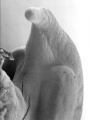
File:Stage14 sem4a.jpg == Human Embryo Carnegie Stage 14== Carnegie Stage 14 day 32 somite 35(752 × 1,000 (79 KB)) - 16:02, 14 May 2011
File:Stage14 sem4b.jpg == Human Embryo Carnegie Stage 14== Carnegie Stage 14 day 32 somite 35(602 × 800 (57 KB)) - 16:02, 14 May 2011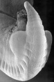
File:Stage14 sem6.jpg == Human Embryo Carnegie Stage 14== Carnegie Stage 14 day 32 somite 35(1,569 × 2,394 (311 KB)) - 16:23, 14 May 2011
File:Stage14 sem6a.jpg == Human Embryo Carnegie Stage 14== Carnegie Stage 14 day 32 somite 35(655 × 1,000 (87 KB)) - 16:23, 14 May 2011
File:Stage14 sem6b.jpg == Human Embryo Carnegie Stage 14== Carnegie Stage 14 day 32 somite 35(524 × 800 (61 KB)) - 16:23, 14 May 2011
File:Stage14 sem3.jpg == Human Embryo Carnegie Stage 14== Carnegie Stage 14 day 32 somite 35(2,114 × 2,997 (443 KB)) - 15:51, 14 May 2011
File:Stage14 sem6c.jpg == Human Embryo Carnegie Stage 14== Carnegie Stage 14 day 32 somite 35(393 × 600 (39 KB)) - 16:24, 14 May 2011
File:Stage14 sem3a.jpg == Human Embryo Carnegie Stage 14== Carnegie Stage 14 day 32 somite 35(705 × 1,000 (94 KB)) - 15:51, 14 May 2011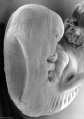
File:Stage14 sem3b.jpg == Human Embryo Carnegie Stage 14== Carnegie Stage 14 day 32 somite 35(564 × 800 (67 KB)) - 15:51, 14 May 2011
File:Stage14 sem3c.jpg == Human Embryo Carnegie Stage 14== Carnegie Stage 14 day 32 somite 35(423 × 600 (42 KB)) - 15:52, 14 May 2011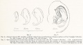
File:Streeter1922-06-07.jpg ...ns of human embryos of the Carnegie Collection: No. 460 (21 mm.), No. 417 (32 mm.), No. 886 (43 mm.). X14. ...'']] Reconstruction of left auricular cartilage of a 50 mm. fetus (No. 84, Carnegie Collection). X 14. A model of the external form of the auricle was made, in(1,200 × 653 (107 KB)) - 11:00, 31 October 2015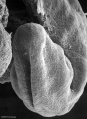
File:Stage11 sem7a.jpg ==Human Embryo Carnegie stage 11== [[Carnegie stage 11]] 25 days, 19 somite pairs(729 × 1,000 (141 KB)) - 14:20, 16 August 2016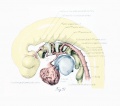
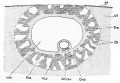
File:Congdon1922-32.jpg ==Figs. 32. Embryo 4 mm length== ...outgrowths for the fourth and probably for the pulmonary arch are present. Embryo No. {{CE836}}, length 4 mm.(1,133 × 1,000 (132 KB)) - 07:28, 22 January 2020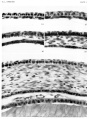
File:Streeter1957 plate01.jpg A segment of the cornea near the mid-line is shown in an embryo of each horizon. The mesoclermal component is a thin layer one or two cells Fig. 14. [[:Category:Carnegie Embryo 5609|'''No. 5609''']], xix, I5-I-I x 800.(1,500 × 2,009 (486 KB)) - 17:37, 6 November 2016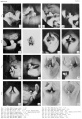
File:Spaulding-plate02.jpg File:Spaulding-fig07.jpg|Fig. 7. Carnegie Embryo No. {{CE1936}} File:Spaulding-fig08.jpg|Fig. 8. Carnegie Embryo No. {{CE955}}(1,373 × 2,000 (449 KB)) - 17:53, 7 November 2017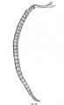
File:Williams1908-fig20.jpg ...of a median section of the notochord and chondrostyle of a Human Embryo of 32 mm== [[Category:Human]][[Category:Human Embryo]] [[Category:Carnegie Stage 23]](920 × 1,500 (96 KB)) - 16:57, 7 December 2021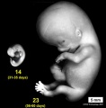
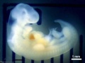
File:Stage14compare23.jpg ==Human Embryo Size Comparison== ...esent approximately the middle and at the end of the embryonic period. The embryo images have been scaled to the same magnification (5 mm scale bar).(593 × 601 (32 KB)) - 00:12, 11 December 2013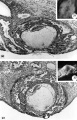
File:Hertig1956 plate05.jpg ...n at the margin of the ovum at the right. [[:Category:Carnegie Embryo 8558|Carnegie 8558]], Section 10-1-4. X 100. ...nce is not shown, arose from this defect. [[:Category:Carnegie Embryo 8558|Carnegie 8558]], Sequence 5. X22.(1,280 × 2,002 (466 KB)) - 21:28, 23 February 2017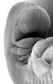
File:Stage14 sem2.jpg ==Human Embryo Carnegie stage 14== Carnegie stage 14, 32 day, 35 somite pairs(1,261 × 2,000 (234 KB)) - 16:39, 9 June 2011
File:Stage14 sem2a.jpg ==Human Embryo Carnegie stage 14== Carnegie stage 14, 32 day, 35 somite pairs(631 × 1,000 (83 KB)) - 16:39, 9 June 2011
File:Stage14 sem2b.jpg ==Human Embryo Carnegie stage 14== Carnegie stage 14, 32 day, 35 somite pairs(505 × 800 (58 KB)) - 16:39, 9 June 2011- ...This embryo was later numbered Carnegie Embryo {{CE1399}} and described as Carnegie stage {{CS8)). ...rner1920 fig29-30.jpg|fig 29-30]] | [[:File:Turner1920 fig31-32.jpg|fig 31-32]] | [[:File:Turner1920 fig33-34.jpg|fig 33-34]] | [[:File:Turner1920 fig35-1 KB (176 words) - 12:39, 10 August 2018
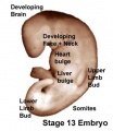
File:Stage13 surface bulges.jpg ==Human Embryo Carnegie Stage 13== Carnegie Stage 13 (day 28)(337 × 389 (14 KB)) - 06:08, 12 August 2011- ...ection Embryo No. {{CE397}}. This embryo was {{Tubal}} and classified as [[Carnegie stage 16]] occurring during [[Week 6]]. {{Carnegie stage 16 links}}3 members (0 subcategories, 2 files) - 16:11, 7 November 2017
- ! [[Carnegie stage 11]] <br>23 - 26 days <gallery caption="Human Embryo (stage 11)">2 KB (252 words) - 07:05, 7 April 2016
- ...he Carnegie Collection Embryo No. CE417}}. This embryo was classified as [[Carnegie stage 23]] occurring during [[Week 8]]. {{Carnegie stage 23 links}}2 members (0 subcategories, 2 files) - 13:47, 22 November 2017
- ...an embryo''']]. (1948) Carnegie Inst. Wash. Pub. 575, Contrib. to Embryol. 32: 205-261.<noinclude>[[Category:Template]][[Category:Reference]][[Category:H384 bytes (47 words) - 20:00, 1 April 2019
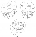
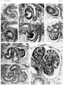
File:Mall1912-fig31-33.jpg ==Fig. 31 - 33. Outline drawings of the heart of an embryo 10 mm long== '''Fig. 32''' The same heart, viewed from below.(963 × 1,000 (94 KB)) - 17:32, 31 July 2017- ...ges that relate to the [[Carnegie Collection]] Embryo No. {{CE449}}. This embryo would be early fetal development [[Week 9]] based upon the CRL 36 mm. {{Carnegie Collection fetal table}}1 member (0 subcategories, 0 files) - 14:05, 18 November 2017

File:Stage14 sem2al.jpg ==Human Embryo Carnegie stage 14== Carnegie stage 14, day 32, 35 somites, week 5, right ventrolateral view upper embryo.(504 × 800 (68 KB)) - 10:06, 5 August 2012
File:Stage14 sem2cl.jpg ==Human Embryo Carnegie stage 14== Carnegie stage 14, day 32, 35 somites, week 5, right ventrolateral view upper embryo.(252 × 400 (22 KB)) - 10:06, 5 August 2012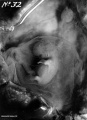
File:Hubrecht Homo32 fig01.jpg ==Human Embryo Carnegie Stage 20== Based on the external appearance and limb development this embryo appears to be Stage 20.(872 × 1,200 (159 KB)) - 06:11, 30 September 2013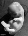
File:Hubrecht Homo32 fig02.jpg ==Human Embryo Carnegie Stage 20== Based on the external appearance and limb development this embryo appears to be Stage 20.(914 × 1,200 (127 KB)) - 06:11, 30 September 2013- ==Stage 13 Embryo Sagittal== ...EFIC movie shows sagittal sections through the Stage 13 week 4 to 5 human embryo. Shown below on this page is also the [[#Unlabelled Version|unlabelled movi1 KB (198 words) - 15:20, 4 April 2017
- ==Stage 13 Embryo Sagittal== ...part (pharyngeal arches and heart) of the Stage {{CS13}} week 4 to 5 human embryo. Shown below on this page is also the [[#Unlabelled Version|unlabelled movi1 KB (204 words) - 13:00, 27 April 2018
File:Stage14 sem2l.jpg ==Human Embryo Carnegie stage 14== Carnegie stage 14, day 32, 35 somites, week 5, right ventrolateral view upper embryo.(630 × 1,000 (96 KB)) - 12:11, 10 September 2017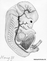
File:Hubrecht Homo32 fig03.jpg ==Human Embryo Carnegie Stage 20== ...catalogue page. Based on the external appearance and limb development this embryo appears to be Stage 20.(618 × 800 (114 KB)) - 06:10, 30 September 2013
File:Stage14 bf4.jpg ==Human Embryo Carnegie stage 14== Carnegie stage 14, 32 day, 35 somite pairs(2,650 × 2,000 (352 KB)) - 17:55, 30 May 2011
File:Stage14 bf4a.jpg ==Human Embryo Carnegie stage 14== Carnegie stage 14, 32 day, 35 somite pairs(1,325 × 1,000 (137 KB)) - 17:56, 30 May 2011
File:Stage14 bf4b.jpg ==Human Embryo Carnegie stage 14== Carnegie stage 14, 32 day, 35 somite pairs(1,060 × 800 (99 KB)) - 17:56, 30 May 2011
File:Stage13 bf2.jpg ==Human Embryo Carnegie Stage 13== Carnegie Stage 13 (day 28)(1,000 × 750 (61 KB)) - 04:58, 13 April 2011
File:Stage13 bf2a.jpg ==Human Embryo Carnegie Stage 13== Carnegie Stage 13 (day 28)(800 × 600 (40 KB)) - 04:58, 13 April 2011
File:Stage13 bf2b.jpg ==Human Embryo Carnegie Stage 13== Carnegie Stage 13 (day 28)(600 × 450 (24 KB)) - 04:58, 13 April 2011
File:Stage13 bf2c.jpg ==Human Embryo Carnegie Stage 13== Carnegie Stage 13 (day 28)(400 × 300 (12 KB)) - 04:55, 13 April 2011- ...arnegie Collection]] Embryo No. {{CE721}}. This embryo was classified as [[Carnegie stage 15|Stage 15]] occurring during [[Week 5]]. {{Carnegie stage 15 links}}15 members (0 subcategories, 7 files) - 16:09, 7 November 2017

File:Stage14 sem1c.jpg ==Human Embryo Carnegie stage 14== Carnegie stage 14, 32 day, 35 somite pairs(400 × 327 (23 KB)) - 09:37, 3 June 2014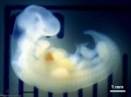
File:Stage14 bf1.jpg ==Human Embryo Carnegie Stage 14== Carnegie stage 14, 32 day, 35 somite pairs(1,000 × 743 (27 KB)) - 09:38, 3 June 2014- ...Carnegie Collection Embryo No. {{CE389}}. This embryo was classified as [[Carnegie stage 15|Stage 15]] occurring during [[Week 5]]. {{Carnegie stage 15 links}}1 member (0 subcategories, 0 files) - 16:07, 7 November 2017
File:Stage14 bf1c.jpg ==Human Embryo Carnegie stage 14== Carnegie stage 14, 32 day, 35 somite pairs(400 × 297 (8 KB)) - 20:49, 25 October 2013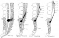
File:Streeter1919-fig02.jpg ..., irrespective of form, as shown in stipple. The fetuses are listed in the Carnegie Collection follows: No. {{CE75}}, 30 mm.; No. {{CE1656}}, 67 mm.; No. {{CE1 ...67-mm. fetus, 4.75 mm. long; 111-mm. fetus, 12.25 mm. long; 221-mm. fetus, 32 mm. long.(2,023 × 1,318 (499 KB)) - 22:24, 31 January 2019- ...egory includes pages and images that relate to the [[Carnegie Collection]] Embryo No. {{CE431}}. {{Carnegie stage 20 links}}4 members (0 subcategories, 2 files) - 22:53, 14 November 2017
- ! colspan=6|[[Carnegie Collection|Carnegie Embryos]] - [[Paper - The early development of the meninges of the spinal c ! width=100px|Embryo<br>no.8 KB (659 words) - 23:19, 31 January 2019
- ! width=100px|Embryo<br>no. | [[Carnegie stage 10|'''X''']] (20-22 days)6 KB (582 words) - 11:45, 4 October 2017
- ! colspan=6|[[Carnegie Collection|Carnegie Embryos]] - [[Paper - The early development of the meninges of the spinal c ! width=100px|Embryo<br>no.8 KB (650 words) - 12:04, 4 October 2017
- ! colspan=6|[[Carnegie Collection|Carnegie Embryos]] - [[Paper - The early development of the meninges of the spinal c ! width=100px|Embryo<br>no.6 KB (591 words) - 11:41, 4 October 2017
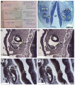
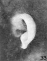
File:Human CS13-15 otic vesicle 01.jpg ==Human Otic Vesicle Carnegie Stages 13 to 15== '''Carnegie Stages''' : {{CS13}} 32 post ovulatory days, {{CS14}} 33 post ovulatory days, {{CS15} 35 post ovula(1,574 × 1,779 (364 KB)) - 12:31, 6 April 2018- :[[Carnegie stage table|'''Carnegie Stages''']]: [[Carnegie_stage_14|14]] | [[Carnegie_stage_15|15]] ==Carnegie Stages==3 KB (354 words) - 10:32, 16 March 2020
File:Bartelmez1922-fig02.jpg a. Mall embryo no. {{CE391}} — eight somites—section 45. d. N.Y.U. embryo no. 4 — fourteen somites — section 32.(1,203 × 1,700 (317 KB)) - 18:45, 23 June 2019
File:Stage14 sem1.jpg ==Human Embryo== Carnegie stage 14, 32 day, 35 somite pairs(1,000 × 819 (94 KB)) - 20:41, 25 October 2013- Embryo 1951-01-09 CRL 11 mm - [[Week 6]] [[Carnegie stage 16|stage 16]] [[Carnegie stage 17|stage 17]] ...]] | [[:File:Embryo-1951-09-01-Slide-60 Scene11-7.jpg|Zoom x40]] | [[:File:Embryo-1951-09-01-Slide-60 Scene11-8.jpg|Zoom x80]]2 KB (236 words) - 14:03, 19 June 2017

File:Stage14 bf3b.jpg ==Human Embryo Carnegie stage 14== Carnegie stage 14, 32 day, 35 somite pairs(547 × 800 (43 KB)) - 17:43, 30 May 2011
File:Stage14 bf3.jpg ==Human Embryo Carnegie stage 14== Carnegie stage 14, 32 day, 35 somite pairs(1,368 × 2,000 (159 KB)) - 17:43, 30 May 2011
File:Stage14 bf3a.jpg ==Human Embryo Carnegie stage 14== Carnegie stage 14, 32 day, 35 somite pairs(684 × 1,000 (61 KB)) - 17:43, 30 May 2011
File:Stage14 bf1a.jpg '''Human Embryo''' Carnegie stage 14, 32 day, 35 somite pairs(800 × 594 (20 KB)) - 20:48, 25 October 2013
File:Stage14 bf1b.jpg '''Human Embryo''' Carnegie stage 14, 32 day, 35 somite pairs(600 × 446 (13 KB)) - 20:48, 25 October 2013- ...egory includes pages and images that relate to the [[Carnegie Collection]] Embryo No. {{CE584a}. ===Embryo No. {{CE584a}}, 2.5 mm Crown-Rump Length===1 member (0 subcategories, 0 files) - 16:43, 7 November 2017
- ...egory includes pages and images that relate to the [[Carnegie Collection]] Embryo No. {{CE453}}. ===Embryo No. {{CE453}}, 23 mm Crown-Rump Length===1 member (0 subcategories, 0 files) - 16:39, 7 November 2017

File:Stage14 bf2l.jpg '''Human Embryo''' Carnegie stage 14, 32 day, 35 somite pairs(1,000 × 834 (66 KB)) - 20:51, 25 October 2013- ...Carnegie Collection Embryo No. {{CE836}}. This embryo was classified as [[Carnegie stage 13]] occurring during [[Week 4]]. {{Carnegie stage 13 links}}10 members (0 subcategories, 4 files) - 15:54, 7 November 2017

File:Stage14 bf2cl.jpg '''Human Embryo''' Carnegie stage 14, 32 day, 35 somite pairs(400 × 333 (17 KB)) - 14:11, 21 October 2010- |+ '''Human Embryo Somite Number''' ! width=120px|Carnegie Stage864 bytes (109 words) - 23:30, 29 October 2018
- ...llection), with Capsular Attachment of the Connecting Stalk|An Early Human Embryo (No. 1285)]][[Category:Jan Florian]] [[Category:Carnegie Stage 7]][[Category:Week 3]]1 KB (149 words) - 12:13, 8 February 2020
- ! width=120px|Carnegie Stage | (about 32 days) the lens placode is indented by the lens pit.2 KB (240 words) - 17:12, 8 January 2019
- File:Streeter1922-fig32.jpg|Fig. 32. No. {{CE2075}} 40 mm (R) </gallery><noinclude>[[Category:Carnegie Embryo]][[Category:Gallery]][[Category:Template]][[Category:1920's]][[Category:Hea1 KB (142 words) - 14:18, 22 November 2017
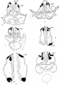
File:Streeter1922-fig33.jpg ...tively thick and swollen character of the surrounding parts. Figures 30 to 32 differ from the preceding ones only in the increasing prominence of the ear These are indicated by the letter R. All specimens are from the Carnegie Collection, and length given is crown-rump.(435 × 554 (45 KB)) - 01:23, 28 January 2013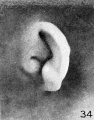
File:Streeter1922-fig34.jpg ...tively thick and swollen character of the surrounding parts. Figures 30 to 32 differ from the preceding ones only in the increasing prominence of the ear These are indicated by the letter R. All specimens are from the Carnegie Collection, and length given is crown-rump.(437 × 556 (47 KB)) - 01:23, 28 January 2013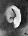
File:Streeter1922-fig35.jpg ...tively thick and swollen character of the surrounding parts. Figures 30 to 32 differ from the preceding ones only in the increasing prominence of the ear These are indicated by the letter R. All specimens are from the Carnegie Collection, and length given is crown-rump.(430 × 556 (41 KB)) - 01:22, 28 January 2013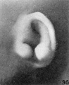
File:Streeter1922-fig36.jpg ...tively thick and swollen character of the surrounding parts. Figures 30 to 32 differ from the preceding ones only in the increasing prominence of the ear These are indicated by the letter R. All specimens are from the Carnegie Collection, and length given is crown-rump.(560 × 686 (66 KB)) - 01:22, 28 January 2013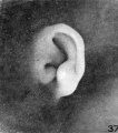
File:Streeter1922-fig37.jpg ...tively thick and swollen character of the surrounding parts. Figures 30 to 32 differ from the preceding ones only in the increasing prominence of the ear These are indicated by the letter R. All specimens are from the Carnegie Collection, and length given is crown-rump.(606 × 685 (82 KB)) - 01:22, 28 January 2013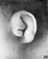
File:Streeter1922-fig38.jpg ...tively thick and swollen character of the surrounding parts. Figures 30 to 32 differ from the preceding ones only in the increasing prominence of the ear These are indicated by the letter R. All specimens are from the Carnegie Collection, and length given is crown-rump.(560 × 687 (69 KB)) - 01:21, 28 January 2013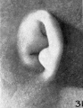
File:Streeter1922-fig31.jpg ...tively thick and swollen character of the surrounding parts. Figures 30 to 32 differ from the preceding ones only in the increasing prominence of the ear These are indicated by the letter R. All specimens are from the Carnegie Collection, and length given is crown-rump.(428 × 552 (42 KB)) - 01:23, 28 January 2013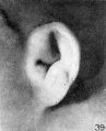
File:Streeter1922-fig39.jpg ...tively thick and swollen character of the surrounding parts. Figures 30 to 32 differ from the preceding ones only in the increasing prominence of the ear These are indicated by the letter R. All specimens are from the Carnegie Collection, and length given is crown-rump.(553 × 680 (67 KB)) - 01:21, 28 January 2013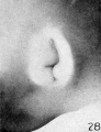
File:Streeter1922-fig28.jpg ...tively thick and swollen character of the surrounding parts. Figures 30 to 32 differ from the preceding ones only in the increasing prominence of the ear These are indicated by the letter R. All specimens are from the Carnegie Collection, and length given is crown-rump.(426 × 554 (36 KB)) - 01:24, 28 January 2013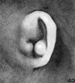
File:Streeter1922-fig40.jpg ...tively thick and swollen character of the surrounding parts. Figures 30 to 32 differ from the preceding ones only in the increasing prominence of the ear These are indicated by the letter R. All specimens are from the Carnegie Collection, and length given is crown-rump.(610 × 677 (81 KB)) - 01:18, 28 January 2013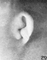
File:Streeter1922-fig29.jpg ...tively thick and swollen character of the surrounding parts. Figures 30 to 32 differ from the preceding ones only in the increasing prominence of the ear These are indicated by the letter R. All specimens are from the Carnegie Collection, and length given is crown-rump.(433 × 554 (42 KB)) - 01:24, 28 January 2013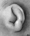
File:Streeter1922-fig41.jpg ...tively thick and swollen character of the surrounding parts. Figures 30 to 32 differ from the preceding ones only in the increasing prominence of the ear These are indicated by the letter R. All specimens are from the Carnegie Collection, and length given is crown-rump.(559 × 677 (76 KB)) - 01:19, 28 January 2013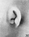
File:Streeter1922-fig30.jpg ...tively thick and swollen character of the surrounding parts. Figures 30 to 32 differ from the preceding ones only in the increasing prominence of the ear These are indicated by the letter R. All specimens are from the Carnegie Collection, and length given is crown-rump.(435 × 551 (41 KB)) - 01:23, 28 January 2013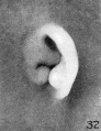
File:Streeter1922-fig32.jpg ...tively thick and swollen character of the surrounding parts. Figures 30 to 32 differ from the preceding ones only in the increasing prominence of the ear These are indicated by the letter R. All specimens are from the Carnegie Collection, and length given is crown-rump.(426 × 552 (44 KB)) - 01:23, 28 January 2013- ...ave a crown rump length (CRL) of 18 - 22 mm. This page also shows specific embryo examples in various collections. ...Carnegie stage 20''']] resource page. See also [[Carnegie stage 20#Events|Carnegie Stage 20 - Events]]175 members (24 subcategories, 94 files) - 22:51, 31 January 2019
- | [[Carnegie stage 2|II]] Segmenting egg 2 cells | [[Carnegie stage 2|II]] 12 ± cells4 KB (514 words) - 14:12, 28 September 2018

File:Stage14 sem1a.jpg '''Human Embryo''' Carnegie stage 14, 32 day, 35 somite pairs(800 × 655 (66 KB)) - 20:42, 25 October 2013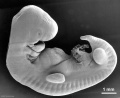
File:Stage14 sem1b.jpg '''Human Embryo''' Carnegie stage 14, 32 day, 35 somite pairs(600 × 491 (41 KB)) - 20:42, 25 October 2013- | [[Carnegie stage 2|II]] Segmenting egg 2 cells | [[Carnegie stage 2|II]] 12 ± cells5 KB (525 words) - 13:35, 9 August 2017
- ...arnegie Collection]] Embryo No. {{CE810}}. This embryo was classified as [[Carnegie stage 15]] occurring during [[Week 5]]. {{Carnegie Collection stage 15 table}}6 members (0 subcategories, 2 files) - 15:58, 7 November 2017
- ...2-fig58.jpg|58. Embryo 1708, 154 mm]] | [[:File:Streeter1922-fig59.jpg|59. Embryo 1742, 191.2 mm]] | [[Book_-_Contributions_to_Embryology_Carnegie_Institutio ...Development|Outer Ear Development]] | [[Book_-_Contributions_to_Embryology|Carnegie Contributions to Embryology]]5 KB (522 words) - 22:33, 25 April 2016
- ...pment. The models include whole surface views as well as detailed views of embryo internal structures. ...sets were incorporated in 1972 into the [[Carnegie Collection]] (assigned Carnegie Nos. 10315-10434), but have since been returned to the University of Goetti4 KB (528 words) - 09:21, 12 May 2020
- ! colspan=3|[[Week 5]] - Human Embryo Stages and Events ({{GA}} week 7) | <center>32</center>2 KB (306 words) - 09:50, 26 July 2019
- ! colspan=12| [[Carnegie Collection]] - [[Carnegie stage 23|Stage 23]] | {{CE417}} || E,32 Ch., 0x60x40 || Good || {{Formalin}} || P || {{Transverse}} || 100 || Al4 KB (415 words) - 19:50, 5 October 2020
- | [[Carnegie stage 13|13]] | [[Carnegie stage 13|13]]13 KB (1,092 words) - 11:29, 4 October 2017
- {{Carnegie stage somites table}} {{Carnegie stage table 1}}4 KB (413 words) - 23:09, 29 October 2018
- '''00:32''' - re-expansion ...ida]] | {{implantation}} | [[Carnegie stage 3]] | [[Carnegie stage 4]] | [[Carnegie stage 5]]3 KB (385 words) - 16:46, 24 April 2018
- | [[Carnegie stage 13|13]] | [[Carnegie stage 13|13]]14 KB (1,101 words) - 11:36, 4 October 2017
- '''Modern Notes:''' [[Carnegie stage 2]] | [[Week 2]] [[:Category:Carnegie Embryo 8190|'''Carnegie, No. 8190''']]3 KB (382 words) - 11:34, 26 July 2020
- == Carnegie Stage 13 == ...d media related to embryonic development in [[week 4]] to [[week 5]], 28 - 32 days, {{GA}} week 6-7. The embryos have a crown rump length (CRL) of 4 - 6312 members (42 subcategories, 189 files) - 17:10, 22 May 2018

File:Stage14 bf2al.jpg '''Human Embryo''' Carnegie stage 14, 32 day, 35 somite pairs(800 × 667 (48 KB)) - 11:25, 4 September 2009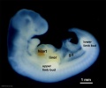
File:Stage14 bf2bl.jpg '''Human Embryo''' Carnegie stage 14, 32 day, 35 somite pairs(600 × 500 (31 KB)) - 11:25, 4 September 2009- ==Carnegie Collection== * 6mm Pig Embryo - approximately Human day 32 (Carnegie Stage 13/14 embryo) Stage13_3d01.flv4 KB (586 words) - 22:20, 4 May 2014
- ! colspan=12| [[Carnegie Collection]] - [[Carnegie stage 21|Stage 21]] ...|| Bichlor, acetic, formol || P || {{Transverse}} || 20 || Al. coch. || 32 || {{Male}} || 1920 ||4 KB (373 words) - 19:44, 5 October 2020
- ...images. (6 mm pig embryo, approximately Human day 32, Carnegie Stage 13/14 embryo) {{Carnegie stage 13 links}}3 KB (431 words) - 12:23, 21 May 2017
- [[Image:Stage9sm.jpg|thumb|Carnegie stage 9 showing somite formation]] [[Image:Stage 9 SEM1.jpg|thumb|Carnegie stage 9 scanning electron microscope image showing somite formation]]5 KB (773 words) - 12:04, 9 August 2010
- | [[:Category:Carnegie Embryo 8698|8698]] | [[Carnegie stage 2|II]] Segmenting egg 2 cells10 KB (1,243 words) - 17:01, 25 February 2017
- ...| [[:File:Spaulding-fig31.jpg|Fig. 31]] | [[:File:Spaulding-fig32.jpg|Fig. 32]] |[[:File:Spaulding-fig33.jpg|Fig. 33]] | [[:File:Spaulding-fig34.jpg|Fig. ...oric Embryology]][[Category:Human]][[Category:Genital]][[Category:Carnegie Embryo]]3 KB (281 words) - 16:49, 22 April 2016
- ...n either age or size. The human embryonic period proper is divided into 23 Carnegie stages. Criteria beyond morphological features include age in days, number ...an take from as little as 10 days in chickens to nearly 60 days in humans. Carnegie is the name of a historical US Institute that historically categorised thes5 KB (566 words) - 12:46, 22 May 2018
- ...images. (6 mm pig embryo, approximately Human day 32, Carnegie Stage 13/14 embryo) ...day 18. It is an ectodermal structure, extending along the surface of the embryo for much of its length. During the middle of the fourth week, an indentatio3 KB (393 words) - 02:13, 8 March 2013
- ...images. (6 mm pig embryo, approximately Human day 32, Carnegie Stage 13/14 embryo) ...day 18. It is an ectodermal structure, extending along the surface of the embryo for much of its length. During the middle of the fourth week, an indentatio3 KB (396 words) - 02:13, 8 March 2013
- ...images. (6 mm pig embryo, approximately Human day 32, Carnegie Stage 13/14 embryo) {{Carnegie stage 13 links}}3 KB (502 words) - 12:22, 21 May 2017
- ! width=150px|Carnegie Stage | (about 32 days) Lens placode is indented by the lens pit, cup-shaped and still commun3 KB (384 words) - 00:40, 1 June 2018
File:Minot1904 fig01.jpg ...en was the Peters embryo, and included in the list were 92 embryos of 0.19-32 mm, as well as 17 fetuses of 33-210 mm." [[Carnegie stage 6]][[Category:Charles Minot]][[Category:Historic Embryology]](800 × 555 (100 KB)) - 12:15, 9 February 2017- {{Carnegie Collection stage 22 table}} ...Category:Carnegie Embryo 6832|No. 6832]]. Bottom row, [[:Category:Carnegie Embryo 8394|No. 8394]]. All views are at the same magnification.4 KB (653 words) - 15:34, 26 June 2019
- --[[User:Z3186755|3186755]] 23:32, 1 September 2010 (UTC) 1. What Carnegie stages occur during week 3 and week 4?5 KB (641 words) - 08:22, 21 October 2010
- ...ovascular_System_-_Spleen_Development|Spleen]] | [[Cardiovascular System - Carnegie Stage 22|Stage 22]] | [[Cardiovascular System - Abnormalities|Abnormalities ...43 mm]] | [[Book_-_Contributions_to_Embryology_Carnegie_Institution_No.24|Carnegie No.24]] | [[Embryology_History_-_George_Streeter|George Streeter]]8 KB (986 words) - 14:55, 10 April 2018
- :[[Carnegie_stage_table|'''Carnegie Stages''']]: [[Carnegie_stage_1|1]] | [[Carnegie_stage_2|2]] | [[Carnegie_s | 324 KB (504 words) - 11:17, 25 October 2012
- ...images. (6 mm pig embryo, approximately Human day 32, Carnegie Stage 13/14 embryo) The vascular system of the embryo is formed from blood islands that appear in the extraembryonic mesoderm of3 KB (424 words) - 02:23, 8 March 2013
- ...images. (6 mm pig embryo, approximately Human day 32, Carnegie Stage 13/14 embryo) The vascular system of the embryo is formed from blood islands that appear in the extraembryonic mesoderm of3 KB (485 words) - 02:22, 8 March 2013
- ! colspan=11| [[Carnegie Collection|Carnegie Collection Embryos]] - [[Carnegie stage 12|Stage 12]] | {{CE6488}} || 28 || E, 32 Ch,22 || Good || {{Formalin}} || C—P || {{Transverse}} || 10 || Al. c5 KB (558 words) - 20:00, 5 October 2020
- ...dentify the internal and external developmental process of the embryo. The Carnegie stages are covered over 8 weeks (56 days). ...Carnegie stage does the human neural tube normally completely close??? At Carnegie stage 13 (4 weeks) the neural tube closes completely.5 KB (755 words) - 14:12, 31 October 2009
- ...images. (6 mm pig embryo, approximately Human day 32, Carnegie Stage 13/14 embryo, week 5) {{Carnegie stage 13 links}}4 KB (632 words) - 04:03, 12 May 2020
- See also [[Carnegie stage 14#Events|'''Carnegie stage 14 Events''']] {{Carnegie stage 14 links}}8 KB (1,167 words) - 17:53, 16 March 2020
- [[week 4]] to [[week 5]], 28 - 32 days. See also [[Carnegie stage 13#Events|'''Carnegie stage 13 Events''']]9 KB (1,189 words) - 17:55, 16 March 2020
- ...there are many different organs and tissues differentiating throughout the embryo, only a few select systems will be discussed. [[Media:BGD2010-Embryo Lab 170510-603.mp3|listen Part 3]] | [[:File:BGD2010-Embryo Lab 170510-603.mp3|download]] (2.1 Mb MP3 16:14)6 KB (865 words) - 12:19, 18 May 2010
- [[File:Ziegler_model_05.jpg|thumb|An example of a Ziegler embryo model.]] ...based upon their own collection. (More? [[Carnegie_Stages#Osborne_O._Heard|Carnegie Models]])7 KB (1,113 words) - 17:34, 25 October 2020
- The following data is from a study of human embryonic carnegie stages{{#pmid:7364662|PMID7364662}} and other sources. * Week 5 - [[Carnegie_stage_14|Stage 14]] - (about 32 days) the lens placode is indented by the lens pit, cup-shaped and still co3 KB (415 words) - 11:01, 12 March 2018
- ...opment olfactory and related structures in staged human embryos from the [[Carnegie Collection]]. ...es has been established from the serially sectioned human embryos of the [[Carnegie Collection]], from stage {{CS11}} to stage {{CS23}}.5 KB (716 words) - 16:42, 26 February 2022
- == Carnegie Stage Table == | width="65" |<center> 28 - 32 </center>([[week 5|'''week 5''']])9 KB (1,026 words) - 09:42, 25 November 2015
- ==Embryo Images Links== ==Embryo Stage 13==13 KB (1,908 words) - 12:55, 9 February 2024
- ...images. (6 mm pig embryo, approximately Human day 32, Carnegie Stage 13/14 embryo, week 5) ...ing down to the cloaca, this becomes known as the mesonephric duct, in the embryo. The next stage is the formation of the mesonephros, also in week 4. Its di4 KB (588 words) - 02:20, 8 March 2013
- ...images. (6 mm pig embryo, approximately Human day 32, Carnegie Stage 13/14 embryo) ...ing down to the cloaca, this becomes known as the mesonephric duct, in the embryo. The next stage is the formation of the mesonephros, also in week 4. Its di4 KB (592 words) - 02:20, 8 March 2013
- ==Carnegie Stages== Historic [[Carnegie Stages]] 1 to 4.15 KB (2,010 words) - 10:31, 16 March 2020
- [[Carnegie stage 3]] ...e coalescence of intercellular spaces when the organism has acquired about 32 cells. In in vitro studies, a cavity formed in some human embryos at 16-2013 KB (1,877 words) - 15:40, 26 June 2019
- ...re are also sets of [[Carnegie stage 22 - selected serial sections]] and [[Carnegie stage 13 - serial sections]]. | [[:File:Stage_22_image_032.jpg|32]]8 KB (1,005 words) - 10:19, 13 March 2014
- ...epage&q=rabbit%20embryo%20stages&f=false - good for what studies in rabbit embryo has been used for. And has a good table for embryological stages!! http://books.google.com/books?id=ljAKtC-iIrIC&pg=PA264&dq=rabbit+embryo+stages&as_brr=3#v=onepage&q=rabbit%20embryo%20stages&f=false5 KB (710 words) - 14:05, 31 October 2009
- ...he genu and anterior midbody showed the least maturation by postnatal week 32, attaining 50% and 49% of the average adult area. Thus, the CC of baboons s ...t not significantly increased by letrozole and estradiol (99 +/- 11 ng/ml; 32.7 +/- 5.2). In contrast, the number of LH-positive fetal pituitary cells de10 KB (1,340 words) - 10:17, 22 March 2012
- The "Mateer Embryo" was later catalogued as Carnegie Embryo {{CE1399}} Carnegie stage {{CS8)). ...Poor || Formol || P || Trans. || 10 || {{HE}} etc. || 1916 || "Mateer embryo" described by Streeter (1920)13 KB (2,015 words) - 10:01, 18 August 2020
- ==Carnegie Collection== --[[User:S8600021|Mark Hill]] 13:21, 22 August 2009 (EST) Updating carnegie stage pages with different format and new sets of images.9 KB (1,246 words) - 11:09, 3 April 2020
- [[File:Carnegie_Institute_of_Washington_logo.jpg|thumb|Carnegie Institute of Washington]] ...The papers documented not only early human development, using mainly the [[Carnegie Collection]] of embryos, but also that in animal models of development.13 KB (1,652 words) - 13:55, 11 August 2020
- {{Carnegie No.20 Header}} Frontal section through the region of the ear in a human embryo 4 mm. long (Carnegie Collection, No. 588, slide 6, row 6, section 6). The section is 15 um thick18 KB (2,849 words) - 22:04, 22 April 2012
- ...hilly 1987|link=Embryology History - Ronan O'Rahilly|Ronan O'Rahilly (1987 Carnegie Labs)]] ...t study,<ref name=Weller1933>{{Ref-Weller1933}}</ref> used the following [[Carnegie Collection]] embryos: stage {{CS9}} (No. {{CE1878}}), {{CS10}} ({{CE391}};8 KB (1,113 words) - 18:19, 16 March 2020
- [[File:Kyoto Embryo Collection - cover.jpg|200px|link=http://itunes.apple.com/us/book/id1143922 [[File:AvailableBook.png|150px|link=https://itunes.apple.com/au/book/kyoto-embryo-collection/id1143922693?mt=11]]12 KB (1,631 words) - 17:18, 11 December 2017
- During week 4 a number of features appear visible on the embryo surface: ==Carnegie Stage 12 to 14==7 KB (981 words) - 07:53, 15 October 2015
- ...]]) 17:55, 22 December 2014 (EST) Added [[Model Embryo 7.5mm Movie 1|Human Embryo 7.5mm model]] (Stage 15) from the [[Blechschmidt Collection]]. Added draft ...ll]] ([[User talk:Z8600021|talk]]) 19:38, 15 December 2014 (EST) Added new embryo images [[Carnegie_stage_13#Hill_Collection|Stage 13]] and [[Carnegie_stage_20 KB (2,538 words) - 11:26, 6 July 2015
- --[[User:Z8600021|Mark Hill]] 08:32, 24 December 2012 (EST) Updated [[Animal_Development|animal]] talk pages wi ...y Models]] page with information about [[Embryology_Models#Carnegie_Models|Carnegie Models]].24 KB (3,058 words) - 00:17, 19 January 2015
- ...ke this a [[Carnegie stage 10|stage 10]] or [[Carnegie stage 11|stage 11]] embryo. =Photographs of a Series of Sections of an Early Human Embryo=8 KB (1,225 words) - 11:40, 17 April 2018
- ...sion]] and [[Movie_-_Model_Embryo_to_128_Cell_Stage|Flash version]]. Added Carnegie collection [[Carnegie_stage_8#Carnegie_Collection|stage 8 images]]. ...mbryo to 32 Cell Stage|Quicktime version]] and [[Movie - Model Embryo to 32 Cell Stage|Flash version]].26 KB (3,399 words) - 23:53, 20 August 2013
- ...l|Maternal changes video]] and table of [[Fetal_Development#Carnegie_Fetal|Carnegie embryos at fetal stage]]. [[File:Stage15lefthead7207.jpg|thumb|150px|link=Carnegie stage 15|Human Embryo (stage 15}]]12 KB (1,556 words) - 14:07, 6 March 2018
- [[File:Human embryo day 2.jpg|thumb|300px|Human morula (day 2){{#pmid:19924284|PMID19924284}}]] A key event prior to morula formation is "compaction", where the 8 cell embryo undergoes changes in cell morphology and cell-cell adhesion that initiates10 KB (1,311 words) - 15:45, 8 June 2020
- The embryo is now 0.4 mm diameter in size. The initial images are displayed unlabeled to allow you to explore the embryo for yourself, linked labeled versions are also available for some images.18 KB (2,462 words) - 11:08, 3 April 2020
- [[Media:BGD2010-Embryo Lab 120510-305.mp3|listen Part 5]] | [[:File:BGD2010-Embryo Lab 120510-305.mp3|download]] (1.2 Mb MP3 9:26) | [[File:Human embryo day 2.jpg|200px|Stage 2 Day 2]]7 KB (1,027 words) - 14:31, 13 May 2010
- --[[User:S8600021|Mark Hill]] 15:32, 2 December 2010 (EST) Began adding the template links required for [[Medi ...System Development]] added [[:File:Human- Stage 22 integument 01.jpg|human embryo stage 22 skin image]] and movie of melanoblast migration in muse skin [[Qui23 KB (2,948 words) - 23:52, 20 August 2013
- ...should never be assigned merely on the basis of embryonic length. A 20-mm embryo, for example, could belong to any of three stages. Measurements, however, a ...out 5 mm in length and the chorion about 25 mm in diameter. At 8 weeks the embryo is about 30 mm in length, and the chorion is about 65 mm in diameter.7 KB (1,051 words) - 22:21, 4 February 2016
- == Carnegie Stage Table 1== <center> 28 - 32 </center>('''week 5''')39 KB (4,806 words) - 23:27, 21 November 2013
- ...ula; fusion of chorio-amniotic folds, chorio-amniotic stalk; neural plate; embryo bent dorsally; bud of allantoic stalk ...y; ectochorionic cyst collapsing; allantoic stalk projects into exocoelom; embryo bent dorsally8 KB (1,073 words) - 13:05, 4 May 2018
- ...lar layer separating ectoderm and endoderm, mesoderm also lies outside the embryo as '''extra-embryonic mesoderm''' (covered in placenta lecture). Embryonic | {{Embryo logocitation}}15 KB (1,947 words) - 16:05, 1 October 2019
- ...nt and Gaunt (1978). Florian, who used graphic reconstruction of the human embryo to great advantage, elaborated the mathematical background in Czech in 1928 ==The Carnegie Collection==17 KB (2,622 words) - 08:15, 25 March 2020
- '''Lab 5''' - Present --[[User:Z3252833|z3252833]] 00:32, 26 August 2010 (UTC) ===Lab 3 - Trilaminar Embryo to Early Embryo===12 KB (2,006 words) - 10:19, 21 October 2010
- {{Carnegie Collection stage 23 table}} ...[:Category:Carnegie Embryo 7425|No 7425]]. Lower row, [[:Category:Carnegie Embryo 4570|No. 4570]]. All views are at the same magnification.11 KB (1,723 words) - 15:37, 26 June 2019
- ! Carnegie Stage | 3221 KB (2,334 words) - 06:10, 15 September 2019
- ...es, blastocyst formation and hatching, and the generation of the bilaminar embryo. There will also be an introduction to the uterine changes at implantation | valign="bottom"|{{Embryo 1.6mm movie 1}}20 KB (2,612 words) - 15:45, 24 September 2019
- [[Image:Stage9sm.jpg|thumb|Carnegie stage 9 showing somite formation]] [[Image:Stage 9 SEM1.jpg|thumb|Carnegie stage 9 scanning electron microscope image showing somite formation]]16 KB (2,146 words) - 01:53, 28 August 2010
- This {{Embryology}} category shows pages and media related to Carnegie stage 7 of embryonic development. In human development this stage occurs du {{Carnegie stage 7 links}}148 members (9 subcategories, 84 files) - 15:32, 6 August 2017
- ...etimes referred to as the Minot Collection, now forms part of the larger [[Carnegie Collection]]. The collection was described in detail by Minot (1905).<ref n [[Carnegie Collection]] - HDAC 7 Charles Sedgwick Minot Embryological Collection18 KB (2,541 words) - 14:05, 9 November 2019
- == Data For Carnegie Stages Comparison Graph (Species/Days) == | 3213 KB (1,641 words) - 13:46, 16 October 2014
- ! 1910-30 Carnegie Stages | [[File:Human Carnegie stage 10-23.jpg|600px|link=Carnegie Stages]]18 KB (2,335 words) - 21:41, 30 June 2017
- ! 1910-30 Carnegie Stages | [[File:Human Carnegie stage 10-23.jpg|600px|link=Carnegie Stages]]15 KB (2,038 words) - 13:45, 19 June 2017
- 15:32, October 8, 2009 Trigone_001.flv (file) 321 KB 15:32, October 8, 2009 Testis_001.flv (file) 253 KB44 KB (5,947 words) - 08:56, 20 June 2016
- Department of EmbryologyThe Carnegie Institution Of Washington, Baltimore, Maryland ...empt was made to recover a specimen comparable to the youngest known human embryo.24 KB (3,966 words) - 16:21, 2 April 2017
- https://archive.org/details/carnegieinstitu07washgoog/page/n14/mode/2up Carnegie Trust Deed https://archive.org/search.php?query=Carnegie%20Institution%20of%20Washington20 KB (2,926 words) - 12:55, 11 August 2020
- ==Carnegie Collection== ===Carnegie No. 8905===17 KB (2,245 words) - 19:12, 23 April 2012
- In the embryo, the majority of the vertebrate skeleton is initially formed as a {{cartila ...iction of {{BMP}} signaling in developing joints is perturbed upon loss of embryo movement'''{{#pmid:29467244|PMID29467244}} "Dynamic mechanical loading of s17 KB (2,291 words) - 10:49, 14 February 2020
- [[File:Kyoto940 stage21-07.jpg|thumb|150px|Kyoto embryo (940) [[Carnegie stage 21|stage 21]] histology]] ...:Z8600021|talk]]) 16:03, 1 November 2016 (AEDT) Added Kyoto embryo (940) [[Carnegie stage 21|stage 21]] histology images.16 KB (2,061 words) - 14:50, 31 December 2016
- '''1. What Carnegie stages occur during week 3 and week 4?''' The Carnegie states that occur during Week 3 are:15 KB (2,529 words) - 15:28, 21 October 2010
- | [[File:Human embryo day 2.jpg|200px|Stage 2 Day 2]] | [[File:Human embryo day 3.jpg|200px|Stage 2 Day 3]]9 KB (1,371 words) - 10:04, 8 May 2019
- ...to age fetuses based upon their bone ossification using embryos from the [[Carnegie Collection]]. ...ths) group (millimeters) (millimeters) 2 3 Up to so 69 3 19 81-135 115 ‘ 4 32 136-175 157 5 54 176-215 194 6 74 216-255 233 41 M 33 F 7 ’ 67 256-285 2722 KB (3,279 words) - 22:35, 27 May 2018
- [[File:Stage 22 image 221.jpg|thumb|Developing Pituitary - Human Embryo Carnegie stage 22]] * 2 sets of teeth: 20 deciduous teeth, 32 permanent teeth6 KB (786 words) - 14:19, 5 October 2011
- ...2, 1989). There is also a newer 1993 Downs and Davies staging of the mouse embryo included in each Theiler stage (Staging of gastrulating mouse embryos by mo * One-cell stage embryo (fertilised egg)19 KB (2,778 words) - 08:20, 24 April 2018
- ...and 13'''{{#pmid:26995337|PMID26995337}} "We selected 37 human embryos at Carnegie Stage (CS) {{CS11}}-{{CS13}} (28-33 days after fertilization) and three-dim Human embryo small intestine secondary loops (week 7 to 8).{{#pmid:26297675|PMID2629767513 KB (1,698 words) - 09:49, 3 May 2020
- ! 1910-30 Carnegie Stages | [[File:Human Carnegie stage 10-23.jpg|600px|link=Carnegie Stages]]16 KB (2,167 words) - 01:46, 11 December 2017
- ==Embryo/Fetus Size Comparison== [[File:Size comparison embryo-fetus.jpg|left]] This is an enlarged image of the actual size comparison sh9 KB (1,322 words) - 09:46, 1 April 2019
- ! Carnegie Stage | 3241 KB (4,622 words) - 05:56, 15 September 2019
- [[Carnegie stage 2]] ...olated from the mammalian 2-cell organism is capable of forming a complete embryo. Separation of the early blastomeres is believed to account for about one-t13 KB (1,803 words) - 00:07, 7 June 2018
- ...as intact, and its development was classified according to the widely used Carnegie stages (CSs). The CS of the specimen was identified as the later half of CS {{Embryo 3.4mm movie 1}}9 KB (1,095 words) - 09:20, 12 May 2020
- ...of the human embryo between Carnegie stage 19 to 23 in week 8 using the [[Carnegie Collection]] embryos. {{Carnegie stage 19 links}}34 KB (5,269 words) - 14:20, 3 December 2021
- ...id=4-u1.0-B978-0-443-06811-9..10004-1 Chapter 4 - Fourth Week: Forming the Embryo] ...umber of antenatal visits were available, 97.3% of women who gave birth at 32 weeks or more gestation attended at least one antenatal visit, with 91.9% a26 KB (3,884 words) - 13:04, 16 August 2013
- ...id=4-u1.0-B978-0-443-06811-9..10004-1 Chapter 4 - Fourth Week: Forming the Embryo] ...umber of antenatal visits were available, 97.3% of women who gave birth at 32 weeks or more gestation attended at least one antenatal visit, with 91.9% a26 KB (3,900 words) - 12:12, 19 June 2013
- ...these are not the Carnegie stages, that can be identified by the Carnegie embryo number. File:Spaulding-fig05.jpg|Fig. 5. Carnegie Embryo {{CE1936}} 14 mm {{male}}22 KB (3,254 words) - 13:19, 7 February 2020
- ...e show that these stages correlate precisely with the growth of the entire embryo and its organs. Applying the new staging system to phenotype analyses of E1 Deciphering the Mechanisms of Developmental Disorders; embryo; high-resolution episcopic microscopy; knock out; morphology; mutant20 KB (2,944 words) - 18:05, 17 February 2017
- | The bilaminar ({{epiblast}} and {{hypoblast}}) embryo is now about 0.2 mm diameter in size. The three extra embryonic spaces (amn {{Carnegie stage 6 links}}20 KB (2,609 words) - 10:00, 5 October 2018
- Embryo Liverpool II was later described in - {{Ref-HarrisonJeffcoate1953}} =A Presomite Human Embryo showing an early stage of the Primitive Streak=16 KB (2,452 words) - 16:24, 7 August 2017
- ...embryo of given Streeter horizon or Carnegie stage; 2. the age of a fresh embryo, or one of undisputed Streeter staging, by comparison with mean figures of ...an embryo with morphological criteria that enable it to be allocated to a Carnegie stage may be the first indication that developmental arrest has occurred. A16 KB (2,044 words) - 19:20, 21 December 2017
- This {{Embryology}} category shows pages and media related to [[Carnegie stage 6]] occurring at the end of [[Week 2]] (13-14 days) of embryonic deve {{Carnegie stage 6 links}}108 members (22 subcategories, 49 files) - 10:04, 5 October 2018
- ...that of the aortic sac, shown in red. The reconstruction was made from an embryo in which myocardium was identified and coloured silver. The entirety of the '''Fig. 38.''' The image shows a reconstructed episcopic dataset from a mouse embryo early during [[:Category:Mouse E11.5|E11.5]]. The reconstruction is viewed34 KB (5,265 words) - 15:49, 18 February 2017
- ...blood development, including the fact that the red corpuscles in the early embryo are nucleated and that the later cells lack a nucleus. Specific informatio ...red blood cells begins to be evident during the second month in the human embryo and (2) that few nucleated red cells are found by the middle of the third m13 KB (2,081 words) - 21:13, 31 May 2018
- From The Department Of Embryology, Carnegie Institution of Washington, Baltimore, Maryland. ...ral adjustments that occur in many regions in the development of the human embryo.21 KB (3,433 words) - 22:47, 31 January 2019
- ...crown-rump length of 14.5 mm. The embryo was in Streeter’s Horizon early [[Carnegie stage 18|XVIII]]. Several factors led to the selection of this particular h ...arious levels in a plane coinciding with the transverse plane in which the embryo was sectioned. Figure 3 illustrates the internal structure that is evident21 KB (3,340 words) - 12:00, 31 January 2020
- ...iscovered the "Yale" embryo and was a member of the Carnegie Institute's [[Carnegie Collection|embryology department]] research group from 1932 to 1971 when sh ...were carried out in the [[Carnegie Collection|Department of Embryology]], Carnegie Institution of Washington at Baltimore.) Subsequent checking of the {{monke17 KB (2,515 words) - 11:22, 31 May 2019
- ...uman embryo'''{{#pmid:22296782|PMID22296782}} "In all, 171 samples between Carnegie stage (CS) 17 and CS 23 were selected from MR image datasets of human embry File:Streeter1922-fig09.jpg|Embryo 6mm14 KB (2,012 words) - 12:52, 14 May 2018
- [[File:HillH5 Stage 16 bf16.gif|right|HillH5 "Baxter" embryo (G.L.) CRL 9.5 mm ventral view detail animation (stereo pair [[:File:HillH5 ...l cord}}) is the connecting region between the functional placenta and the embryo/fetal umbilical region. The human cord varies greatly in overall length inc14 KB (1,862 words) - 15:57, 1 June 2019
- ...the development of the human cranium (skull). Also describes a ''Tatusia'' embryo, an armadillo. ...ruction of the Head of a Thirty-Millimetre Embryo|Fawcett - Head of a 30mm Embryo]] | [[ Paper - On the Development, Ossification, and Growth of the Palate B13 KB (2,200 words) - 20:16, 24 August 2020
- 2 This twin specimen belongs in the collection of the Carnegie Institution, where it is listed as no. {{CE1126}}. Acknowledgment is due th ...Schwalbe’s (’06) well-known reconstructions, based upon the Spee 1.54-mm. embryo, are purely hypothetical. Beside the present case, the only other illustrat9 KB (1,486 words) - 10:44, 5 May 2019
- ...}</ref> Later in 1921 along with Mall published a review of abnormal human embryo development.<ref>{{Ref-Mall1921}}</ref> ...lips of the blastopore (in the late gastrula stage) to other parts of the embryo and found that as expected they differentiated into structures characterist26 KB (3,787 words) - 12:53, 12 September 2017
- 2 This twin specimen belongs in the collection of the Carnegie Institution, where it is listed as no. {{CE1126}}. Acknowledgment is due th ...Schwalbe’s (’06) well-known reconstructions, based upon the Spee 1.54-mm. embryo, are purely hypothetical. Beside the present case, the only other illustrat9 KB (1,493 words) - 12:30, 18 January 2020
- ...arnegie Collection]] Embryo No. {{CE43}}. This embryo was classified as [[Carnegie stage 19]] occurring during [[Week 7]]. {{Carnegie stage 19 links}}19 members (0 subcategories, 14 files) - 09:43, 20 October 2020
- ==Appendix 1 - Embryos In The Carnegie Collection== The Carnegie specimens of stages 2-23 are listed in the following tables.68 KB (7,342 words) - 09:26, 2 October 2020
- ...int Louis University, Saint Louis, Missouri, and Department of Embryology, Carnegie Institution of Washington, Baltimore, Maryland ...identified by a number of external and internal characteristics, and each embryo at a given stage has a similar degree of organization and differentiation t20 KB (2,901 words) - 14:06, 3 December 2021
- ...cess of implantation and early differentiation of cells that will form the embryo and the placenta. ...to all material derived from this fertilized zygote and includes both the embryo and the non-embryonic tissues (placenta, fetal membranes).17 KB (2,289 words) - 09:41, 30 July 2016


