Placenta - Cord: Difference between revisions
mNo edit summary |
mNo edit summary |
||
| Line 16: | Line 16: | ||
|-bgcolor="F5FAFF" | |-bgcolor="F5FAFF" | ||
| | | | ||
* '''Persistent right umbilical vein: a study using serial sections of human embryos and fetuses'''{{#pmid:30310717|PMID30310717}} "Persistent right umbilical vein (PRUV) is a common anomaly of the venous system. Although candidates for future PRUV were expected to occur more frequently in earlier specimens, evaluation of serial horizontal sections from 58 embryos and fetuses of gestational age 5-7 weeks found that only two of these embryos and fetuses were candidates for anomalies. In a specimen, a degenerating right umbilical vein (UV) joined the thick left UV in a narrow peritoneal space between the liver and abdominal cavity, and in the other specimen, a degenerating left UV joined a thick right UV in the abdominal wall near the liver. In these two specimens, the UV drained into the normal, umbilical portion of the left liver. These results strongly suggested that, other than the usual PRUV draining into the right liver, another type of PRUV was likely to consist of the right UV draining into the left liver." | |||
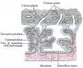
* '''Human Chorionic Gonadotropin Induces Human Macrophages to Form Intracytoplasmic Vacuoles Mimicking Hofbauer Cells in Human Chorionic Villi'''{{#pmid:23128164|PMID23128164}} The most characteristic morphological feature of macrophages in the stroma of placental villi, known as Hofbauer cells, is their highly vacuolated appearance. They also show positive immunostaining for human chorionic gonadotropin (hCG)." | * '''Human Chorionic Gonadotropin Induces Human Macrophages to Form Intracytoplasmic Vacuoles Mimicking Hofbauer Cells in Human Chorionic Villi'''{{#pmid:23128164|PMID23128164}} The most characteristic morphological feature of macrophages in the stroma of placental villi, known as Hofbauer cells, is their highly vacuolated appearance. They also show positive immunostaining for human chorionic gonadotropin (hCG)." | ||
|} | |} | ||
| Line 29: | Line 29: | ||
<pubmed limit=5>Placental Cord</pubmed> | <pubmed limit=5>Placental Cord</pubmed> | ||
|} | |||
{| class="wikitable mw-collapsible mw-collapsed" | |||
! Older papers | |||
|- | |||
| | |||
* '''Hofbauer cells in early human placenta: possible implications in vasculogenesis and angiogenesis'''{{#pmid:17350092|PMID17350092}} "The stroma of the placental villi contain numerous macrophages, so-called Hofbauer cells which are of mesenchymal origin and are thought to function in many processes. ...Double immunohistochemistry staining with CD31/PECAM1 and CD68 was applied to placental tissues. In placental villous core, majority of the Hofbauer cells were found to be either in close contact with angiogenic cell cords and primitive vascular tubes or located in between them. Moreover, the number of Hofbauer cells and vasculogenic structures were found to be significantly correlated. The findings of this study suggest for the first time that Hofbauer cells might be involved in the processes of vasculogenesis and angiogenesis in the placenta." | |||
|} | |} | ||
==Hofbauer Cells== | ==Hofbauer Cells== | ||
Revision as of 15:34, 22 October 2018
| Embryology - 21 May 2024 |
|---|
| Google Translate - select your language from the list shown below (this will open a new external page) |
|
العربية | català | 中文 | 中國傳統的 | français | Deutsche | עִברִית | हिंदी | bahasa Indonesia | italiano | 日本語 | 한국어 | မြန်မာ | Pilipino | Polskie | português | ਪੰਜਾਬੀ ਦੇ | Română | русский | Español | Swahili | Svensk | ไทย | Türkçe | اردو | ייִדיש | Tiếng Việt These external translations are automated and may not be accurate. (More? About Translations) |
Introduction


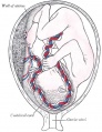
The placenta (Greek, plakuos = flat cake) named on the basis of this organs appearance. The placental cord (umbilical cord) is the connecting region between the functional placenta and the embryo/fetal umbilical region. The human cord varies greatly in overall length increasing to about 60 to 70 cm at term. This extraembryonic structure contains the placental blood vessels and allantois.
There are essentially 3 separate aortic/venous circulatory systems: umbilical, systemic and vitelline. The umbilical system is lost at birth, the vitelline contributes to the portal system and the systemic (embryonic) is extensively remodelled to fom the the cardiovascular system.
Some Recent Findings

|
| More recent papers |
|---|
 This table allows an automated computer search of the external PubMed database using the listed "Search term" text link.
More? References | Discussion Page | Journal Searches | 2019 References | 2020 References Search term: Placental Cord <pubmed limit=5>Placental Cord</pubmed> |
| Older papers |
|---|
|
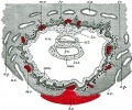
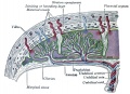
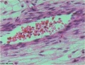
Hofbauer Cells

Hofbauer Cells (red asterisks)[4] |
|
| Historic Embryology - Hofbauer Cells | ||
|---|---|---|
Chapter 14. Hofbauer Cells in Normal and Pathologic Conceptuses Contributions to Embryology Carnegie Institution No.56 (1921)
|


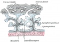
Wharton's Jelly

Placental cord cross-section showing Wharton's Jelly |
First described and named after Thomas Wharton (1614–1673) an English physician and anatomist.
|
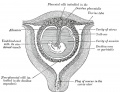
Placental Cord Histology

Placental cord cross-section

Placental vein

Placental artery

Placental allantois


Human placental cord (3.5 month) cross-section.
Persistent Right Umbilical Vein
A fairly rare anomaly, a study of 15,237 obstetric ultrasound examinations performed after 15 weeks' gestation identified only 33 cases of persistent right umbilical vein.[5] Some studies have identified associated fetal anomalies with this condition[6], including cardiac abnormalities.[7]
Cord Length
The following are lengths and classifications at term.
- Normal range - 50 to 60 cm.
- Short cord - less than 35 cm.
- Long cords - over 70 cm can be associated with wrapping around the fetus and other abnormalities.[8]
Cord Coiling
A recent review of the published literature on cord coiling[9] states: "Previous studies that draw a link between abnormal cord coiling and clinical outcome are generally too small and/or selective to allow meaningful conclusions or applicability to low-risk populations."
The following suggested associations[10] should therefore be reconsidered.
- Hypocoiling - associated with increased incidence of fetal demise, intrapartum fetal heart rate decelerations, operative delivery for fetal distress, anatomic-karyotypic abnormalities and chorio-amnionitis.
- Hypercoiling - associated with increased incidence of fetal growth restriction, intrapartum fetal heart rate decelerations, vascular thrombosis and cord stenosis.
Placental Cord Ultrasound

There are a number of analyses that can be made by ultrasound scanning of the fetal placental cord. Some detected abnormalities (blood vessel number, blood flow[11]) have been associated with adverse developmental outcomes.
- Quantification of cord length, diameter, structural abnormalities.
- Quantification of placental blood vessel number and size.
- Quantification of uterine artery blood flow (doppler analysis).
Ultrasound image of transverse scan through the cord show the method of estimation of the cross-sectional area.

Cord Abnormalities
Cord Vessel Number

Persistent Right Umbilical Vein
A fairly rare anomaly, a study of 15,237 obstetric ultrasound examinations performed after 15 weeks' gestation identified only 33 cases of persistent right umbilical vein.[5] Some studies have identified associated fetal anomalies with this condition[6], including cardiac abnormalities.[7]
Cord Knotting

There are few abnormalities associated with umbilical cord development, other that abnormally short or long cords, which in most cases do not cause difficulties.
In some cases though, long cords can wrap around limbs or the fetus neck, which can then restrict blood flow or lead to tissue or nerve damage, and therefore effect develoment.
Cord knotting can also occur (1%) in most cases these knots have no effect, in some cases of severe knotting this can prevents the passage of placental blood.
Umbilical Cord Torsion
Rare umbilical cord torsion, even without knot formation can also affect placental blood flow, even leading to fetal demise.[12]
Cord Length
References
- ↑ Kim JH, Jin ZW, Murakami G, Chai OH & Rodríguez-Vázquez JF. (2018). Persistent right umbilical vein: a study using serial sections of human embryos and fetuses. Anat Cell Biol , 51, 218-222. PMID: 30310717 DOI.
- ↑ Yamaguchi M, Ohba T, Tashiro H, Yamada G & Katabuchi H. (2013). Human chorionic gonadotropin induces human macrophages to form intracytoplasmic vacuoles mimicking Hofbauer cells in human chorionic villi. Cells Tissues Organs (Print) , 197, 127-35. PMID: 23128164 DOI.
- ↑ Seval Y, Korgun ET & Demir R. (2007). Hofbauer cells in early human placenta: possible implications in vasculogenesis and angiogenesis. Placenta , 28, 841-5. PMID: 17350092 DOI.
- ↑ Lorenzi T, Turi A, Lorenzi M, Paolinelli F, Mancioli F, La Sala L, Morroni M, Ciarmela P, Mantovani A, Tranquilli AL, Castellucci M & Marzioni D. (2012). Placental expression of CD100, CD72 and CD45 is dysregulated in human miscarriage. PLoS ONE , 7, e35232. PMID: 22606231 DOI.
- ↑ 5.0 5.1 Hill LM, Mills A, Peterson C & Boyles D. (1994). Persistent right umbilical vein: sonographic detection and subsequent neonatal outcome. Obstet Gynecol , 84, 923-5. PMID: 7970470
- ↑ 6.0 6.1 Weichert J, Hartge D, Germer U, Axt-Fliedner R & Gembruch U. (2011). Persistent right umbilical vein: a prenatal condition worth mentioning?. Ultrasound Obstet Gynecol , 37, 543-8. PMID: 20922781 DOI.
- ↑ 7.0 7.1 Lide B, Lindsley W, Foster MJ, Hale R & Haeri S. (2016). Intrahepatic Persistent Right Umbilical Vein and Associated Outcomes: A Systematic Review of the Literature. J Ultrasound Med , 35, 1-5. PMID: 26635256 DOI.
- ↑ <pubmed>11178630</pubmed>
- ↑ Jessop FA, Lees CC, Pathak S, Hook CE & Sebire NJ. (2014). Umbilical cord coiling: clinical outcomes in an unselected population and systematic review. Virchows Arch. , 464, 105-12. PMID: 24259031 DOI.
- ↑ de Laat MW, Franx A, van Alderen ED, Nikkels PG & Visser GH. (2005). The umbilical coiling index, a review of the literature. J. Matern. Fetal. Neonatal. Med. , 17, 93-100. PMID: 16076615 DOI.
- ↑ Shwarzman P, Waintraub AY, Frieger M, Bashiri A, Mazor M & Hershkovitz R. (2013). Third-trimester abnormal uterine artery Doppler findings are associated with adverse pregnancy outcomes. J Ultrasound Med , 32, 2107-13. PMID: 24277892 DOI.
- ↑ Hallak M, Pryde PG, Qureshi F, Johnson MP, Jacques SM & Evans MI. (1994). Constriction of the umbilical cord leading to fetal death. A report of three cases. J Reprod Med , 39, 561-5. PMID: 7966052
Reviews
Articles
Search PubMed
May 2010 search "Placental Cord Development]" All (650) Review (91) Free Full Text (119)
Search Pubmed: Placental Cord | Umbilical Cord | Placental Cord Development | Umbilical Cord Development | Hofbauer cells
Additional Images
see all online Placental materials
Historic

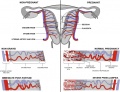
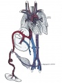
Fetal circulation overview


Glossary Links
- Glossary: A | B | C | D | E | F | G | H | I | J | K | L | M | N | O | P | Q | R | S | T | U | V | W | X | Y | Z | Numbers | Symbols | Term Link
Cite this page: Hill, M.A. (2024, May 21) Embryology Placenta - Cord. Retrieved from https://embryology.med.unsw.edu.au/embryology/index.php/Placenta_-_Cord
- © Dr Mark Hill 2024, UNSW Embryology ISBN: 978 0 7334 2609 4 - UNSW CRICOS Provider Code No. 00098G