Hamburger Hamilton Stages: Difference between revisions
mNo edit summary |
mNo edit summary |
||
| Line 8: | Line 8: | ||
:[[Hamburger Hamilton Stages|'''HH Links''']]: [[:File:HHstage1-4.jpg|stage 1-4]] | [[:File:HHstage5-10.jpg|stages 5-10]] | [[:File:HHstage11-14.jpg|stages 11-14]] | [[:File:HHstage15-18.jpg|stages 15-18]] | [[:File:HHstage19-21.jpg|stages 19-21]] | [[:File:HHstage22-25.jpg|stages 22-25]] | [[:File:HHstage26-28.jpg|stages 26-28]] | [[:File:HHstage29-32.jpg|stages 29-32]] | [[:File:HHstage33-35.jpg|stages 33-35]] | [[:File:HHstage36-37.jpg|stages 36-37]] | [[:File:HHstage38-39.jpg|stages 38-39]] | [[:File:HHstage40-41.jpg|stages 40-41]] | [[:File:HHstage42-43.jpg|stages 42-43]] | [[:File:HHstage44-45.jpg|stages 44-45]] | [[Hamburger Hamilton Stages]] | :[[Hamburger Hamilton Stages|'''HH Links''']]: [[:File:HHstage1-4.jpg|stage 1-4]] | [[:File:HHstage5-10.jpg|stages 5-10]] | [[:File:HHstage11-14.jpg|stages 11-14]] | [[:File:HHstage15-18.jpg|stages 15-18]] | [[:File:HHstage19-21.jpg|stages 19-21]] | [[:File:HHstage22-25.jpg|stages 22-25]] | [[:File:HHstage26-28.jpg|stages 26-28]] | [[:File:HHstage29-32.jpg|stages 29-32]] | [[:File:HHstage33-35.jpg|stages 33-35]] | [[:File:HHstage36-37.jpg|stages 36-37]] | [[:File:HHstage38-39.jpg|stages 38-39]] | [[:File:HHstage40-41.jpg|stages 40-41]] | [[:File:HHstage42-43.jpg|stages 42-43]] | [[:File:HHstage44-45.jpg|stages 44-45]] | [[Hamburger Hamilton Stages]] | ||
{{Hamburger Hamilton stages table}} | |||
Original paper<ref name=HamburgerHamilton1951>{{Ref-HamburgerHamilton1951}}</ref>(and all data) was republished in 1992.<ref><pubmed>1304821</pubmed>| [http://onlinelibrary.wiley.com/doi/10.1002/aja.1001950404/pdf PDF]</ref> | |||
<gallery> | <gallery> | ||
| Line 25: | Line 32: | ||
File:HHstage44-45.jpg|stages 44-45 | File:HHstage44-45.jpg|stages 44-45 | ||
</gallery> | </gallery> | ||
Revision as of 09:47, 2 June 2017
| Embryology - 26 Apr 2024 |
|---|
| Google Translate - select your language from the list shown below (this will open a new external page) |
|
العربية | català | 中文 | 中國傳統的 | français | Deutsche | עִברִית | हिंदी | bahasa Indonesia | italiano | 日本語 | 한국어 | မြန်မာ | Pilipino | Polskie | português | ਪੰਜਾਬੀ ਦੇ | Română | русский | Español | Swahili | Svensk | ไทย | Türkçe | اردو | ייִדיש | Tiếng Việt These external translations are automated and may not be accurate. (More? About Translations) |
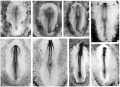
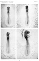
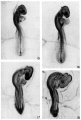
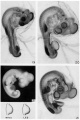
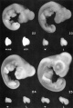
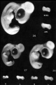
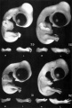
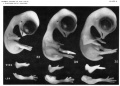
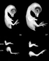
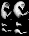
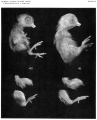
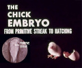
The 1951 Hamburger Hamilton Stages[1] are most commonly used for chicken staging.
Note that there was also an earlier Witschi staging and an even older 1900 Normentafeln zur Entwicklungsgeschichte der Wirbeltiere - Gallus domesticus (Normal Plates of the Development of the Chicken Embryo} by Franz Keibel and Karl Abraham.
- HH Links: stage 1-4 | stages 5-10 | stages 11-14 | stages 15-18 | stages 19-21 | stages 22-25 | stages 26-28 | stages 29-32 | stages 33-35 | stages 36-37 | stages 38-39 | stages 40-41 | stages 42-43 | stages 44-45 | Hamburger Hamilton Stages
| 3.5-4.5 hr | Shell membrane of egg formed in isthmus of oviduct | ||
| Germ wall formed from marginal periblast | |||
| 4.5-24.0 hr | Shell of egg formed in uterus | ||
| Preprimitive streak (embryonic shield) | |||
| 6-7 hr | Initial primitive streak, 0.3-0.5 mm long | ||
| 12-13 hr | Intermediate primitive streak | ||
| 18-19 hr | Definitive primitive streak, ±1.88 mm long | ||
| 19-22 hr | Head process (notochord) | ||
| 23-25 hr | Head fold | ||
| 23-26 hr | 1 somite; neural folds | ||
| ca. 23-26 hr | 1-3 somites; coelom | ||
| 26-29 hr | 4 somites; blood islands | ||
| 29-33 hr | 7 somites; primary optic vesicles | ||
| ca. 33 hr | 8-9 somites; anterior amniotic fold | ||
| 33-38 hr | 10 somites; 3 primary brain vesicles | ||
| 40-45 hr | 13 somites; 5 neuromeres of hindbrain | ||
| 45-49 hr | 16 somites; telencephalon | ||
| 48-52 hr | 19 somites; atrioventricular canal | ||
| ca. 50-52 hr | 20-21 somites; tail bud | ||
| 50-53 hr | 22 somites; trunk flexure; visceral arches I and II, clefts 1 and 2 | ||
| ca. 50-54 hr | 23 somites; premandibular head cavities | ||
| 50-55 hr | 24-27 somites; visceral arch III, cleft 3 | ||
| 51-56 hr | 26-28 somites; wing bud; posterior amniotic fold | ||
| 52-64 hr | 29-32 somites; leg bud; epiphysis | ||
| 3 da | 30-36 somites extending beyond level of leg bud; allantois | ||
| 3.0-3.5 da | 37- 40 somites extending into tail; maxillary process | ||
| 3.0-3.5 da | 40-43 somites; rotation completed; eye pigment | ||
| 3.5 da | 43-44 somites; visceral arch IV, cleft 4 | ||
| 3.5-4.0 da | Somites extend to tip of tail | ||
| 4 da | Dorsal contour from hindbrain to tail is a curved line | ||
| 4.5 da | Toe plate | ||
| 4.5-5.0 da | Elbow and knee joints | ||
| 5 da | 1st 3 toes | ||
| 5.0-5.5 da | Beak | ||
| 5.5-6.0 da | 3 digits, 4 toes | ||
| 6.0-6.5 da | Rudiment of 5th toe | ||
| 6.5-7.0 da | Feather germs; scleral papillae; egg tooth | ||
| 7.0-7.5 da | Web between 1st and 2nd digits | ||
| 7.5 da | Anterior tip of mandible has reached beak | ||
| 7.5-8.0 da | Web on radial margin of wing and 1st digit | ||
| 8 da | Nictitating membrane | ||
| 8.5-9.0 da | Phalanges in toes | ||
| 10 da | Length of 3rd toe from tip to middle of metatarsal joint = 5.4 ±0.3 mm; length of beak from anterior angle of nostril to tip of bill = 2.5mm; primordium of comb; labial groove; uropygial gland | ||
| 11 da | Length of 3rd toe = 7.4 ±0.3mm; length of beak = 3.0 mm | ||
| 12 da | Length of 3rd toe = 8.4 ± 0.3 mm; length of beak = 3.1 mm | ||
| 13 da | Length of 3rd toe = 9.8 ± 0.3 mm; length of beak = 3.5 mm | ||
| 14 da | Length of beak = 4.0 mm; length of 3rd toe = 12.7 ± 0.5 mm | ||
| 15 da | Length of beak from anterior angle of nostril to tip of upper bill = 4.5 mm; length of 3rd toe = 14.9 ± 0.8 mm | ||
| 16 da | Length of beak = 4.8 mm; length of 3rd toe = 16.7 ± 0.8 mm | ||
| 17 da | Length of beak = 5.0 mm; length of 3rd toe = 18.6 ± 0.8 mm | ||
| 18 da | Length of beak = 5.7 mm; length of 3rd toe = 20.4 ± 0.8 mm | ||
| 19-20 da | Yolk sac half enclosed in body cavity; chorio-allantoic membrane contains less blood and is "sticky" in living embryo | ||
| 20-21 da | Newly-hatched chick | ||
Original paper[2](and all data) was republished in 1992.[3]
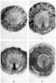
stage 1-4
stages 5-10
stages 11-14
stages 15-18
stages 19-21
stages 22-25
stages 26-28
stage 29-32
stage 33-35
stages 36-37
stages 38-39
stages 40-41
stages 42-43
stages 44-45


| Hamburger Hamilton Stages (1951) | ||
|---|---|---|
| 3.5-4.5 hr | Shell membrane of egg formed in isthmus of oviduct | |
| Germ wall formed from marginal periblast | ||
| 4.5-24.0 hr | Shell of egg formed in uterus | |
| Preprimitive streak (embryonic shield) | ||
| 6-7 hr | Initial primitive streak, 0.3-0.5 mm long | |
| 12-13 hr | Intermediate primitive streak | |
| 18-19 hr | Definitive primitive streak, ±1.88 mm long | |
| 19-22 hr | Head process (notochord) | |
| 23-25 hr | Head fold | |
| 23-26 hr | 1 somite; neural folds | |
| ca. 23-26 hr | 1-3 somites; coelom | |
| 26-29 hr | 4 somites; blood islands | |
| 29-33 hr | 7 somites; primary optic vesicles | |
| ca. 33 hr | 8-9 somites; anterior amniotic fold | |
| 33-38 hr | 10 somites; 3 primary brain vesicles | |
| 40-45 hr | 13 somites; 5 neuromeres of hindbrain | |
| 45-49 hr | 16 somites; telencephalon | |
| 48-52 hr | 19 somites; atrioventricular canal | |
| ca. 50-52 hr | 20-21 somites; tail bud | |
| 50-53 hr | 22 somites; trunk flexure; visceral arches I and II, clefts 1 and 2 | |
| ca. 50-54 hr | 23 somites; premandibular head cavities | |
| 50-55 hr | 24-27 somites; visceral arch III, cleft 3 | |
| 51-56 hr | 26-28 somites; wing bud; posterior amniotic fold | |
| 52-64 hr | 29-32 somites; leg bud; epiphysis | |
| 3 da | 30-36 somites extending beyond level of leg bud; allantois | |
| 3.0-3.5 da | 37- 40 somites extending into tail; maxillary process | |
| 3.0-3.5 da | 40-43 somites; rotation completed; eye pigment | |
| 3.5 da | 43-44 somites; visceral arch IV, cleft 4 | |
| 3.5-4.0 da | Somites extend to tip of tail | |
| 4 da | Dorsal contour from hindbrain to tail is a curved line | |
| 4.5 da | Toe plate | |
| 4.5-5.0 da | Elbow and knee joints | |
| 5 da | 1st 3 toes | |
| 5.0-5.5 da | Beak | |
| 5.5-6.0 da | 3 digits, 4 toes | |
| 6.0-6.5 da | Rudiment of 5th toe | |
| 6.5-7.0 da | Feather germs; scleral papillae; egg tooth | |
| 7.0-7.5 da | Web between 1st and 2nd digits | |
| 7.5 da | Anterior tip of mandible has reached beak | |
| 7.5-8.0 da | Web on radial margin of wing and 1st digit | |
| 8 da | Nictitating membrane | |
| 8.5-9.0 da | Phalanges in toes | |
| 10 da | Length of 3rd toe from tip to middle of metatarsal joint = 5.4 ±0.3 mm; length of beak from anterior angle of nostril to tip of bill = 2.5mm; primordium of comb; labial groove; uropygial gland | |
| 11 da | Length of 3rd toe = 7.4 ±0.3mm; length of beak = 3.0 mm | |
| 12 da | Length of 3rd toe = 8.4 ± 0.3 mm; length of beak = 3.1 mm | |
| 13 da | Length of 3rd toe = 9.8 ± 0.3 mm; length of beak = 3.5 mm | |
| 14 da | Length of beak = 4.0 mm; length of 3rd toe = 12.7 ± 0.5 mm | |
| 15 da | Length of beak from anterior angle of nostril to tip of upper bill = 4.5 mm; length of 3rd toe = 14.9 ± 0.8 mm | |
| 16 da | Length of beak = 4.8 mm; length of 3rd toe = 16.7 ± 0.8 mm | |
| 17 da | Length of beak = 5.0 mm; length of 3rd toe = 18.6 ± 0.8 mm | |
| 18 da | Length of beak = 5.7 mm; length of 3rd toe = 20.4 ± 0.8 mm | |
| 19-20 da | Yolk sac half enclosed in body cavity; chorio-allantoic membrane contains less blood and is "sticky" in living embryo | |
| 20-21 da | Newly-hatched chick | |
|
Hamburger V. and Hamilton HL. A series of normal stages in the development of the chick embryo. (1951) J Morphol. 88(1): 49-92. PMID 24539719 PDF Original 1951 paper (and all data) was republished in 1992. <pubmed>1304821</pubmed> PDF | ||
References
- ↑ Hamburger V. and Hamilton HL. A series of normal stages in the development of the chick embryo. (1951) J Morphol. 88(1): 49-92. PMID 24539719 PDF
- ↑ Hamburger V. and Hamilton HL. A series of normal stages in the development of the chick embryo. (1951) J Morphol. 88(1): 49-92. PMID 24539719 PDF
- ↑ <pubmed>1304821</pubmed>| PDF
Additional Media
Movies
|
|
|
|
Images
HH4 and HH10 endoderm

HH20 head muscle MyoR
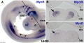
HH20 head muscle MyoR and MyoD


Glossary Links
- Glossary: A | B | C | D | E | F | G | H | I | J | K | L | M | N | O | P | Q | R | S | T | U | V | W | X | Y | Z | Numbers | Symbols | Term Link
Cite this page: Hill, M.A. (2024, April 26) Embryology Hamburger Hamilton Stages. Retrieved from https://embryology.med.unsw.edu.au/embryology/index.php/Hamburger_Hamilton_Stages
- © Dr Mark Hill 2024, UNSW Embryology ISBN: 978 0 7334 2609 4 - UNSW CRICOS Provider Code No. 00098G