Gastrointestinal Tract - Gall Bladder Development: Difference between revisions
No edit summary |
|||
| Line 9: | Line 9: | ||
The pars cystica vacuolates and expands, the stalk becoming the cystic duct. This structure is initially hollow, then solid (by proliferation of epithelial lining), and then recanalized occurs by vacuolation of this expanded epithelium. (some text above modified from Bani-Hani KE., 2005) | The pars cystica vacuolates and expands, the stalk becoming the cystic duct. This structure is initially hollow, then solid (by proliferation of epithelial lining), and then recanalized occurs by vacuolation of this expanded epithelium. (some text above modified from Bani-Hani KE., 2005) | ||
:{{Template:Gastrointestinal Tract Links}} | [[Gastrointestinal Tract - Gall Bladder Histology|Gall Bladder Histology]] | [http://embryology.med.unsw.edu.au/Notes/git7a.htm original page] | |||
== Some Recent Findings == | |||
{| | |||
|-bgcolor="F5FAFF" | |||
| | |||
* '''Embryology of the biliary tract'''<ref><pubmed>20551648</pubmed></ref> | |||
|} | |||
==Embryonic Development== | ==Embryonic Development== | ||
Revision as of 16:11, 22 August 2011
Introduction

This section of notes gives an overview of Gall Bladder development, histology and abnormalities associated with the biliary system. In the adult, the gall bladder is a site of bile salt storage and concentration, to then be released into the small intestine where they act to solubilize dietary lipids by their detergent effect. Bile salts are a cholesterol derivative (breakdown product).
The transverse septum differentiates to form the hepatic diverticulum and the hepatic primordium, these two structures together will go on to form different components of the mature liver and gall bladder.
The hepatic diverticulum divides into two parts: pars hepatica (larger cranial part, primordium of the liver) and pars cystica (smaller ventral invagination, primordium of gall bladder).
The pars cystica vacuolates and expands, the stalk becoming the cystic duct. This structure is initially hollow, then solid (by proliferation of epithelial lining), and then recanalized occurs by vacuolation of this expanded epithelium. (some text above modified from Bani-Hani KE., 2005)
| Gall Bladder Histology | original page
Some Recent Findings
|
Embryonic Development

Early embryonic gall bladder (Carnegie stage 13, Week 4)

Late embryonic gall bladder (Carnegie stage 22, Week 8)
Additional Images
See also Gall Bladder Histology
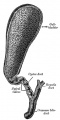
Historic drawing gall bladder anatomy
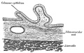
Historic drawing gall bladder transverse section
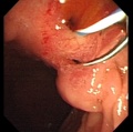
Abnormality - ectopic opening of the common bile duct (CBD) into the duodenal bulb.


References
- ↑ <pubmed>20551648</pubmed>
Reviews
<pubmed>20551648</pubmed> <pubmed>20152372</pubmed> <pubmed>15853977</pubmed> <pubmed>15382016</pubmed>
Articles
<pubmed>21078254</pubmed> <pubmed>20191134</pubmed> <pubmed>16273658</pubmed>
Search Pubmed
July 2010
Search Bookshelf Gall Bladder Development
Search Pubmed Now: Gall Bladder Development
Glossary Links
- Glossary: A | B | C | D | E | F | G | H | I | J | K | L | M | N | O | P | Q | R | S | T | U | V | W | X | Y | Z | Numbers | Symbols | Term Link
Cite this page: Hill, M.A. (2024, May 7) Embryology Gastrointestinal Tract - Gall Bladder Development. Retrieved from https://embryology.med.unsw.edu.au/embryology/index.php/Gastrointestinal_Tract_-_Gall_Bladder_Development
- © Dr Mark Hill 2024, UNSW Embryology ISBN: 978 0 7334 2609 4 - UNSW CRICOS Provider Code No. 00098G