Category:Respiratory
From Embryology
This Embryology category shows content related to respiratory development.
- Note the section of notes include the musculoskeletal diaphragm development.
- The sense of smell development is covered separately under Sensory Development.
Subcategories
This category has the following 2 subcategories, out of 2 total.
Pages in category 'Respiratory'
The following 148 pages are in this category, out of 148 total.
2
A
B
- Book - A History of Science 9
- Book - Contributions to Embryology Carnegie Institution No.12
- Book - Contributions to Embryology Carnegie Institution No.38
- Book - Human Embryology and Morphology 2
- Book - Manual of Human Embryology 17-10
- Book - Manual of Human Embryology 17-9
- Book - Text-Book of Embryology 13
C
E
F
H
L
M
P
- Paper - A case of atresia of the esophagus combined with traoheoesophageal fistula in a 9 mm human embryo, and its embryological explanation
- Paper - A further communication on the formation of the nasal cavities (1912)
- Paper - A histological investigation of the development and structure of the human lung
- Paper - A preliminary communication on the formation of the nasal cavities (1911)
- Paper - Conditions of foetal respiration in the goat (1934)
- Paper - Congenital defects in the diaphragm (1940)
- Paper - Cor biloculare, with a note on the development of the pulmonary veins (1937)
- Paper - Development of olfactory and related structures in staged human embryos
- Paper - Development of the human diaphragm and pleural sacs
- Paper - Development of the larynx (1910)
- Paper - Early developmental stages of the human lung
- Paper - Malformation of the diaphragm in a dog (1924)
- Paper - Normal development of the trachea and esophagus in man
- Paper - Note on a case of defective development of the diaphragm, accompanied by stenosis of the anal canal (1918)
- Paper - On the genesis of air cells in the conchae nasales (1910)
- Paper - On the origin of the pulmonary arteries in mammals
- Paper - On the origin of the pulmonary arteries in mammals 2
- Paper - On the prenatal and neonatal lung (1913)
- Paper - Origin of the pulmonary vessels in the chick (1922)
- Paper - Studies on the development of the human larynx (1911)
- Paper - The Course of the Phrenic Nerve in the Embryo
- Paper - The development of nerve-endings in the respiratory muscles of the sheep (1940)
- Paper - The development of the bronchopulmonary segments in human embryos of horizons XVII to XIX
- Paper - The development of the lungs
- Paper - The Development of the Nose and of the Pharynx and its Derivatives in Man
- Paper - The development of the terminal air passages of the human lung
- Paper - The embryology of the bird's lung 2 (1916)
- Paper - The functional history of the coelom and the diaphragm (1913)
- Paper - The genesis and development of the nasolacrimal passages in man
- Paper - The lateral wall of the cavum nasi in man, with especial reference to the various developmental stages
- Paper - The lung of a human foetus of 170 mm C.R. length
- Paper - The right lung of a human foetus of 152 millimeters CRL
- Paper - The sinus maxillarus and its relations in the embryo, child, and adult man
- Paper - The terminals of the human bronchiole (1922)
- Paper - True congenital diverticulum of the trachea in a subject showing also right aortic arch (1929)
- Paper - Two anomalies in the construction of the diaphragm (1924)
- Template:Pseudoglandular stage
R
- Template:Ref-AddisonHow1913
- Template:Ref-Amin1914
- Template:Ref-BarcroftElliottFlexner1934
- Template:Ref-BarnardDay1937
- Template:Ref-Bremer1902
- Template:Ref-Bremer1909
- Template:Ref-Cooper1938
- Template:Ref-Dickson1940
- Template:Ref-Flint1906
- Template:Ref-Frazer1910
- Template:Ref-Frazer1911a
- Template:Ref-Frazer1911b
- Template:Ref-Frazer1912
- Template:Ref-Goodrich1918
- Template:Ref-Grosser1912
- Template:Ref-GrosserLewisMcMurrich1912
- Template:Ref-Guinane1924
- Template:Ref-Huntington1919
- Template:Ref-LiebowMiller1940
- Template:Ref-LocyLarsell1916b
- Template:Ref-Mall1901c
- Template:Ref-Miller1919
- Template:Ref-Palmer1936a
- Template:Ref-Palmer1936b
- Template:Ref-Schaeffer1910c
- Template:Ref-Stibbe1929
- Template:Ref-Wells1954
- Template:Ref-WellsBoyden1954
- Template:Ref-Willson1922
- Template:Respiratory
- Template:Respiratory abnormalities
- Template:Respiratory Links
- Template:Respiratory Postnatal Timeline table
- Respiratory Quiz
- Template:Respiratory Species Comparison collapsetable
- Template:Respiratory Species Comparison table
- Respiratory System - Abnormalities
- Respiratory System - Carnegie Stage 13
- Respiratory System - Carnegie Stage 22
- Respiratory System - Diaphragm
- Respiratory System - Histology
- Respiratory System - Molecular
- Respiratory System - Postnatal
- Respiratory System - Upper Respiratory Tract
- Respiratory System Development
- Template:Respiratory terms
S
Media in category 'Respiratory'
The following 75 files are in this category, out of 275 total.
(previous page) (next page)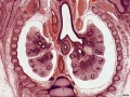 ME54 002.jpg 800 × 601; 157 KB
 ME54 003.jpg 800 × 601; 169 KB
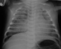 Meconium aspiration syndrome 01.jpg 640 × 515; 24 KB
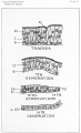
 Minot1897 463.jpg 731 × 600; 73 KB
 Model Sox9 lung development 01.jpg 600 × 576; 43 KB
 Mouse E14.5 Titf1 gene expression.jpg 481 × 739; 33 KB
 Mouse HOXA5 expression E12.5.jpg 1,000 × 577; 192 KB
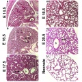
 Mouse lung development 01.jpg 1,000 × 1,254; 791 KB
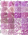 Mouse lung development 01a.jpg 800 × 1,003; 495 KB
 Mouse lung development 02.jpg 922 × 922; 239 KB
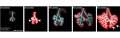
 Mouse lung development 03.jpg 540 × 1,200; 349 KB
 Mouse lung E14.5 Sox9.jpg 600 × 594; 48 KB
 Mouse lung histology E18.5.jpg 600 × 594; 54 KB
 Mouse lung micro-CT 01.jpg 650 × 668; 113 KB
 Mouse respiratory 36 somites.jpg 394 × 432; 15 KB
 Mouse respiratory 36 to 60 somites.gif 394 × 432; 154 KB
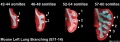
 Mouse respiratory 36 to 60 somites.jpg 1,200 × 383; 58 KB
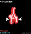 Mouse respiratory 44 somites.jpg 394 × 432; 19 KB
 Mouse respiratory 44 to 60 somites.jpg 1,000 × 347; 52 KB
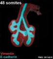 Mouse respiratory 48 somites.jpg 394 × 432; 23 KB
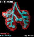 Mouse respiratory 54 somites.jpg 394 × 432; 30 KB
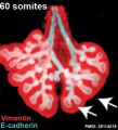 Mouse respiratory 60 somites.jpg 394 × 432; 34 KB
 Mouse respiratory Tbx4 and Tbx5.jpg 889 × 800; 112 KB
 Mouse whole lung E12.5.jpg 600 × 594; 17 KB
 Mouse whole lung E14.5.jpg 600 × 594; 17 KB
 Mouse- respiratory development 01.jpg 1,000 × 571; 125 KB
 Mouse- respiratory development 04.jpg 800 × 728; 73 KB
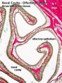
 Nasal cavities.jpg 500 × 307; 19 KB
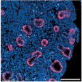
 Neonatal human pulmonary neuroendocrine cell EM01.jpg 836 × 1,200; 405 KB
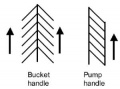 Neonatal rib orientation.jpg 300 × 223; 8 KB
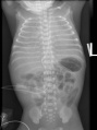 NRDS.jpg 481 × 645; 155 KB
 Palmer1934 plate01.jpg 1,000 × 1,482; 252 KB
 Palmer1934 plate02.jpg 1,000 × 1,639; 226 KB
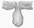
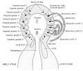
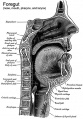 Pharynx cartoon.jpg 418 × 600; 88 KB
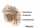 Pharynx.jpg 434 × 340; 13 KB
 Pig lung alveolarization.jpg 600 × 389; 38 KB
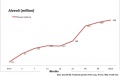 Postnatal alveoli number.jpg 800 × 504; 26 KB
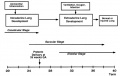 Preterm delivery and lung development.jpg 701 × 444; 41 KB
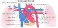 Pulmonary circulation cartoon.jpg 800 × 385; 65 KB
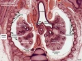
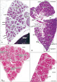 Pulmonary Pleura - pseudoglandular and canalicular stages 01.jpg 1,280 × 1,863; 707 KB
 Pulmonary Pleura - pseudoglandular and canalicular stages 02.jpg 671 × 853; 233 KB
 Rat respiratory 01.mp4 ; 3.63 MB
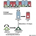 Respiratory epithelium cells cartoon.jpg 500 × 500; 44 KB
 Respiratory histology 01.jpg 450 × 600; 86 KB
 Respiratory histology 02.jpg 450 × 600; 37 KB
 Respiratory histology 03.jpg 450 × 600; 29 KB
 Respiratory histology 04.jpg 450 × 600; 31 KB
 Respiratory histology 05.jpg 450 × 600; 96 KB
 Respiratory histology 06.jpg 450 × 600; 95 KB
 Respiratory histology 07.jpg 1,280 × 1,024; 255 KB
 Respiratory histology 08.jpg 1,280 × 1,024; 263 KB
 Respiratory histology 09.jpg 1,280 × 1,024; 236 KB
 Respiratory histology 11.jpg 450 × 600; 65 KB
 Respiratory histology 12.jpg 450 × 600; 88 KB
 Respiratory histology 13.jpg 450 × 600; 102 KB
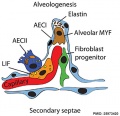 Respiratory secondary septum 01.jpg 500 × 482; 45 KB
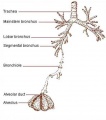 Respiratory tract.jpg 500 × 567; 27 KB
 Rugh 088.jpg 943 × 800; 141 KB
 Stage 13 image 022.jpg 1,000 × 473; 101 KB
 Stage 13 image 023.jpg 1,000 × 544; 110 KB
 Stage 13 image 072.jpg 1,000 × 614; 116 KB
 Stage 22 image 166.jpg 1,000 × 658; 197 KB
 Stage 22 image 167.jpg 1,000 × 662; 194 KB
 Stage 22 image 170.jpg 1,000 × 657; 117 KB
 Stage 22 image 171.jpg 1,000 × 653; 206 KB
 Stage 22 image 200.jpg 1,200 × 877; 563 KB
 Stage 22 image 209.jpg 1,200 × 808; 305 KB
 Stage 22 vomeronasal organ.jpg 600 × 554; 125 KB
 Stage14 respiratory tract.jpg 406 × 431; 76 KB
 Stage14-22 lungs.jpg 450 × 377; 55 KB
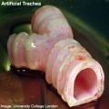 Stem cell artificial trachea and bronchi.jpg 400 × 400; 31 KB
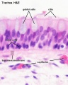 Trachea histology 01.jpg 480 × 600; 47 KB
 Windle1940 fig32.jpg 1,000 × 847; 100 KB


