Category:Musculoskeletal
From Embryology
This Embryology category covers content related to both muscle (skeletal muscle), cartilage, bone (skeleton) and joint development.
Subcategories
This category has the following 12 subcategories, out of 12 total.
Pages in category 'Musculoskeletal'
The following 159 pages are in this category, out of 159 total.
2
A
B
- Bone Development
- Bone Histology
- Template:Bone histology
- Template:Bone timeline
- Book - Handbook of Pathological Anatomy 2
- Book - Handbook of Pathological Anatomy 2.1
- Book - Handbook of Pathological Anatomy 2.10
- Book - Handbook of Pathological Anatomy 2.11
- Book - Handbook of Pathological Anatomy 2.12
- Book - Handbook of Pathological Anatomy 2.13
- Book - Handbook of Pathological Anatomy 2.14
- Book - Handbook of Pathological Anatomy 2.15
- Book - Handbook of Pathological Anatomy 2.2
- Book - Handbook of Pathological Anatomy 2.3
- Book - Handbook of Pathological Anatomy 2.4
- Book - Handbook of Pathological Anatomy 2.5
- Book - Handbook of Pathological Anatomy 2.6
- Book - Handbook of Pathological Anatomy 2.7
- Book - Handbook of Pathological Anatomy 2.8
F
L
M
- McMurrich1914 Chapter 7
- Template:Meckel1812-2 header
- File talk:Mesoderm cartoon 04.jpg
- Mouse Gene Expression Movie
- Template:Mouse limb links
- Template:Muscle timeline
- Template:Musculoskeletal
- Template:Musculoskeletal abnormalities
- Template:Musculoskeletal Links
- Musculoskeletal System - Abnormalities
- Musculoskeletal System - Appendicular Skeleton Development
- Musculoskeletal System - Axial Skeleton Development
- Musculoskeletal System - Bone Development
- Musculoskeletal System - Bone Development Timeline
- Musculoskeletal System - Cartilage Development
- Musculoskeletal System - Joint Development
- Musculoskeletal System - Limb Abnormalities
- Musculoskeletal System - Limb Development
- Musculoskeletal System - Muscle Development
- Musculoskeletal System - Muscle Development Timeline
- Musculoskeletal System - Pelvis Development
- Musculoskeletal System - Shoulder Development
- Musculoskeletal System - Skull Development
- Musculoskeletal System - Tendon Development
- Musculoskeletal System Development
P
- Paper - A contribution to the embryology of the fore-limb
- Paper - A model of the left half of the human mandible at the 17 mm CRL stage
- Paper - A study of the development of the mammalian pelvis
- Paper - Abnormal ribs and vertebrae in a human foetus (1919)
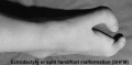
- Paper - An infrequent developmental abnormality of the foot (1928)
- Paper - Chondrification in the hands and feet of staged human embryos
- Paper - Congenital absence of ventrolateral abdominal musculature (1946)
- Paper - Congenital variation of the pectoral muscles, with report of a case (1915)
- Paper - Demonstration of the cartilaginous skeleton in mammalian fetuses
- Paper - Development and variation of the nerves and the musculature of the inferior extremity and of the neighboring regions of the trunk in man
- Paper - Development of the rectus abdominis and its sheath in the human fetus
- Paper - Development of the thoracic vertebrae in man (1905)
- Paper - Development of the ventral abdominal walls in man
- Paper - Developmental horizons in human embryos- A review of the histogenesis of cartilage and bone
- Paper - Difference in the ossification of the male and female skeleton (1928)
- Paper - Early development of the cervical vertebrae and the base of the occipital bone in man
- Paper - Extra digits and digital reductions
- Paper - Factors leading to the development of a joint between the manubrium and the mesosternum
- Paper - Fetal age assessment by centers of ossification
- Paper - Multiple malformations of the limbs (1928)
- Paper - Observations on the development of the human vertebral column
- Paper - Observations on the structure and development of bone
- Paper - On Ossification Centers in Human Embryos
- Paper - On the development and morphology of the human sphenoid bone
- Paper - On the mechanism controlling the growth in length of the long bones (1934)
- Paper - Pads on the palm and sole of the human foetus
- Paper - Principles of growth and repair in membrane bones (1946)
- Paper - Rare congenital malformation of hands and feet (1924)
- Paper - Roentgenographic observations of the times of appearance of epiphyses and their fusion with the diaphyses (1932)
- Paper - Some Gross Structural and Quantitative Aspects of the Developmental Anatomy of the Human Embryonic, Fetal and Circumnatal Skeleton
- Paper - Some rare muscular anomalies (1915)
- Paper - Studies of the development of the human skeleton (1905)
- Paper - The arrangement of the bursae in the superior extremities of the full-time foetus
- Paper - The development and ossification of the human clavicle
- Paper - The development of joints
- Paper - The development of muscle in the human foetus
- Paper - The development of nerve endings in the human foetus
- Paper - The development of synovial joints
- Paper - The development of the arm in man (1902)
- Paper - The development of the arteries of the human lower extremity
- Paper - The development of the human femoral artery, a correction
- Paper - The development of the limbs, body-wall and back
- Paper - The development of the limbs, body-wall and back (1901)
- Paper - The development of the patella
- Paper - The early development of the knee joint in staged human embryos
- Paper - The early development of the mammalian sternum
- Paper - The embryology of the human hip joint (1943)
- Paper - The epiphysis of the head of the femur (1915)
- Paper - The growth of the human foot
- Paper - The growth of the long bones in foetal life, as exemplified by a case of foetal syphilis (1929)
- Paper - The history of the earliest stages in the human clavicle
- Paper - The measurement of diaphysial growth in proximal and distal directions (1916)
- Paper - The ontogeny and phylogeny of the sternum (1919)
- Paper - The origin of the osteoblast and of the osteoclast (1913)
- Paper - The origin of the vertebrate limb (1912)
- Paper - The origin, growth, and fate of osteoclasts and their relation to bone resorption (1920)
- Paper - The proximo-distal sequence of origin of the parts of the chick wing and the role of the ectoderm
- Paper - The relation of the myotomes to the ventrolateral musculature and to the anterior limbs in amblystoma (1910)
- Paper - The relations of endogenous and exogenous factors in bone and tooth development (1937)
- Paper - The ribs in the second month of development (1912)
- Paper - The sexual differences of the fetal pelvis
- Paper - The sexual differences of the fetal pelvis (1899)
- Paper - The sternalis muscle in the anencephalous foetus (1936)
- Paper - The sternum - its early development and ossification in man and mammals
- Paper - Vertebral Regional Determination in Young Human Embryos
- Template:Pelvis
R
S
Media in category 'Musculoskeletal'
The following 200 files are in this category, out of 262 total.
(previous page) (next page) 226.jpg 565 × 705; 71 KB
 231.jpg 747 × 493; 69 KB
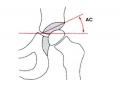 Acetabular angle.jpg 600 × 433; 22 KB
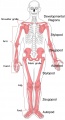 Appendicular skeleton developmental regions.jpg 1,000 × 1,849; 143 KB
 Appendicular skeleton small.jpg 220 × 407; 12 KB
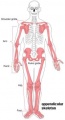
 Appendicular skeleton.jpg 1,000 × 1,849; 125 KB
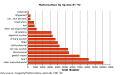 Australian abnormalities graph allsystem.png 509 × 320; 7 KB
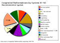 Australian abnormalities pie skmus.png 481 × 344; 9 KB
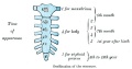
 Axial skeleton.jpg 1,000 × 1,434; 111 KB
 Bailey223.jpg 875 × 638; 108 KB
 Bailey224.jpg 823 × 883; 114 KB
 Bailey225.jpg 749 × 499; 100 KB
 Bailey226.jpg 565 × 705; 71 KB
 Bailey227.jpg 952 × 847; 226 KB
 Bailey228.jpg 907 × 832; 134 KB
 Bailey229.jpg 877 × 790; 86 KB
 Bailey230.jpg 894 × 918; 145 KB
 Bailey231.jpg 747 × 493; 69 KB
 Bailey232.jpg 753 × 491; 61 KB
 Bailey233.jpg 752 × 773; 106 KB
 Bailey234.jpg 741 × 717; 127 KB
 Bailey235.jpg 871 × 759; 119 KB
 Bailey236.jpg 962 × 675; 89 KB
 Bailey237.jpg 940 × 499; 62 KB
 Bailey238.jpg 786 × 330; 33 KB
 Bailey239 240.jpg 944 × 496; 128 KB
 Bailey239+240.jpg 944 × 496; 128 KB
 Bailey241.jpg 658 × 539; 80 KB
 Bailey242.jpg 434 × 611; 76 KB
 Bailey243.jpg 605 × 320; 70 KB
 Bailey298.jpg 342 × 713; 56 KB
 Bladder exstrophy - female.jpg 1,200 × 900; 112 KB
 Bladder exstrophy - male.jpg 637 × 480; 31 KB
 Bladder Exstrophy.jpg 600 × 383; 37 KB
 BMP syndactyly.jpg 417 × 1,073; 70 KB
 Bone structure cartoon.jpg 520 × 300; 46 KB
 Bone-femur.jpg 798 × 1,000; 150 KB
 Bone-structure.jpg 450 × 600; 27 KB
 Braune 1877 plate 30 fig1.jpg 805 × 1,200; 205 KB
 Caudal duplication syndrome.jpg 700 × 599; 47 KB
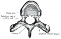
 Cervical vertebra.jpg 767 × 514; 71 KB
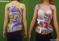 Cheneau brace.jpg 800 × 565; 79 KB
 Clefthand-apical-defect.jpg 500 × 568; 19 KB
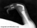 Congenital limb reduction xray.jpg 793 × 600; 18 KB
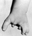
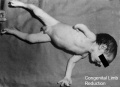 Congenital limb reduction.jpg 400 × 289; 11 KB
 Craniofrontonasal syndrome.jpg 1,280 × 543; 130 KB
 Developing joint.jpg 414 × 600; 33 KB
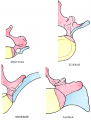
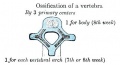
 Developing vertebra.jpg 558 × 428; 93 KB
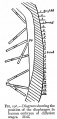
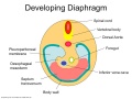 Diaphragm components.jpg 600 × 450; 41 KB
 Ectrodactyly 01.jpg 1,200 × 619; 113 KB
 Ectrodactyly.jpg 500 × 244; 9 KB
 Endochondral bone cartoon.jpg 946 × 513; 127 KB
 Endochondral bone.jpg 600 × 451; 89 KB
 Endochondral ossification 2.jpg 400 × 533; 99 KB
 Endochondral ossification.jpg 400 × 533; 91 KB
 Fawcett1930 fig01.jpg 800 × 669; 69 KB
 Femoral hernia repair.jpg 800 × 596; 52 KB
 Fetal 10wk urogenital 1.jpg 800 × 600; 109 KB
 Fetal 10wk urogenital 2.jpg 800 × 600; 110 KB
 Fetal 10wk urogenital 3.jpg 800 × 600; 107 KB
 Fetal 10wk urogenital 4.jpg 800 × 600; 105 KB
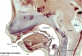
 Fetal head lateral.jpg 632 × 447; 34 KB
 Fetal head medial.jpg 632 × 447; 34 KB
 Fetal head section 01.jpg 1,200 × 821; 186 KB
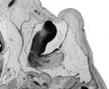
 Fetal head section.jpg 1,200 × 821; 167 KB
 Fetal week 10 hard palate 01.jpg 800 × 532; 77 KB
 Fetal week 10 hard palate 02.jpg 398 × 633; 66 KB
 Fetal week 10 hard palate 03.jpg 600 × 450; 122 KB
 Fetal week 10 hard palate 04.jpg 1,198 × 795; 196 KB
 Fetal week 10 hard palate 06.jpg 534 × 778; 88 KB
 Fetal week 10 hard palate 07.jpg 534 × 778; 97 KB
 Fetal week 10 palate 01.gif 534 × 778; 1.14 MB
 Fetal week 10 palate 01.mp4 ; 427 KB
 Fetal week 10 palate icon.jpg 534 × 778; 100 KB
 Fetal week 10 soft palate 01.jpg 571 × 784; 95 KB
 Fetal week 10 soft palate 02.jpg 534 × 778; 87 KB
 Fetal week 10 soft palate 03.jpg 534 × 778; 95 KB
 Fetal week 14 head bone lateral 01.jpg 1,000 × 773; 107 KB
 Fetal week 9 hard palate fusion 01.jpg 661 × 400; 51 KB
 Fetal week 9 head lateral 01.jpg 700 × 600; 78 KB
 Foster134.jpg 342 × 503; 37 KB
 Foster135.jpg 965 × 815; 230 KB
 Gray0065.jpg 600 × 345; 39 KB
 Gray0067.png 350 × 463; 46 KB
 Gray0082.jpg 450 × 287; 30 KB
 Gray0101.jpg 350 × 190; 17 KB
 Gray0118.jpg 480 × 252; 22 KB
 Gray0119.jpg 450 × 307; 22 KB
 Gray0379.jpg 713 × 600; 114 KB
 Gray0391.jpg 600 × 530; 79 KB
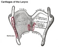
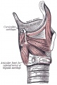
 Gray0950 arytenoid cartilage.jpg 600 × 500; 39 KB
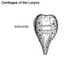
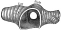
 Gray0950 cricoid cartilage.jpg 600 × 500; 42 KB
 Gray0950 epiglottis cartilage.jpg 600 × 500; 24 KB
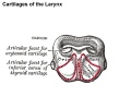
 Gray0950 thyroid cartilage.jpg 600 × 500; 54 KB
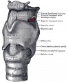
 Gray0950.jpg 500 × 1,000; 106 KB
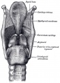
 Gray0951.jpg 600 × 724; 100 KB
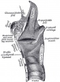
 Gray0952.jpg 588 × 800; 102 KB
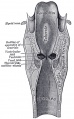
 Gray0953.jpg 588 × 800; 106 KB
 Gray0954.jpg 497 × 800; 94 KB
 Gray0955.jpg 616 × 600; 91 KB
 Gray0956.jpg 508 × 400; 49 KB
 Gray0957.jpg 421 × 700; 74 KB
 Gray0958.jpg 508 × 700; 85 KB
 Gray0959.jpg 471 × 700; 91 KB
 Gray0960.jpg 530 × 650; 99 KB
 Gray0961.jpg 600 × 769; 66 KB
 Gray0963.jpg 600 × 300; 48 KB
 Gray1028.jpg 800 × 960; 202 KB
 Head and heart muscle cartoon.jpg 874 × 800; 129 KB
 Hematopoietic and stromal cell differentiation.jpg 1,000 × 617; 107 KB
 Human embryonic shoulder girdle 01.jpg 1,000 × 726; 81 KB
 Human embryonic shoulder girdle 02.jpg 1,025 × 713; 109 KB
 Human embryonic shoulder girdle 04.jpg 1,000 × 755; 71 KB
 Human fetal temporal bone and mandible 01.jpg 1,200 × 805; 170 KB
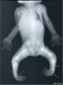
 Human fetus skeleton x-ray 01.jpg 662 × 910; 54 KB
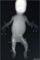
 Human fetus skeleton x-ray 02.jpg 686 × 928; 70 KB
 Human fetus skeleton x-ray 03.jpg 628 × 934; 81 KB
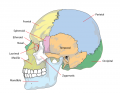 Human skull lateral simplified.png 740 × 576; 138 KB
 Human- fetal week 10 cerebellum A.jpg 347 × 284; 24 KB
 Human- fetal week 10 cerebellum B.jpg 347 × 284; 21 KB
 Human- fetal week 10 cerebellum C.jpg 347 × 284; 25 KB
 Human- fetal week 10 cerebellum D.jpg 347 × 284; 23 KB
 Human- fetal week 10 head A.jpg 600 × 544; 113 KB
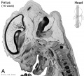 Human- fetal week 10 head A1.jpg 1,200 × 1,088; 159 KB
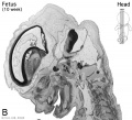 Human- fetal week 10 head B.jpg 600 × 544; 66 KB
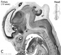 Human- fetal week 10 head C.jpg 600 × 544; 118 KB
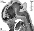 Human- fetal week 10 head D.jpg 600 × 544; 111 KB
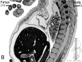
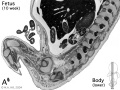 Human- fetal week 10 lower body A.jpg 600 × 450; 96 KB
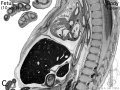
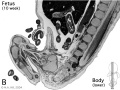 Human- fetal week 10 lower body B.jpg 600 × 450; 93 KB
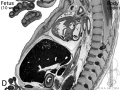
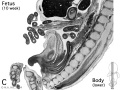 Human- fetal week 10 lower body C.jpg 600 × 450; 94 KB
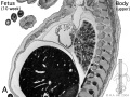 Human- fetal week 10 upper body A.jpg 600 × 450; 104 KB
 Human- fetal week 10 upper body B.jpg 600 × 450; 105 KB
 Human- fetal week 10 upper body C.jpg 600 × 450; 109 KB
 Human- fetal week 10 upper body D.jpg 600 × 450; 106 KB
 Human- fetal week 10 urogenital A.jpg 600 × 450; 109 KB
 Human- fetal week 10 urogenital B.jpg 600 × 450; 109 KB
 Human- fetal week 10 urogenital C.jpg 600 × 450; 105 KB
 Human- fetal week 10 urogenital D.jpg 600 × 450; 101 KB
 Inguinal hernia repair 2.jpg 800 × 718; 53 KB
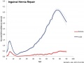 Inguinal hernia repair.jpg 800 × 597; 46 KB
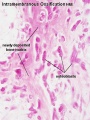 Intramembranous ossification centre.jpg 450 × 600; 69 KB
 Joint development 02.jpg 454 × 403; 22 KB
 Keibel Mall 231-232.jpg 627 × 919; 70 KB
 Keibel Mall 308.jpg 677 × 578; 43 KB
 Keibel Mall 309.jpg 687 × 479; 52 KB
 Keibel Mall 310.jpg 686 × 760; 161 KB
 Keibel Mall 311.jpg 700 × 906; 181 KB
 Keibel Mall 312.jpg 704 × 677; 79 KB
 Keibel Mall 313.jpg 702 × 652; 84 KB
 Keibel Mall 314.jpg 701 × 625; 89 KB
 Keibel Mall 315.jpg 736 × 433; 84 KB
 Keibel Mall 316.jpg 716 × 327; 57 KB
 Keibel Mall 317.jpg 688 × 376; 53 KB
 Keibel Mall 318.jpg 687 × 423; 79 KB
 Keibel Mall 319.jpg 300 × 352; 27 KB
 Keibel Mall 320.jpg 693 × 313; 33 KB
 Keibel Mall 321.jpg 746 × 651; 122 KB
 Keibel Mall 322.jpg 735 × 717; 133 KB
 Keibel Mall 323.jpg 700 × 211; 27 KB
 Keibel Mall 324.jpg 722 × 377; 47 KB
 Keith1902 fig112.jpg 600 × 541; 77 KB
 Keith1902 fig227.jpg 392 × 800; 36 KB
 Keith1902 fig228.jpg 429 × 800; 39 KB
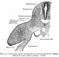
 Keith1902 fig229.jpg 1,000 × 434; 106 KB
 Keith1902 fig230.jpg 479 × 800; 60 KB
 Keith1902 fig231.jpg 586 × 800; 77 KB
 Keith1902 fig232.jpg 924 × 790; 117 KB
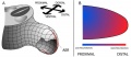
 Limb bud geometry and patterning.jpg 583 × 765; 76 KB
 Limb bud growth model 01.jpg 600 × 261; 30 KB
 Limb bud growth model 02.jpg 600 × 637; 80 KB
 Limb patterning factors 01.jpg 800 × 794; 76 KB
 Limb patterning factors 02.jpg 800 × 794; 73 KB
 Limb patterning factors 03.jpg 800 × 794; 36 KB
 Limb patterning factors 04.jpg 800 × 794; 67 KB
 Limb patterning factors 05.jpg 800 × 794; 37 KB
 Limb patterning factors 06.jpg 800 × 794; 41 KB
 Limb patterning factors 07.jpg 800 × 794; 44 KB
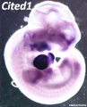 Mouse Cited1.jpg 446 × 550; 37 KB
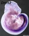 Mouse Dmrt2.jpg 446 × 550; 35 KB
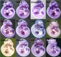
 Mouse E10.5 gene expression.jpg 1,747 × 1,650; 404 KB
 Mouse E11.5 gene expression.jpg 1,760 × 1,650; 396 KB
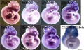 Mouse E12.5 gene expression.jpg 1,823 × 1,100; 272 KB
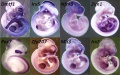 Mouse E13.5 gene expression.jpg 1,764 × 1,100; 266 KB
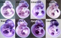 Mouse E9.5 gene expression.jpg 1,757 × 1,100; 269 KB
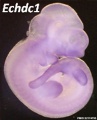 Mouse Echdc1.jpg 446 × 550; 29 KB
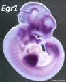 Mouse Egr1.jpg 446 × 550; 34 KB
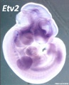 Mouse Etv2.jpg 446 × 550; 33 KB
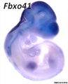 Mouse Fbxo41.jpg 446 × 550; 24 KB
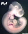 Mouse Figf.jpg 446 × 550; 25 KB
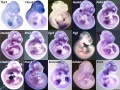 Mouse forelimb gene expression.jpg 2,205 × 1,650; 456 KB
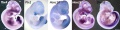 Mouse hindlimb gene expression.jpg 2,237 × 550; 147 KB
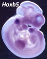 Mouse Hoxb5.jpg 446 × 550; 36 KB
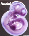 Mouse Hoxb6.jpg 446 × 550; 35 KB
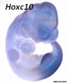 Mouse Hoxc10.jpg 446 × 550; 23 KB
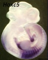 Mouse Hoxc5.jpg 446 × 550; 30 KB
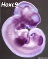 Mouse Hoxc9.jpg 446 × 550; 34 KB
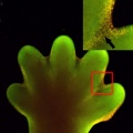 Mouse interdigit apoptosis 01.jpg 800 × 800; 81 KB
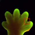 Mouse interdigit apoptosis 02.jpg 764 × 764; 61 KB
 Mouse limb bone development timeline.jpg 1,256 × 469; 107 KB
 Mouse limb gene expression 01.mov ; 1.12 MB


